
Abstract
We analyzed the localization of gold particles, expression of immunogenic protein, and histopathologic changes after vaccinating guinea pigs and mice with a DNA vaccine to the Ebola virus glycoprotein administered by cutaneous particle bombardment. Gold particles were deposited in all layers of the epidermis and in the dermis. Those in the epidermis were lost as the damaged layers sloughed, while those in the dermis were phagocytized by macrophages. Glycoprotein was demonstrated by immunohistochemistry primarily in keratinocytes in the epidermis and hair follicle epithelium and less frequently in dermal macrophages, fibroblasts, sebocytes, and cells that appeared to be Langerhans cells. The number of cells that expressed glycoprotein increased between 4 and 8 hours postvaccination, then decreased to near zero by 48 hours. The vaccine sites were histologically divisible into three zones. The central portion, zone 1, contained the most gold particles in the dermis and epidermis and had extensive tissue damage, including full-thickness epidermal necrosis. Zone 2 contained fewer gold particles in the epidermis and dermis and had less extensive necrosis. The majority of cells in which glycoprotein was expressed were in zone 2. Zone 3 contained gold particles only in the epidermis and had necrosis of only a few scattered cells. Regeneration of the epidermis in damaged areas was evident at 24 hours postvaccination and was essentially complete by day 5 in the mice and day 10 in the guinea pigs. Inflammatory changes were characterized by hemorrhage, edema, and infiltrates of neutrophils initially and by infiltrates of lymphocytes and macrophages at later times. In zone 1, inflammation affected both the epidermis and dermis. Peripherally, inflammation was relatively limited to the epidermis. CD3-positive dendritic epidermal cells were demonstrated in the epidermis and superficial hair follicles of unvaccinated immunocompetent mice and beige mice but not of SCID mice. These cells disappeared from all but the most peripheral portions of the vaccine sites of vaccinated mice within 24 hours. They reappeared slowly, failing to reach numbers comparable with unvaccinated mice by 35 days postvaccination. The epidermis of control guinea pigs also had CD3-positive cells, but they did not have dendrites. These findings should contribute to a better understanding of the mechanisms operating in response to DNA vaccination by particle bombardment.
In recent years, the development of in vivo DNA delivery systems has resulted in novel approaches to vaccination against infectious agents, cancer treatment, and pharmacologic therapy. 9 14 23 36 37 42 44 DNA vaccination involves the delivery of naked DNA encoding an immunogenic polypeptide against which an immune response is desired. Because the foreign DNA serves as a template for the manufacture of the immunogen within the host's own cells, DNA vaccines have properties that fundamentally differ from traditional vaccines. For instance, transfection of mammalian cells with DNA containing viral genes produces viral proteins with native conformation, glycosylation profiles, and other characteristics similar to those produced during natural infections. The result is an immune response that more closely mimics that which follows a natural infection, including both cell-mediated and humoral responses. 1 10 35 43
DNA vaccines can be administered by a variety of routes. 1 26 35 46 A novel method developed recently involves coating the foreign DNA onto microscopic gold beads that are delivered into cells in a variety of tissues, particularly skin, by particle bombardment using the so-called gene gun. 13 32 45 Within cells, the foreign DNA rapidly diffuses away from the gold particle. DNA transcription, translation of messenger RNA, and production of the immunogenic polypedtide are done by the host cell's machinery.
DNA vaccination by particle bombardment has potentially broad application in the prevention of infectious diseases, especially those caused by highly virulent agents. It makes use of neither attenuated pathogens, which may revert to virulence, nor inactivated ones, which can cause disease if they are incompletely inactivated. Further, it does not require handling potentially dangerous pathogens during vaccine production. The accessibility of the skin makes it ideal for DNA vaccination by particle bombardment. The presence of a large population of antigen-presenting cells such as Langerhans cells in the skin provides the basis for a robust immune response after cutaneous DNA vaccination. 32 Cutaneous DNA vaccination by particle bombardment can induce both humoral and cell-mediated immunity with much smaller amounts of DNA than are required for intramuscular injection. 1 47 Cutaneous DNA vaccination favors an IgG1-dominant Th2 immune response, whereas intramuscular vaccination favors an IgG2a-dominant Th1 response. 1 12 30 Presumably, particle bombardment may be safer than injecting DNA into tissues like muscle because exfoliation of the skin within a few days limits the potential for transfection of long-lived cells, which could conceivably lead to adverse reactions such as the induction of tumors or autoimmunity. 2 7 26 30 47 Particle bombardment has also been used to introduce DNA into other tissues, such as the cornea, urinary bladder, oral epithelium, and female reproductive tract. 15 24 38 39
Previous DNA vaccine studies and comparisons of particle bombardment to other delivery methods focused primarily on the molecular and immunologic aspects. The histomorphologic aspects, particularly those regarding cutaneous particle bombardment, have largely been overlooked. In this report, we describe the localization of gold particles, expression of immunogenic protein, and histopathologic changes after vaccinating guinea pigs and mice with a DNA vaccine to the Ebola virus glycoprotein administered by cutaneous particle bombardment. During the course of this study, we found that an antibody to the CD3 molecule labeled numerous dendritic cells in the epidermis of mice. Dendritic epidermal T cells, presumably the cells labeled by the CD3 antibody, may be highly significant with respect to cutaneous homeostasis and immunologic signaling mechanisms. 4–6, 17 18 27 29 However, relatively little is known about the function of these cells in normal and abnormal skin. We are not aware of any reports that describe the localization of CD3-positive dendritic epidermal cells in detail. Therefore, we include a brief description of these cells in both normal and DNA-vaccinated mice.
Materials and Methods
Experimental design
For the DNA vaccine portion of the study, 30 male and female inbred strain 13 guinea pigs (Cavia porcellus), 8 weeks old or older, were administered an Ebola virus envelope glycoprotein (GP) DNA vaccine by particle bombardment of the abdominal skin. Guinea pigs were vaccinated in two groups at different times. The first group consisted of 18 guinea pigs. Three each were killed at 16 (case Nos. 1–3), 24 (case Nos. 4–6), and 48 (case Nos. 7–9) hours and 5 (case Nos. 10–12), 10 (case Nos. 13–15), and 21 (case Nos. 16–18) days postvaccination (PV). Six additional guinea pigs were treated as negative controls and were killed one per timepoint per group (case Nos. 19–24). The second group contained 12 guinea pigs. Three each were killed at 4 (case Nos. 25–27), 8 (case Nos. 28–30), 16 (case Nos. 31–33), and 24 (case Nos. 34–36) hours PV. Three guinea pigs were treated as negative controls and were killed one each at 8, 16, and 24 hours (case Nos. 37–39; the negative control for the 4-hour group was accidentally omitted). Six- to 8-week-old female BALB/c mice (Mus musculus) were similarly vaccinated with the DNA vaccine and were killed two each at 1 (case Nos. 40 and 41), 5 (case Nos. 42 and 43), 10 (case Nos. 44 and 45), and 21 (case Nos. 46 and 47) days PV; only one mouse was vaccinated and then killed at 35 days (case No. 48) PV. Five mice were treated as negative controls and were killed one per timepoint (case Nos. 49–53). After vaccination, all animals were provided food and water ad libidum and were observed daily for evidence of discomfort or other effects of the vaccination procedure. Animals were killed by exposure to carbon dioxide gas in a closed chamber. Necropsies for the collection of tissues were performed immediately after death. Sections of skin and inguinal and axillary lymph nodes from DNA-vaccinated guinea pigs were examined histologically to determine the localization of vaccine particles, histopathologic tissue changes, and expression of Ebola virus GP. Sections of skin from vaccinated mice were analyzed for gold particle localization, histopathologic tissue changes, and changes in CD3-positive dendritic epidermal cells (CD3+ DEC).
For the portion of the study analyzing CD3+ DEC in the skin of normal mice, tissues of adult male and female mice submitted for quality assurance or serving as negative controls in unrelated experimental studies were used. These tissues included haired skin from multiple sites, the skin of the footpad, and lymph nodes of BALB/c (case Nos. 54–56), CD-1 (case Nos. 57–59), C3H/HeN (case Nos. 60–62), natural killer cell-deficient (beige; case Nos. 63 and 64), and severe combined immunodeficient (SCID; case Nos. 65 and 66) mice.
In conducting research using animals, the investigators adhered to the “Guide for the Care and Use of Laboratory Animals” prepared by the Committee on Care and Use of Laboratory Animals of the Institute of Laboratory Animal Resources, National Research Council (NIH Publication 86–23, revised 1996).
Vaccine preparation and administration
The Ebola virus DNA vaccine was prepared as previously described. 37 42 Briefly, the GP gene of the Zaire 76 strain of Ebola virus was amplified by reverse transcriptase-polymerase chain reaction (RT-PCR) and cloned into the BamHI site of pWRG7077. Plasmid DNA was precipitated onto the surface of gold beads at a concentration of 5 µg/mg of gold. Cartridges containing approximately 0.5 mg of gold were loaded into the gene gun apparatus (PowderJect Vaccines, Madison, WI). Animals were anesthetized using a mixture of 1 ml of acepromazine (10 mg/ml) and 10 ml ketamine (100 mg/ml) combined 2:1 with xylazine (20 mg/ml) administered at approximately 0.1 ml/150 gm body weight. The abdominal hair of guinea pigs and mice was removed with commercial hair clippers just before vaccination. Animals were vaccinated with the helium-powered gene gun using a pressure setting of 400 pounds per square inch. One to four nonoverlapping vaccinations were administered along the abdominal midline. As negative controls, guinea pigs were mock-vaccinated with gold beads coated with pWRG7077 lacking the Ebola virus insert and mice were administered gold beads coated with pWRG7077 containing an Ebola virus nucleoprotein gene insert. 42
Pathology
Samples of skin were taken from the DNA vaccine sites and the abdominal flank (normal control) of guinea pigs and mice. Skin samples and samples of the inguinal and axillary lymph nodes of vaccinated animals and nonvaccinated mice were fixed in 10% neutral buffered formalin, processed routinely, and embedded in paraffin blocks. The skin samples from the vaccine sites were hemisectioned through the center of the sites, and both of these sections were embedded face down so that the initial histologic sections included nearly identical samples through the greatest diameter of each vaccine site. Histologic sections were cut at 5 µm, mounted on positively charged glass slides (Superfrost Plus, Fisher Scientific, Pittsburgh, PA), and stained with hematoxylin and eosin (HE).
Serial tissue sections of vaccinated guinea pigs were analyzed by immunohistochemistry for Ebola virus GP expression using a monoclonal antibody directed against Ebola virus GP (M-DA01-A5-B11; obtained under contract from The Centers for Disease Control and Prevention, Atlanta, GA). Tissue sections were deparaffinized, rehydrated, and incubated with proteinase K (DAKO Corporation, Carpinteria, CA) for 6 minutes at room temperature. Immunohistochemistry was performed using a novel immunoperoxidase (IPO)-based, biotinylated-tyramide amplification procedure we developed. For this method, the primary antibodies were diluted 1:2,500 and biotinylated prior to tissue incubation using biotin-labeled anti-mouse immunoglobulin, monovalent Fab fragments (ARK System, DAKO Corporation) according to the manufacturer's instructions. After incubation with the biotinylated primary antibodies, tissue sections were sequentially incubated at room temperature with commercially prepared streptavidin-biotin complex, biotin-labeled tyramide in H2O2 with peroxidase and peroxidase-labeled streptavidin; immunolabeling was completed by immersion in substrate-chromogen solution containing 3,3′-diaminobenzidine with H2O2 (all reagents from CSA System, DAKO Corporation; used according to the manufacturer's instructions). Sections of Ebola virus-infected nonhuman primate tissues served as positive controls.
Skin sections of vaccinated mice and normal mice were analyzed by immunohistochemistry for CD3 using a different IPO method (Envision System, DAKO Corporation). After deparaffinization and proteinase K pretreatment, sections were incubated with rabbit polyclonal antibody to the human CD3 molecule (DAKO Corporation), diluted 1:1,000, for 30 minutes at room temperature. The remainder of the procedure was accomplished according to the manufacturer's instructions. Sections treated with normal rabbit serum served as negative controls. After immunohistochemistry, the tissue sections were counterstained with hematoxylin. Immunohistochemistry for the mouse Thy-1 and T-cell receptor molecules (additional antigens expressed by dendritic epidermal T-cells), 40 as well as the mouse MHC class II molecule (Langerhans cells), was unsuccessful in the formalin-fixed tissues (data not shown).
Results
Gross findings
The gene gun apparatus deposited gold particles in round foci approximately 1 cm in diameter in the skin of guinea pigs (Fig. 1). Erythema developed within a few minutes of vaccination, but it largely dissipated by 24 hours postvaccination (PV). At 48 hours PV, there was a 2–4-mm blanched ring along the periphery of most vaccine sites, and at day 5 PV, the vaccine sites were covered by crusts. The crusts were still loosely adhered at day 10 PV, but by day 21 PV, they had completely sloughed, leaving a pale tan discoloration at the vaccine sites.
Skin; guinea pig; case No. 5. DNA-vaccine sites along the abdominal midline within minutes of particle bombardment. Bar = 0.66 cm.
Histopathologic findings in vaccinated animals
Histologically, the vaccine sites of guinea pigs were divisible into three zones. The central portion, zone 1, contained the most gold particles in the epidermis and dermis and there was extensive tissue damage, including full-thickness epidermal necrosis at early timepoints (Fig. 2). The middle portion, zone 2, contained fewer gold particles in the epidermis and superficial dermis. It had epidermal necrosis that did not affect the full thickness of this layer. The outer portion, zone 3, had a few gold particles in the epidermis only. Necrosis in zone 3 affected only isolated, individual epidermal cells. Along the outermost margins of zone 3, gold particles were present only in the superficial layers of the stratum corneum but did not appear to penetrate into the viable epidermis. Microscopically, gold particles were irregularly round, measured approximately 2 µm in greatest dimension, and were surrounded by an anisotropic halo when viewed under polarized light.
Skin; guinea pig; case No. 4. Full-thickness necrosis of the epidermis (arrows) in the center of a vaccine site (zone 1) at 24 hours postvaccination (PV). HE. Bar = 20 µm.
Gold particles in the epidermis were within keratinocytes and in a few cells of the stratum spinosum with clear cytoplasm and reniform nuclei, features typical of Langerhans cells. Gold particles were also present in the superficial dermis, hair follicles, and sebaceous glands. Zone 1 contained the most deeply deposited particles, extending into the middermis. Those deposited in the outer portions of the vaccine sites were progressively more superficial.
Gold particles in the epidermis of the vaccine sites of guinea pigs were cleared as the damaged layers sloughed. The large majority of gold particles were cleared from the epidermis by day 10 PV. Those in the dermis were engulfed by macrophages. By 24 hours PV, one or more particles were present in dermal macrophages. Later, the number of particles in individual macrophages increased. Individual dermal macrophages often contained 10 or more gold particles from day 5 PV onward. In several axillary and inguinal lymph nodes of the guinea pigs killed at 16 hours PV, a few gold particles were free in the subcapsular sinuses. At 24 hours PV and later timepoints, one to three gold particles were present within the cytoplasm of a few histiocytes in the subcapsular and paracortical sinuses of these lymph nodes.
The most severe histologic skin changes were in the centers of the vaccine sites. Full-thickness epidermal necrosis, disruption of the basement membrane, and fragmentation of collagen bundles in the superficial dermis characterized zone 1 of the vaccine sites of guinea pigs at 4–16 hours PV. In zone 2, the epidermis had less extensive necrosis as well as intracellular and intercellular edema and suprabasilar or dermo-epidermal clefts. By 4 hours PV, these areas were infiltrated by numerous neutrophils and some macrophages and also contained some erythrocytes. There was also neutrophilic exocytosis of zone 1. Similar changes often extended into the epithelium of the superficial hair follicles. The superficial dermis in zone 1 was edematous and contained hemorrhage, necrotic debris, and infiltrates of neutrophils and macrophages. Milder dermal changes were present in zone 2. The only change evident in the deep dermis of guinea pigs was infiltration by a few neutrophils and macrophages.
The most peripheral skin change was necrosis of single cells in the epidermis (Fig. 3). Affected cells, which frequently contained gold particles, were swollen and had condensed, hypereosinophilic cytoplasm and hyperchromatic nuclei. Adjacent cells that did not contain gold particles were morphologically normal. Similar necrotic cells were also present in Huxley's layer of the internal root sheath of hair follicles in more central portions of the vaccine sites. In the skin beyond zone 3, histologic changes were not evident.
Skin; guinea pig; case No. 2. Necrosis of individual cells (arrows) in the epidermis of the peripheral portion of a vaccine site (zone 3) at 16 hours PV. Note the gold particles within the cytoplasm of the affected cells. HE. Bar = 25 µm.
At 24 hours PV, the epidermis of the vaccine sites of guinea pigs exhibited early regeneration. By 48 hours PV, the basement membrane in zone 1 was almost completely covered by hyperplastic epidermis composed of three or more layers of basophilic polygonal cells with scattered mitotic figures. Overlying the hyperplastic epidermis was a layer of necrotic epidermis containing cellular and keratinous debris, degenerate inflammatory cells, and numerous gold particles. In the dermis, more mononuclear cells, including some lymphoid cells, were present, with fewer neutrophils than previously observed. There was also less necrotic debris and only minimal residual hemorrhage in the dermis at 48 hours PV. The epidermis remained hyperplastic and contained a stratum granulosum by day 5 PV (Fig. 4) and a stratum corneum by day 10 PV. A thin layer of necrotic epidermis containing gold particles still loosely adhered at this time. Some mononuclear cells continued to infiltrate the superficial dermis. By day 21 PV, the necrotic layer had sloughed and the epidermis remained slightly thickened. Lymphoid cells infrequently infiltrated the basal layer of epidermis, and a few lymphoid cells and macrophages remained in the dermis.
Skin; guinea pig; case No. 10. Regeneration of the epidermis, including a stratum granulosum, in zone 1 of a vaccine site at day 5 PV. The sloughing epidermal layer contains numerous gold particles. Note the phagocytized gold particles in dermal macrophages (arrow). HE. Bar = 40 µm.
Histologic changes in the lymph nodes draining the vaccine sites of guinea pigs were mild. At 16 and 24 hours PV, the cortical and paracortical sinuses of draining lymph nodes contained erythrocytes admixed with an increased number of neutrophils and macrophages. The number of neutrophils and macrophages was increased through day 5 PV. Otherwise, except for the presence of gold particles, histologic changes were not evident in the draining lymph nodes at later times.
The vaccine sites of mice were divisible into essentially the same three zones as those in the guinea pigs. In the mice, gold particles were deposited into the deepest portion of the dermis in zone 1. Nearly all of the gold particles were cleared from the epidermis of mice by day 5 PV. Those in the dermis were phagocytized by macrophages, as in the guinea pigs. The histopathologic changes in the vaccine sites of mice were qualitatively similar to those in the guinea pigs at the same timepoints. At 24 hours PI, though, there was more extensive epidermal necrosis and the deep dermis of mice contained more abundant cellular infiltrates. Early regeneration of the epidermis was somewhat more prominent at 24 hours PV in the skin of the mice as well, despite the necrosis. By day 5 PV, the damaged layer of epidermis had already sloughed in all mice and there was regeneration and differentiation of all layers of the epidermis, including a stratum corneum. In zone 1 of mice at day 10 PV, the epidermis remained hyperplastic and inflammatory cell infiltrates persisted in the dermis. These changes diminished at later timepoints.
Ebola virus glycoprotein expression
Immunohistochemistry demonstrated Ebola virus glycoprotein (GP) in the skin of guinea pigs as early as 4 hours PV. The number of cells that expressed GP was greater at 8 hours PV but decreased at later timepoints. GP was detected in very few cells by 48 hours PV. The large majority of immunolabeled cells were keratinocytes of the epidermis and superficial hair follicles (Fig. 5). Rarely, sebocytes and dermal cells that appeared to be either macrophages or fibroblasts also expressed GP. A few immunolabeled cells in the epidermis had clear perinuclear cytoplasm (Fig. 6), suggestive of Langerhans cells. About one third to one half of the different types of immunolabeled cells contained a cytoplasmic gold particle. The majority of cells that expressed GP were in zone 2 of the vaccine sites. In zone 1, cells that expressed GP were found in the superficial hair follicles or, rarely, in the dermis but were not found in the epidermis. Immunolabeled cells were not observed in the portions of skin beyond the vaccine sites nor in any sections of lymph nodes or sections of normal skin. The timecourse during which Ebola GP expression occurred in the skin of guinea pigs is summarized in Fig. 7.
Skin; guinea pig; case No. 35. Ebola virus glycoprotein (GP) is expressed in hair follicle epithelial cells (arrows). Immunoperoxidase (IPO) labeling with a mouse monoclonal antibody, hematoxylin counterstain. Bar = 25 µm.
Skin; guinea pig; case No. 35. Ebola virus GP is expressed in an epidermal cell (arrow) with features suggestive of a Langerhans cell. IPO labeling, hematoxylin counterstain. Bar = 20 µm.
The expression of Ebola virus glycoprotein in the skin of guinea pigs determined by immunohistochemistry using a tyramide-amplified immunoperoxidase method. For each timepoint, the mean number of positive cells per histologic section through the greatest diameter of vaccine sites per guinea pig was determined. Entries for 4 and 8 hours were from three guinea pigs each in experiment 2; entries for 48 hours, 5, 10, and 21 days were from three guinea pigs each in experiment 1; entries for 16 and 24 hour included three guinea pigs in experiment 1 and three guinea pigs in experiment 2. The actual mean and range (in parentheses) for each timepoint were 4 hours, 4.83 (3.0–8.0); 8 hours, 8.66 (4.0–11.0); 16 hours, 5.75 (1.5–15.0); 24 hours, 1.83 (0–4.0); 48 hours, 0.33 (0–0.5); 5 days, 0.166 (0–0.166); 10 days, 0; 21 days, 0.
Control animals
The gross and histopathologic changes and gold particle localization in the skin and lymph nodes of negative control animals were identical to those in the vaccinated animals. The only difference was that Ebola virus GP immunolabeling was not evident in the vaccine sites of negative control guinea pigs.
CD3-positive dendritic epidermal cells (CD3+ DEC) in the normal skin of mice
The specificity of the CD3 antibody was demonstrated by its immunohistochemical staining pattern in sections of lymph nodes. This was manifest by strong staining of compact lymphoid cells in the paracortex and medullary cords (T-cell-dependent areas) and staining of scattered lymphoid cells in sinuses and B-cell-dependent areas (Fig. 8). Plasmacytoid, histiocytic, and granulocytic cells were not labeled. In the lymph nodes of SCID mice, very few scattered CD3-positive cells were present (Fig. 9).
Lymph node; BALB/c mouse; case No. 55. CD3 is expressed heavily in T-cell dependent areas (∗) but only in a few scattered cells of B-cell dependent areas. IPO labeling with a rabbit polyclonal antibody, hematoxylin counterstain. Bar = 342 µm.
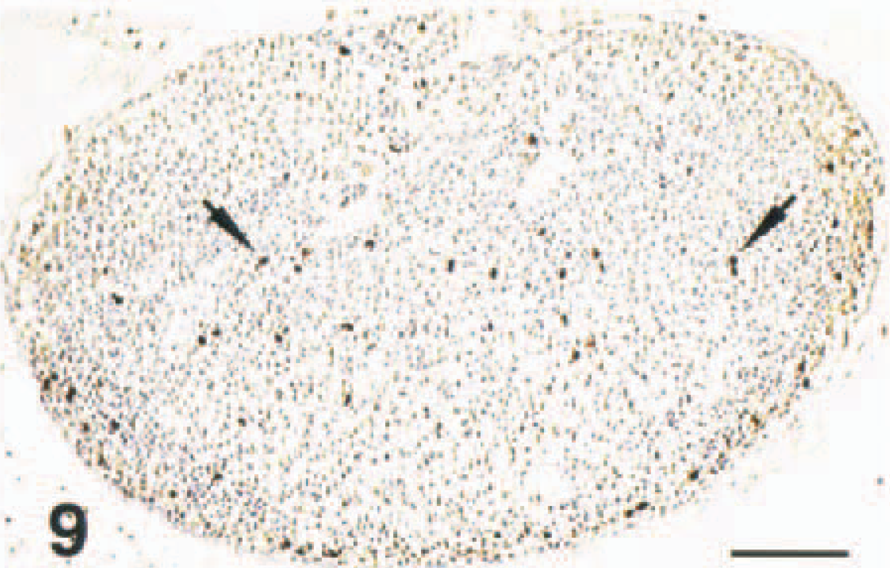
Lymph node; SCID mouse; case No. 66. CD3 is expressed in rare, scattered cells (arrows). IPO labeling, hematoxylin counterstain. Bar = 136 µm.
In the sections of haired skin of immunocompetent mice, CD3-positive cells were scattered throughout the dermis and epidermis. Those in the dermis were generally compact, round, and had scant cytoplasm. In the epidermis, the majority of CD3-positive cells were larger and had prominent dendrites (Fig. 10). The bodies of these cells usually were in the basilar or suprabasilar region of the epidermis.
Skin; BALB/c mouse; case No. 54. CD3-positive dendritic epidermal cell (CD3+ DEC) has multiple, branching dendrites. IPO labeling, hematoxylin counterstain. Bar = 8 µm.
Many CD3+ DEC had four or more dendrites that extended laterally and superficially and coursed in between other epidermal cells. The dendrites occasionally branched and measured up to 50 µm or greater in length. They often projected up to the border of the stratum corneum but appeared to enter it minimally or not at all. The nuclei of CD3+ DEC were typically oval and had dispersed chromatin. Some nuclei were indented. A few smaller, compact CD3-positive cells without dendrites were also scattered throughout the basilar portions of the epidermis. These cells were similar to the CD3-positive cells, i.e., lymphocytes, found in the dermis.
CD3-positive dendritic cells were also present in the basal epithelium of hair follicles. The dendrites of these cells extended inward toward the hair shafts. They were more numerous in the superficial follicles than in the follicles of the middermis. Hair follicles of the deep dermis and panniculus adiposus, i.e., those with prominent inner and outer root sheaths, did not contain any CD3-positive cells. Overall, CD3+ DEC were somewhat more numerous in the superficial hair follicles and the epidermis contiguous to them (Fig. 11) than in the interfollicular regions of the epidermis. CD3-positive cells were also present along the periphery of sebaceous glands, but these generally resembled those of the dermis and seldom had dendrites.
Skin; BALB/c mouse; case No. 54. CD3+ DECs are more numerous in the superficial hair follicles and contiguous epidermis (arrows) than in the intervening epidermis (arrowheads). IPO labeling, hematoxylin counterstain. Bar = 100 µm.
CD3+ DEC were present in the skin of both male and female mice and in all strains of mice used in the study except the SCID mice. There were no obvious differences in the numbers of nor the locations of CD3+ DEC between males and females, between immunocompetent mouse strains or between the different anatomic locations, which included haired skin of the inner forearm, outer thigh, dorsal midline, and pinna. In the haired skin of the SCID mice, there were a few, widely scattered compact CD3-positive cells in the hair follicles and dermis and very rarely in the epidermis. These cells did not have dendrites. CD3-positive dendritic cells were not observed in the nonhaired skin of the footpads of any mice.
CD3+ DEC in vaccinated mice
The sections of skin from vaccinated mice contained normal regions bordering the vaccine sites with appropriate numbers of CD3+ DEC, serving as internal controls. In the centers of the vaccine sites (zone 1), no CD3-positive cells were present within the epidermis at day 1 PV. At days 5 and 10 PV (Fig. 12), rare CD3-positive cells were present in the basal layers of hyperplastic epidermis, but none had dendritic processes. A few CD3-positive cells with dendritic processes were present in the central epidermis of vaccine sites at day 21 PV, and there was further increase in these cells at day 35 PV (Fig. 13); however, fewer CD3+ DEC were present at the later timepoint than in normal skin. In zone 2, there were decreased CD3-positive epidermal cells at day 1 PV, and all of those present lacked dendritic processes. The outer portions of zone 2 contained a few CD3+ DEC at day 5 PV, but there were none in the inner hyperplastic portions. There were progressively more CD3+ DEC in the inner portions of zone 2 as well between days 10 (Fig. 14) and 21 PV (Fig. 15), which correlated with a concurrent decrease in the thickness of the epidermis in this location. By day 35 PV, zone 2 appeared to have relatively normal numbers of CD3+ DEC. In zone 3, the numbers of CD3+ DEC were moderately decreased at day 1 PV; however, they had recovered to near normal by day 10 PV. The numbers of CD3+ DEC in zone 3 remained at normal levels at days 21 and 35 PV.
Skin; DNA-vaccinated mouse; case No. 44. CD3+ DECs are absent from the hyperplastic central epidermis at day 10 PV. Note the degree of dermal inflammation, including infiltrating CD3+ lymphocytes (arrows). IPO labeling, hematoxylin counterstain. Bar = 36 µm.
Skin; DNA-vaccinated mouse; case No. 48. A single CD3+ DEC (arrow) is present in the central epidermis at day 35 PV. IPO labeling, hematoxylin counterstain. Bar = 36 µm.
Skin; DNA-vaccinated mouse; case No. 44. CD3+ DECs are scarce in the slightly thickened epidermis (arrow) of zone 2 at day 10 PV. IPO labeling, hematoxylin counterstain. Bar = 36 µm.
Skin; DNA-vaccinated mouse; case No. 47. The epidermis of zone 2 contains essentially normal numbers of CD3+ DECs (arrow) at day 21 PV. IPO labeling, hematoxylin counterstain. Bar = 36 µm.
The numbers of CD3+ DEC in hair follicles followed the general trends seen in the epidermis. In effect, the loss of these cells at early timepoints was more dramatic centrally than peripherally and was more severe in superficial follicles than in the middle portions of follicles. Likewise, the reappearance of CD3+ DEC occurred progressively from the peripheral to the central zones of the vaccine sites and from the middle to the superfical portions of follicles.
Discussion
In this study, DNA encoding the glycoprotein (GP) gene of Ebola virus Zaire was delivered to the skin of guinea pigs and mice by particle bombardment. This method of DNA immunization was previously shown to induce an immune response against Ebola virus capable of protecting mice against challenge with a lethal dose of the virus. 42 Numerous previous studies described many of the molecular, immunologic, and pharmacologic aspects of DNA administration by particle bombardment. In this report, we focus on the histomorphologic aspects of DNA administration by this method. Mechanisms manifest at the histologic level could have important consequences regarding the expression of immunogenic or pharmacologic proteins and the body's response to them. A particularly unique aspect we describe is the effect of particle bombardment on dendritic epidermal cells of mice that express the CD3 molecule (CD3+ DEC). These cells are likely dendritic epidermal T cells, a previously described but poorly understood cell type.
With a delivery pressure of 400 pounds per square inch, gold particles were deposited in the epidermis, dermis, and adnexa of guinea pigs and mice. Gold particles deposited more deeply in the central portions of vaccine sites (zone 1) than in outlying regions (zones 2 and 3, respectively). Gold particles delivered to the mice deposited more deeply in the dermis in zone 1 than those in the guinea pigs. This finding is not surprising since the skin of mice is thinner. Another factor that could influence the depth of particle delivery is the process of hair removal since intact hairs would impede the penetration of gold particles into the skin. All animals in this study were shaved just before particle delivery with commercial hair clippers.
Because DNA immunization via particle bombardment depends on the transfection of viable cells to manufacture the immunogenic protein, both the location of deposited gold particles and the amount of resulting tissue damage directly affect protein expression. Most of the cells we identified that expressed GP were in the epidermis of zone 2. The epidermis of zone 1 actually contained more gold particles than zone 2, but the full thickness necrosis in this location effectively negated GP production. There was, however, some GP expression in the hair follicle epithelium of zone 1. Zone 3 had the least epidermal necrosis but also had few gold particles. Particles deposited in the relatively acellular dermis had little effect. Only rarely did we observe dermal macrophages or fibroblasts that expressed Ebola virus GP. Our findings indicate that the effectiveness of immunogenic protein expression was therefore partially determined by a balance between gold particle delivery into the epidermis and the amount of epidermal necrosis that results.
The specific types of cells that produce the immunogenic protein might also influence the immune response. In this study, keratinocytes of the epidermis and hair follicles were the primary cell type in which Ebola virus GP was demonstrated, consistent with previous reports. 25 32 33 Other cell types identified morphologically in which GP was infrequently demonstrated included dermal macrophages, fibroblasts, and sebocytes. A few GP-positive epidermal cells had features suggestive of Langerhans cells, but the identity of these cells could not be definitively determined. Langerhans cells, the major antigen-presenting cells of the skin, have been shown to be central to the immune response after DNA vaccination of the skin. 8 32 34 35 They may participate by processing antigen produced in other cells, i.e., keratinocytes, or they may be directly transfected and generate antigen themselves. Langerhans cells present antigen to the immune system by traveling from the skin to the draining lymph nodes or, to a lesser extent, the spleen. We did not identify dendritic cells or other types of cells that expressed Ebola virus GP in the draining lymph nodes. This is not surprising since it was previously shown that lymph node cells expressing DNA-encoded antigen following gene gun vaccination are rare. 32
The duration of protein expression in transfected cells is another factor that could influence the immune response to cutaneous DNA vaccines. It was previously reported that protein expression in mice administered DNA in the skin ceased due to sloughing of the epidermis by 1 week. 7 In this study, however, the number of cells expressing Ebola virus GP in guinea pigs declined to relatively low levels within hours of vaccine delivery, prior to sloughing of the epidermis. The number of GP-expressing cells demonstrated by immunohistochemistry increased between 4 hours postvaccination (PV), the earliest timepoint analyzed, and 8 hours PV. Then the number of GP-expressing cells declined rapidly, reaching nearly zero by 48 hours PV. However, we did not quantify GP production, so the possibility that low-level GP expression that could not be demonstrated by immunohistochemistry occurred after 48 hours PV cannot be ruled out. Also, it is possible that some degradation of GP might have occurred, resulting in loss of the portion of the protein recognized by the monoclonal antibody we used. Nonetheless, our findings indicate that mechanisms other than sloughing of transfected cells may be important in determining the duration of protein expression. For instance, sublethal injury from the penetration of gold particles into cells could modify the protein production machinery and permit only transient expression of protein from the foreign DNA template. Also, the immunogenic protein itself could conceivably alter the cells that produce it. Although we did not observe any histologic differences between the skin of mice or guinea pigs vaccinated with Ebola GP and the skin of negative control animals, Ebola virus GP is thought to have toxic properties 11 and could alter protein expression in the transfected cells that produce it. A third possibility is that different promoters or other gene regulatory elements may affect the duration of immunogenic protein production and the immune response to DNA vaccines. 21 28
A detailed account of the histologic changes in the skin after particle bombardment has not previously been reported, to our knowledge. The most significant histologic change in this study was the presence of full-thickness epidermal necrosis resulting from the impact of numerous gold particles in zone 1 of the vaccine sites of guinea pigs and mice. The impact of fewer gold particles in the epidermis peripherally had milder effects. Some cells containing gold particles had no obvious histologic changes. The demonstration of Ebola virus GP in cells with particles confirmed their viability. Alternatively, other cells with a single gold particle were clearly necrotic. These cells often exhibited very eosinophilic cytoplasm, possibly due to coagulation of cytoplasmic proteins from the heat generated by particle impact. The extensive epidermal necrosis in the central portion of vaccine sites effectively prevented the production of immunogenic protein in this area.
Factors that may determine the amount of necrosis of the critical epidermal layer after particle bombardment include the size and number of gold particles used and the force with which they are applied. A previous study of DNA immunization by particle bombardment found an inverse correlation between the depth of particle penetration and the amount of antigen produced plus the antibody response that occurred. 10 That study provided a very limited histologic description. Our results confirm that particle penetration into the dermis correlates with more extensive necrosis of the overlying epidermis and a lack of protein expression in necrotic foci.
Regeneration of damaged epidermis occurred quickly, beginning within 24 hours of vaccination in both the guinea pigs and mice. Renewal of all layers of the epidermis was present by day 5 PV in the mice and by day 10 PV in the guinea pigs. Inflammation developed soon after vaccination as well. Initially, the inflammation was characterized by hemorrhage, edema, and infiltrates of mainly neutrophils. Later, infiltration by greater numbers of macrophages and lymphocytes occurred. These infiltrating cells may contribute to enhanced immunogenicity. Some infiltrates of mononuclear cells persisted as late as 21 days PV. The macrophages were particularly prominent, as they were often engorged with phagocytized gold particles. Whether these macrophages might have abnormal functional abilities is not known. It is conceivable, however, that alterations in functions such as mobility and the ability to serve as antigen-presenting cells could affect the immune response to booster vaccinations if the same skin sites are used.
The rapid loss of CD3+ DEC from the epidermis of mice after DNA vaccination and the slow recovery of these cells could have important implications regarding the host response to cutaneous DNA administration by particle bombardment. The adult murine epidermis contains a population of dendritic epidermal T cells (DETC) in addition to the dendritic Langerhans cells. DETC develop in the fetal thymus and migrate to the skin. 16 22 31 They express the T-cell receptor (TCR) V-gamma 3+/V-delta 1+ in association with the CD3 molecule, 19 20 but do not express the CD4 and CD8 molecules nor MHC class II antigens. 3 40 41 In contrast, Langerhans cells are of bone marrow origin and express MHC class II antigens but do not express the CD3 molecule. The functions of DETC are not well understood, but they appear to perform a surveillance role in the skin in addition to mediating effector functions through the production of immune-associated soluble factors and a keratinocyte growth factor. 4 6 17–20, 27 29 Because demonstration of the TCR is required as proof of T-cell origin, we make a distinction here between the CD3+ DEC we demonstrated and the previously described DETC. We consider it likely, however, that they are the same cells. This assertion is supported by our finding that CD3+ DEC were present in immunocompetent mice and beige mice, which lack natural killer cells but not T cells, but were not present in SCID mice, which lack both T cells and B cells.
In unvaccinated mice, CD3+ DEC were present in the epidermis and the epithelium of superficial hair follicles. Immunolabeling demonstrated long, branching dendrites capable of contacting numerous neighboring cells. These features appear consistent with roles involving surveillance and intercellular communication that have previously been ascribed to DETC. We also identified CD3-positive epidermal cells in the skin of normal guinea pigs (data not shown); however, they did not have prominent dendrites. Because morphologically it was unclear if these cells represented the same type of cell as the CD3+ DEC in mice, we did not attempt to assess changes in CD3-positive epidermal cells in vaccinated guinea pigs.
Soon after vaccination, CD3+ DEC disappeared from the epidermis and superficial hair follicles of all but the most peripheral portions of the vaccine sites of mice. The numbers of CD3+ DEC gradually returned to normal peripherally. However, there were still reduced numbers of CD3+ DEC in the central portions of vaccine sites at 35 days PV, the latest timepoint examined, even though the epidermis in this location appeared otherwise to have recovered its normal histologic appearance. Therefore, CD3+ DEC appear to recover slowly after severe damage to the epidermis.
It is known that DETC can be induced to manufacture chemokines such as MIP-1a, MIP-1β, RANTES, and lymphotactin, 4 suggesting they could have played a role in mediating the inflammatory cell infiltrates that developed after vaccination. However, we found that cellular infiltrates were more prominent in the heavily damaged areas where CD3+ DEC were lost rather than in the peripheral locations where some CD3+ DEC remained. This indicates that CD3+ DEC probably played a limited role, if any, in mediating the inflammation caused by vaccination. DETC are also known to produce a keratinocyte growth factor, indicating a possible role in cutaneous wound repair. 5 Yet CD3+ DEC were rapidly lost from the damaged portions of epidermis and did not reappear until after the epidermis had largely healed, which indicates they did not play a prominent role in wound repair in the mice of this study.
As with traditional vaccines, DNA vaccines often require booster vaccinations to achieve greater protective immunity. 21 37 42 Depending on the timing of booster vaccinations, lingering effects of previous vaccinations could influence subsequent immune responses if the same sites in the skin are utilized. The lingering effects of a single DNA vaccination demonstrated in this study included epidermal hyperplasia, reduced numbers of CD3+ DEC, and gold particle-laden dermal macrophages. Since relatively little is known about the function of CD3+ DEC, in particular, it is difficult to speculate on what effect decreased numbers of these cells could have on same-site booster vaccinations. Perhaps more importantly, this finding raises the question of what effect particle bombardment may have on Langerhans cells. Because our attempt at immunolabeling Langerhans cells by immunohistochemistry was unsuccessful, we were unable to assess changes in these cells in this study. We are not aware of any previous studies that studied changes in Langerhans cells over time after DNA vaccination of the skin. The effects of particle bombardment on both Langerhans cells and DETC in the skin of rodents given cutaneous DNA vaccines require further study. Finally, the use of immunohistochemistry to label CD3+ DEC offers a simple method that could be useful in assessing changes in DETC in a variety of other cutaneous conditions of rodents.
Footnotes
Acknowledgements
We thank Dr. Connie Schmaljohn and Dr. Al Ruff for vaccinating mice, Dr. Randal Schoepp and Dr. Dana Scott for critical review of the manuscript, and David Custer, Rosie Moxley, Jeff Brubaker, Lynda Miller, Al Thomas, Steve Moon, Anne Castillo, Larry Ostby, and Alan Owens for expert technical assistance. The views, opinions, and/or findings contained herein are those of the authors and should not be construed as official Department of the Army position, policy, or decision unless so designated by other documentation.