
Abstract
Twenty feline vaccine-associated sarcomas were examined by transmission electron microscopy. Tumors contained pleomorphic spindle cells, histiocytoid cells, and giant cells. Most tumors contained myofibroblasts, which had morphologic features similar to those of fibroblasts. These cells were further distinguished by subplasmalemmal dense plaques and thin cytoplasmic actin myofilaments organized as elongated bundles concentrated at irregular intervals forming characteristic dense bodies. Intracellular crystalline particulate material was found in 5 of the 20 tumors. Energy dispersive X-ray spectroscopy was used to identify the crystalline material within one tumor as aluminum-based. One tumor from a feline leukemia virus-infected cat contained budding and immature retroviral particles.
Vaccine-associated sarcomas were first recognized in cats in the early 1990s. 8 Their appearance as distinct lesions appeared to have coincided with increased rabies vaccination frequency and the introduction of the subcutaneous route of vaccine administration. The vaccines incriminated were primarily killed-virus products that contained adjuvant material, some of which contained aluminum compounds. 10
Vaccine-associated sarcomas have unique histologic characteristics when compared with sarcomas unassociated with vaccination. In addition to a broad spectrum of histologic types, there is perivascular infiltration of lymphocytes at the tumor periphery, intratumoral giant cells, a prominent myofibroblastic component, and often the presence of macrophages containing blue-gray intracytoplasmic material. Although histologic and immunohistochemical features of feline vaccine-associated sarcomas have been described, there are few details on the fine structure of these tumors. 7 9 The purpose of this study was to provide ultrastructural details of feline vaccine-associated sarcomas.
Materials and Methods
Tissues were collected from clinical patients with vaccine-associated sarcoma at the time of incisional biopsy or excisional surgery at the University of California Veterinary Medical Teaching Hospital from 1996–1999. Tumors were presumed to be vaccine-associated based on vaccination history and tumor site. For microscopic examination, specimens were fixed in 10% phosphate buffered formalin solution, embedded in paraffin wax, and stained with hematoxylin and eosin. For electron microscopy, tissues were fixed in Karnovsky's fixative, postfixed in 1% osmium tetroxide, dehydrated in acetone, and embedded in epoxy resin. Thick sections cut at about 1.5-µm thickness were stained with methylene blue-azure II for light microscopy. Thin sections of appropriate areas were stained with uranyl acetate and lead citrate and examined in a Philips EM-400 electron microscope. The fine structure analysis focused on cell surfaces, intracytoplasmic organelles and inclusions, and other features such as association with inflammatory cells or collagen.
Energy dispersive X-ray spectroscopy (EDXS) was used for tissue elemental analysis. Spectra were collected with a Hitachi-600 transmission electron microscope (Hitachi America, Ltd., San Diego, CA) equipped with an Oxford Microanalysis Group X-ray detector with beryllium window (Oxford Instruments America, Inc., Concord, MA). The resolution of the detector was 134 eV FWHM at 5.895 keV (Mn). Elemental detection was limited to Z = 11 and greater due to the strong absorption of characteristic X-rays less than 1 keV in energy by the beryllium window.
Medical records were reviewed to determine test results for feline leukemia and feline immunodeficiency virus.
Immunohistochemistry was done on selected tumors, focusing on tumors with and without myofibroblasts, in an effort to characterize thin and intermediate filaments within these specimens. Seven tumors were selected for examination. Immunohistochemistry was done using primary antibodies to the intermediate filaments vimentin (Dako Corp., Carpinteria, CA; 1:200) and desmin (BioGenex, San Ramon, CA, 1:100) and alpha smooth muscle actin thin filaments (BioGenex, 1:200). The label used was streptavidin-biotin horseradish peroxidase (BioGenex) and the chromagen was amino ethyl-carbazol (Zymed Laboratories, South San Francisco, CA).
Results
Twenty tumors were examined. Nineteen tumors were categorized histologically as fibrosarcoma, whereas one had histologic features of a malignant fibrous histiocytoma. By electron microscopy, tumors categorized as fibrosarcomas were composed of irregular, spindle-shaped, plump or stellate fibroblastoid cells arranged in fascicles, with thin cytoplasmic extensions into the collagenous interstitium (Fig. 1). There were variable amounts of collagen fibrils. Cells contained long, tapered or large, irregular nuclei with much euchromatin and prominent nucleoli. Some nuclei that were nearly devoid of chromatin showed nucleolonemae (Fig. 2). Elongated nuclei frequently showed irregular invaginations. Mitochondria, conspicuous Golgi zones, rough endoplasmic reticulum, and polyribosomes were prominent, and some also showed randomly distributed intermediate filaments in the cytoplasm. The endoplasmic reticulum was frequently dilated and contained fibrogranular material. There were rare primitive cell junctions between cells, and when observed, the external lamina was discontinuous. Some cells contained subplasmalemmal thickening (peripheral actin) (Fig. 3).
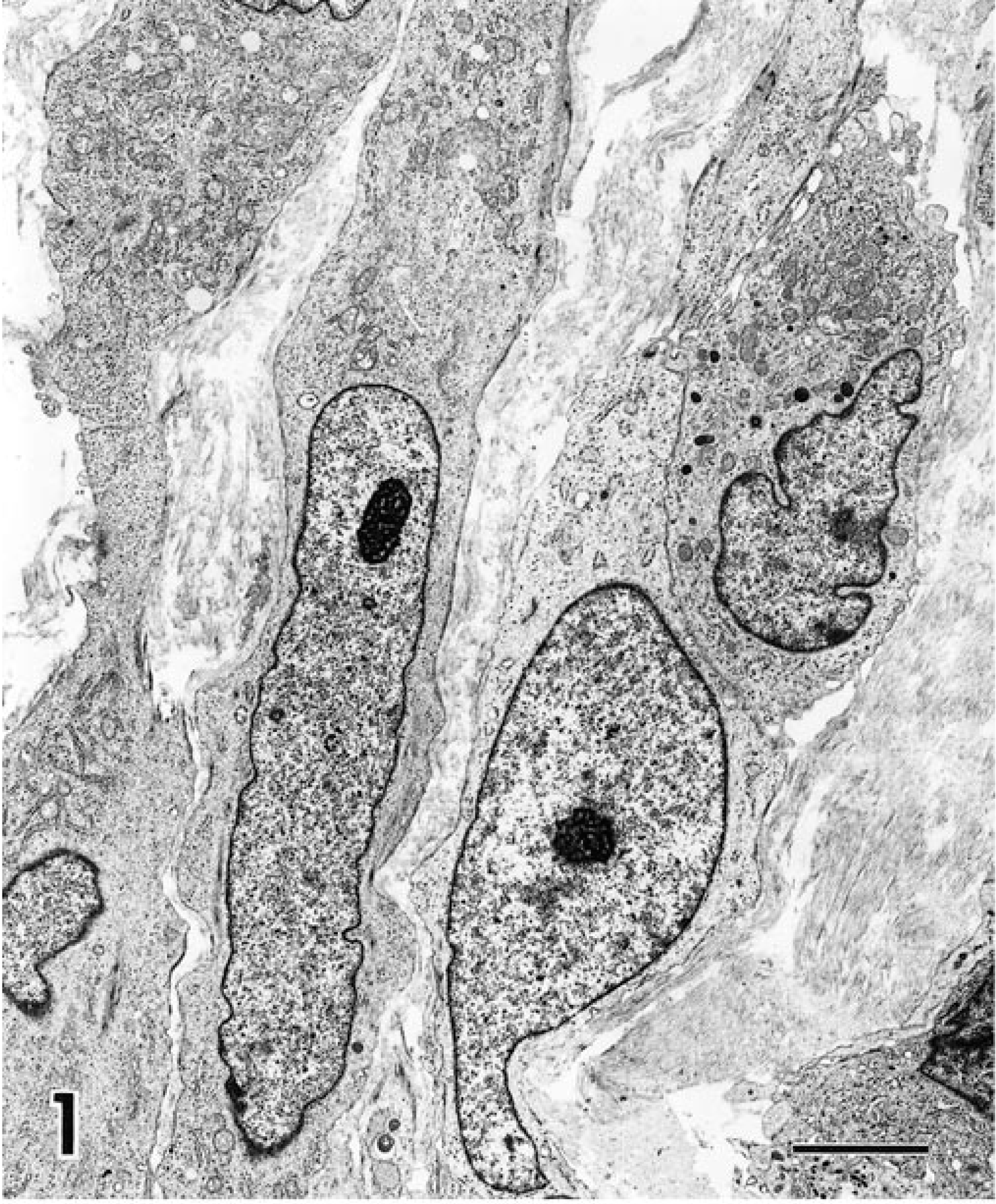
Transmission electron micrograph of feline vaccine-associated fibrosarcoma. Irregular spindle-shaped fibroblastoid cells are surrounded by collagenous interstitium. Bar = 5 µm.
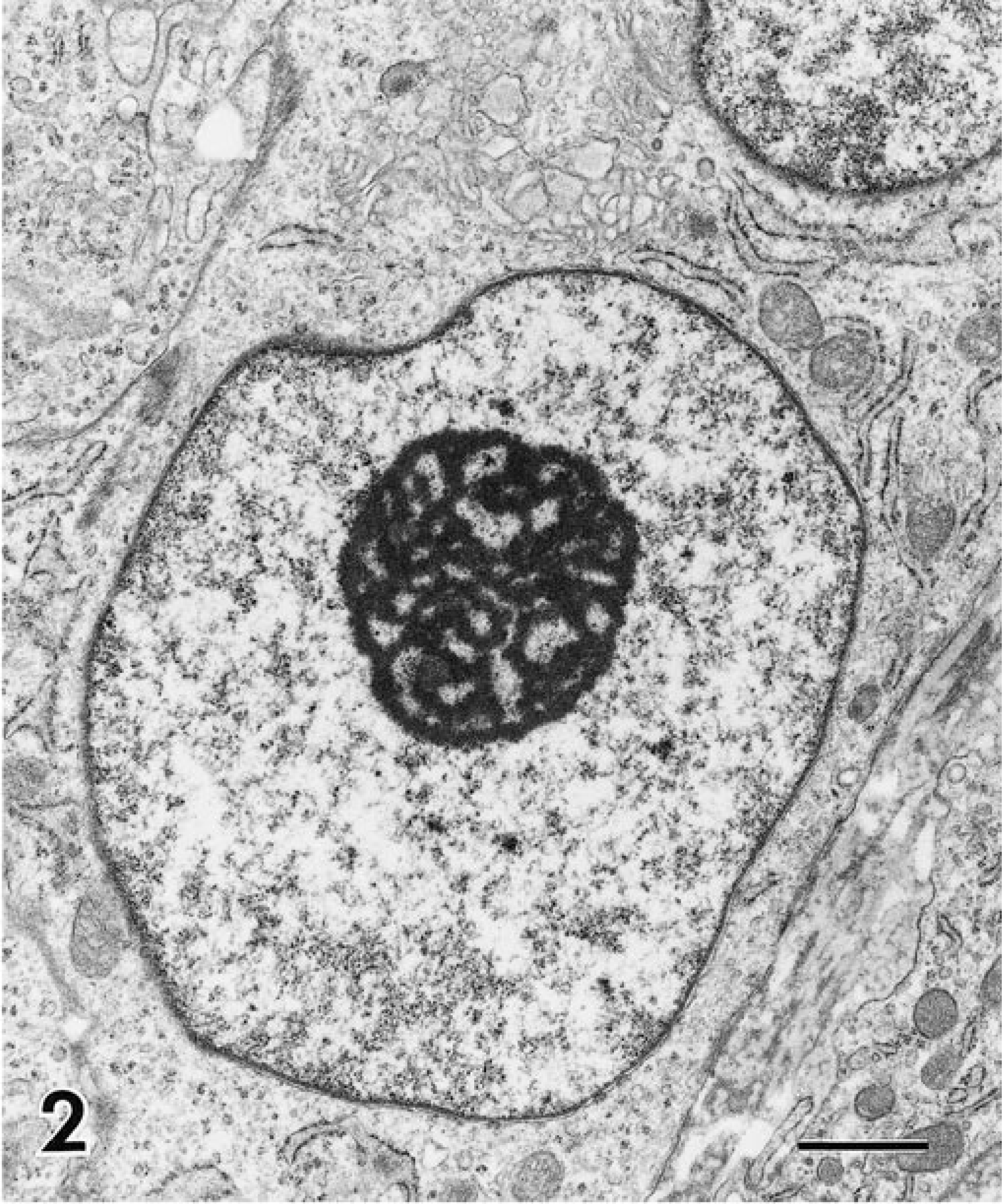
Portion of fibroblastoid cell showing nuclear euchromatin and nucleolonema. Bar = 1 µm.
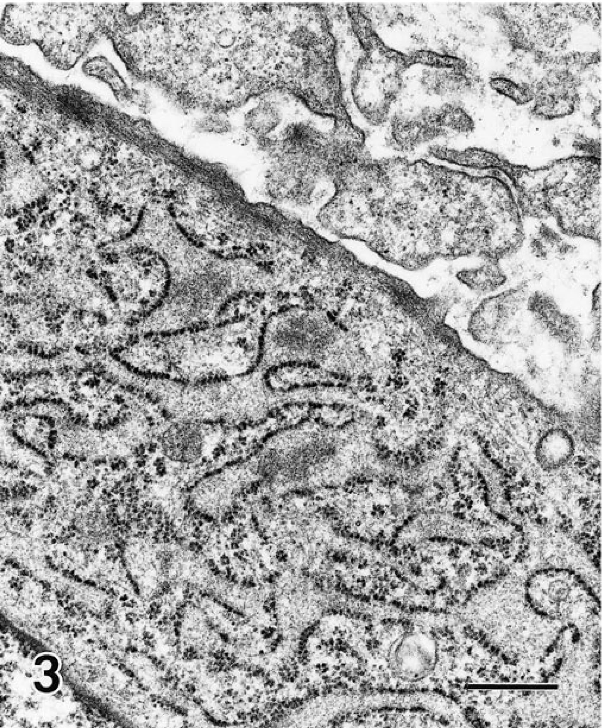
Portion of fibroblastoid cell showing subplasmalemmal thickening or peripheral actin. Bar = 0.5 µm.
Histiocyte-like cells were identified in 14/20 tumors. These cells were round to polygonal in shape and contained abundant mitochondria, variable amounts of rough endoplasmic reticulum, and numerous lysosomes. Giant cells were observed in 4/20 tumors and were a prominent histologic feature of one tumor. Giant cells were large and contained abundant cytoplasm with numerous vacuoles of various sizes. They contained multiple nuclei, often with irregular shapes and folds of the nuclear membranes (Fig. 4). There were prominent nucleoli, and chromatin was scattered diffusely or clumped along the nuclear membrane. There were numerous mitochondria and free ribosomes, scattered profiles of rough endoplasmic reticulum, and multiple indistinct Golgi zones.
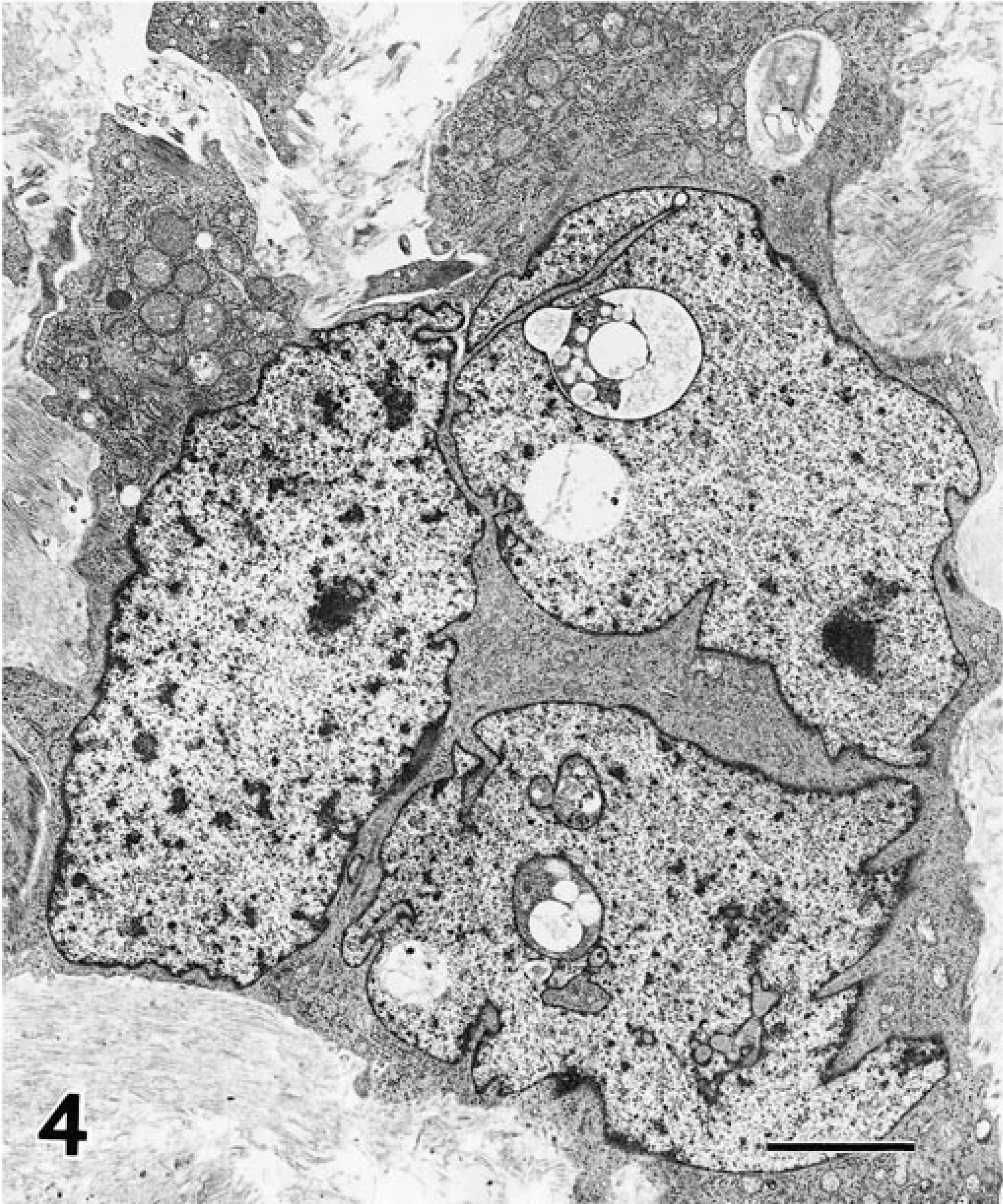
Giant cell contained within a fibrosarcoma showing multiple nuclei, diffuse nuclear chromatin, and irregularly shaped nuclear membrane. Bar = 5 µm.
Cells with myofibroblastic features were frequently observed and were identified in 16/20 tumors. These cells had morphologic features similar to those of fibroblasts but were further distinguished by subplasmalemmal dense attachment plaques and thin cytoplasmic myofilaments organized as elongated bundles concentrated at irregular intervals, forming characteristic dense bodies (Fig. 5). Some cells showed smooth pinocytotic vesicles on the cell surface and rudimentary cell junctions in regions of cell contact (Fig. 6). Although rarely seen, when present, the external lamina was incomplete. Abundant glycogen pools were observed in one tumor that showed myofibroblastic differentiation (Fig. 6).
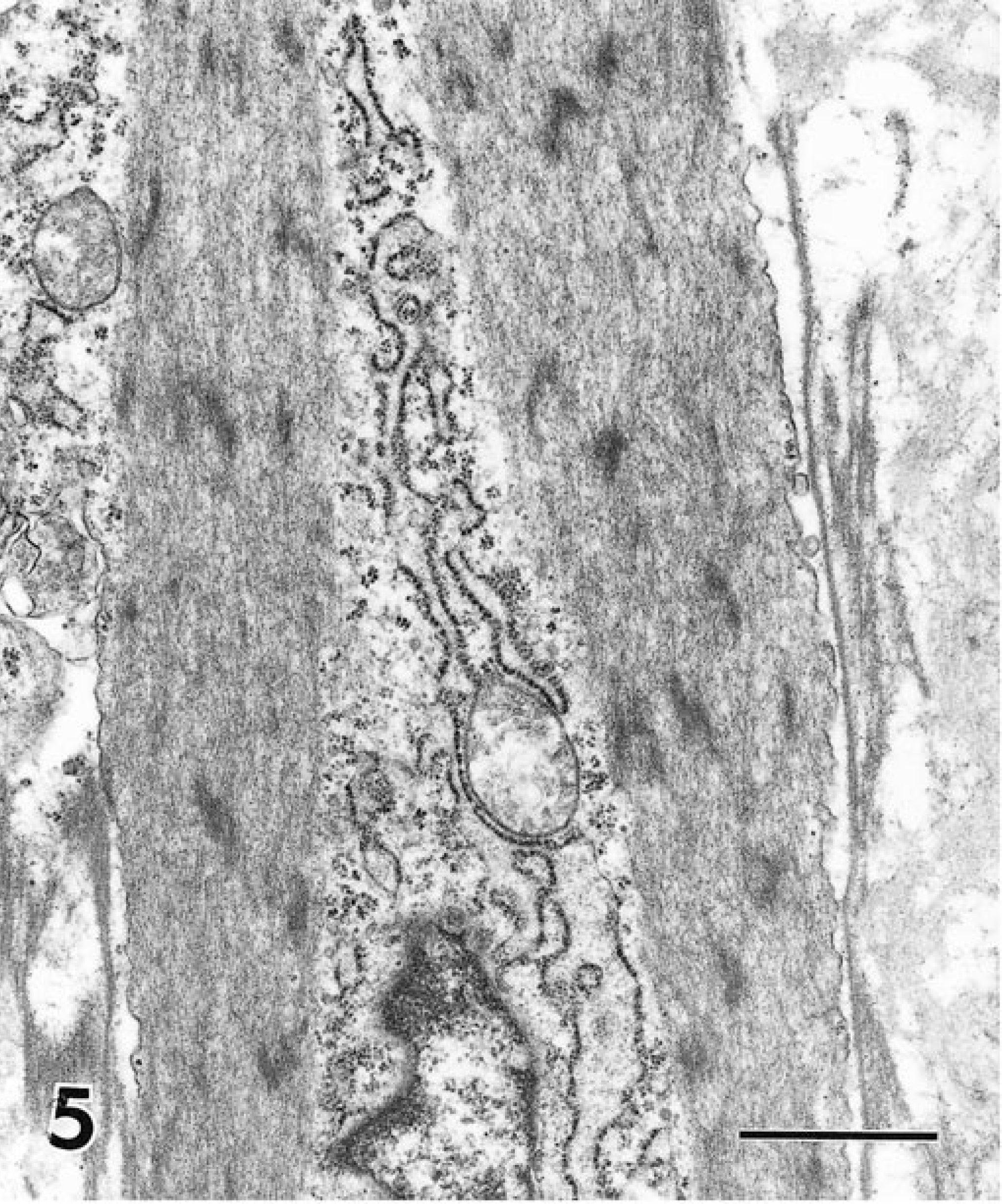
Myofibroblastic cell with thin cyoplasmic actin myofilaments organized as elongated bundles concentrated at irregular intervals as dense bodies. Bar = 1 µm.
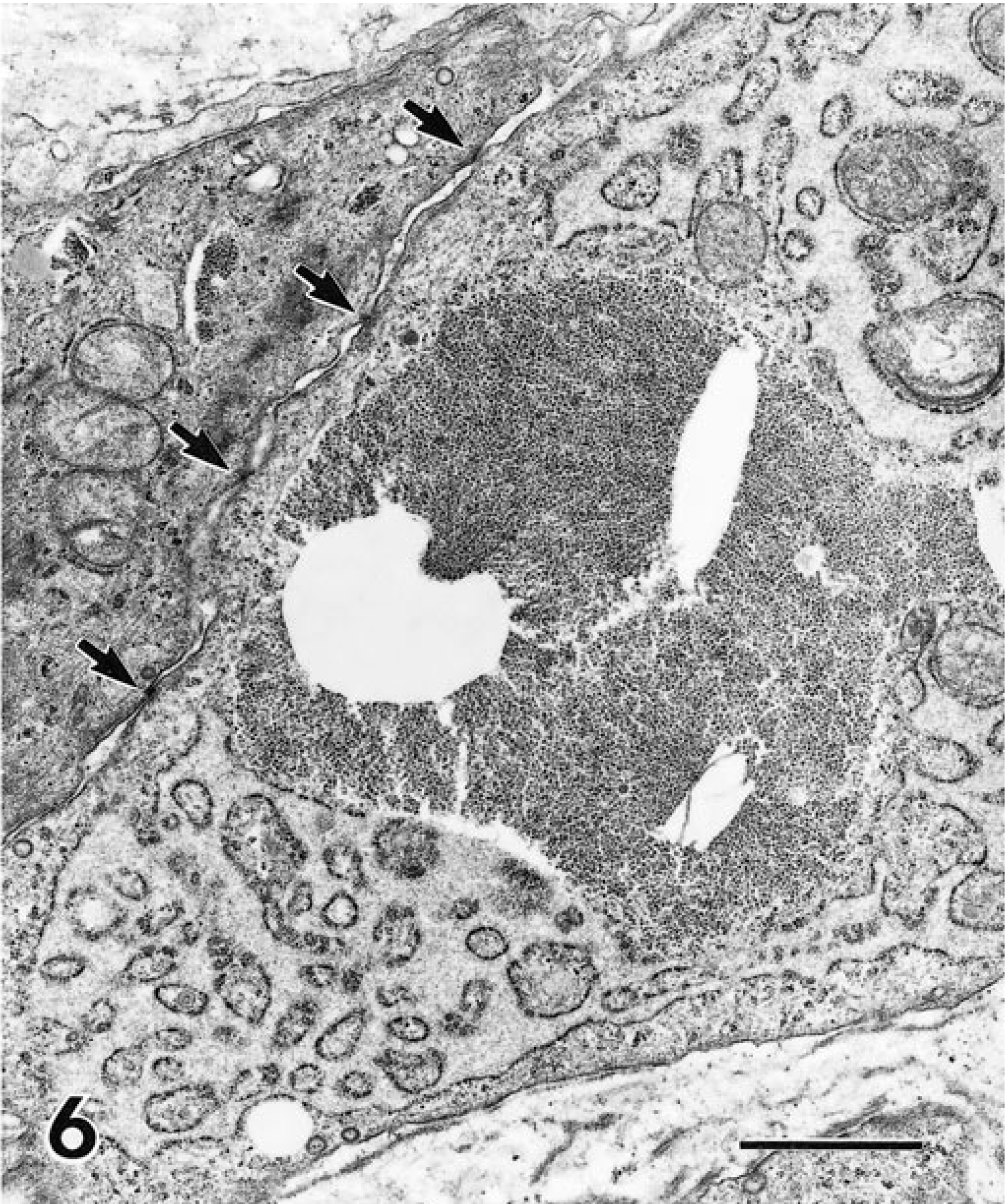
Portion of profile of myofibroblastic cell showing abundant glycogen. Arrows indicate several primitive cell junctions. Bar = 2 µm.
Crystalline particulate material was present within the cytoplasm of histiocyte-like cells or macrophages of 5/20 tumors (Fig. 7), and there was suggestion that particulate material was contained within spindle-shaped tumor cells in one of those specimens. Infiltrating leukocytes, including macrophages, neutrophils, plasma cells, and mast cells, were commonly observed within the vaccine-associated sarcomas.
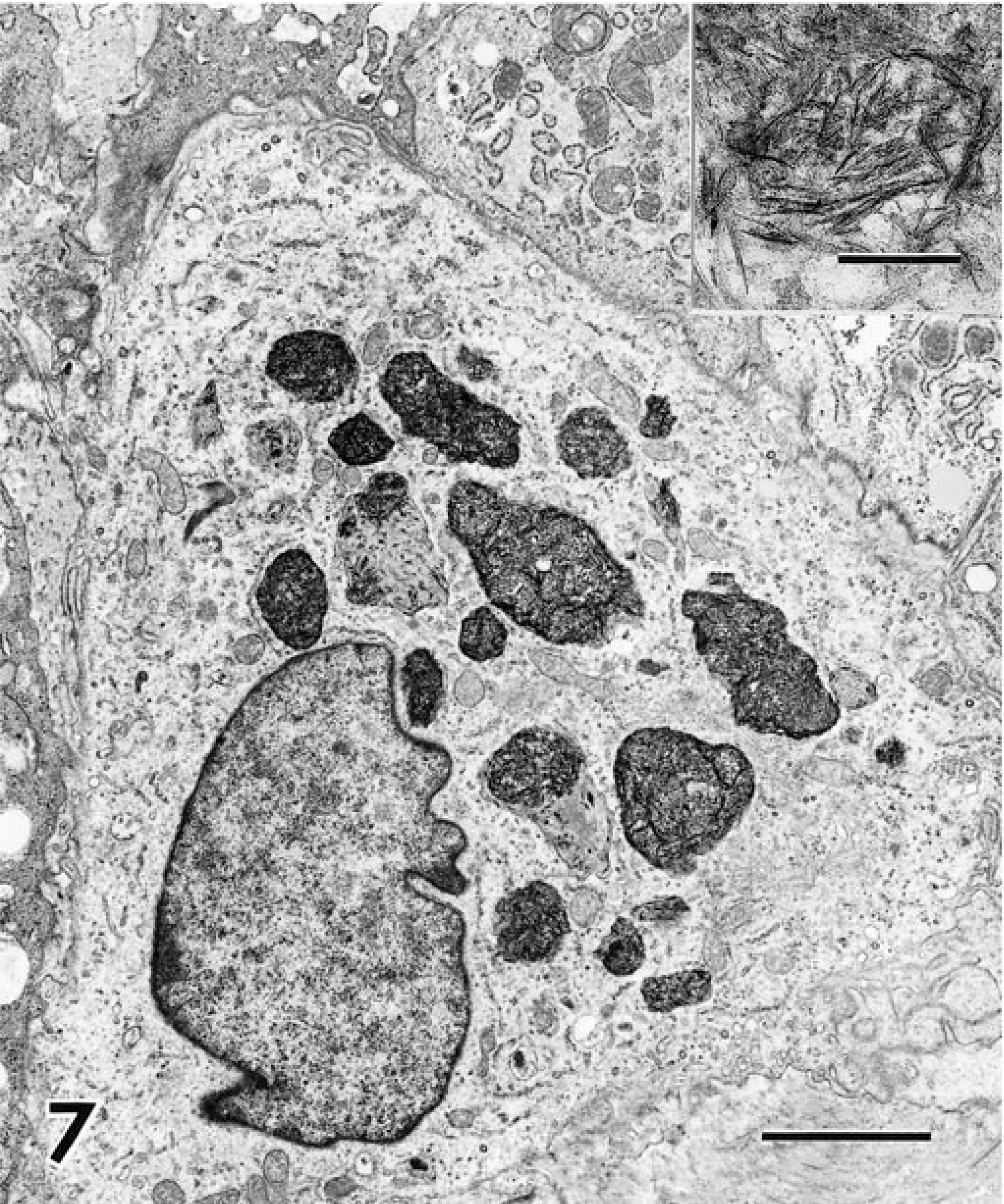
Macrophage showing irregularly shaped intracytoplasmic dense inclusions presumed to contain aluminum. Bar = 4 µm. Inset at higher magnification shows needle-like crystals. Bar = 200 nm.
Other infrequently observed ultrastructural features of the feline vaccine-associated sarcomas were the presence of intratumoral macrophages containing endocytosed erythrocytes, ferritin, or other cellular debris. One tumor contained lipid within neoplastic-appearing fibroblasts (Fig. 8).
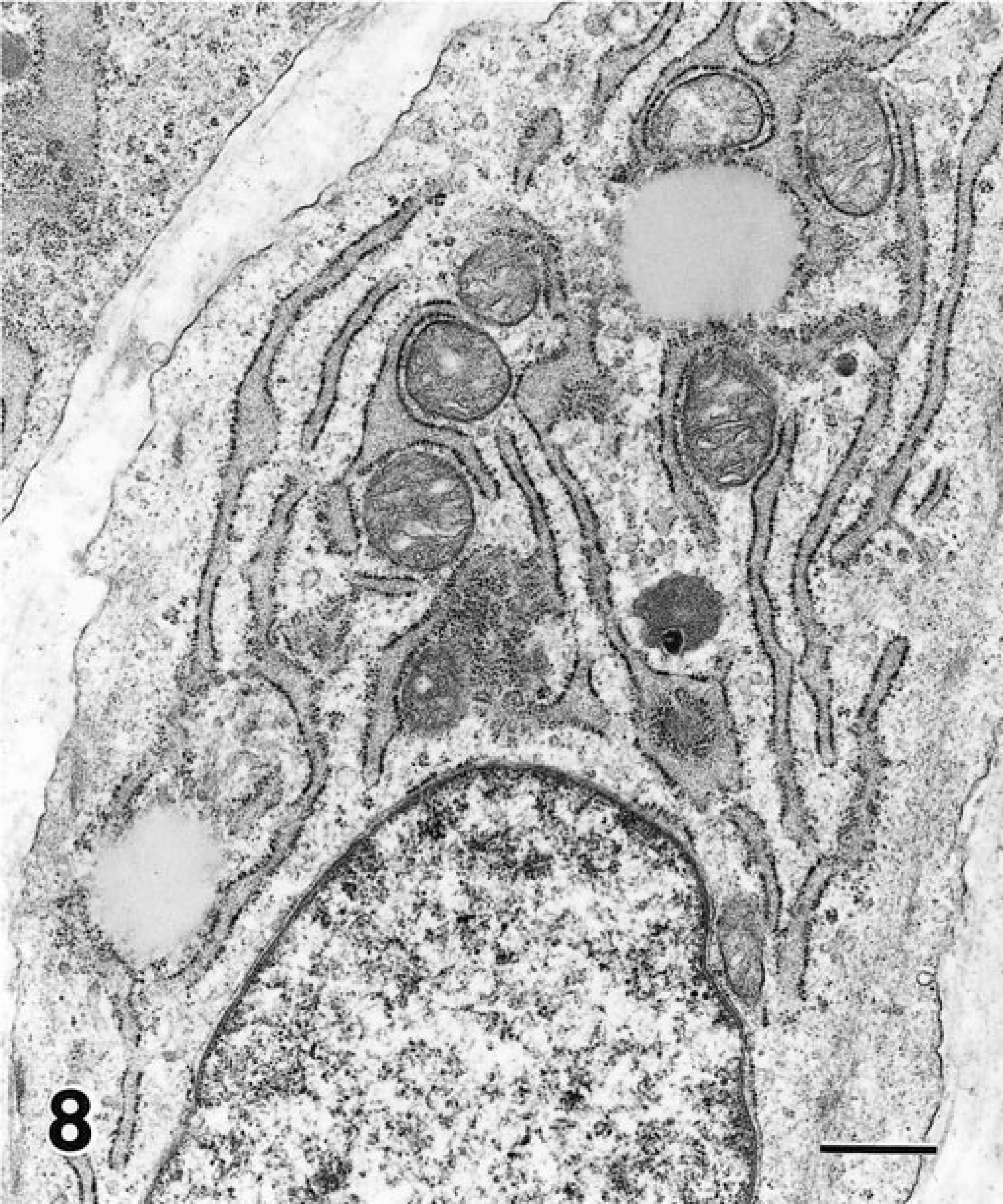
Portion of profile of fibroblastoid cell showing intracytoplasmic lipid. Bar = 1 µm.
Eighteen of 20 cats with tumors had been tested for feline leukemia virus infection and 17 for feline immunodeficiency virus infection. One cat only was serum test positive for feline leukemia virus by an enzyme-linked immunosorbent antibody test. The fibrosarcoma from that cat contained a retrovirus (Fig. 9). The virus appeared as extracellular immature forms that measured 105–120 nm in diameter. Each virus particle contained a partial or complete ring of electron-dense material that was separated from the overlying unit membrane by a distinct, relatively electron-lucent zone. No definite mature forms were observed. Two particles had attachment stalks that connected the virus to apparent tumor cells.
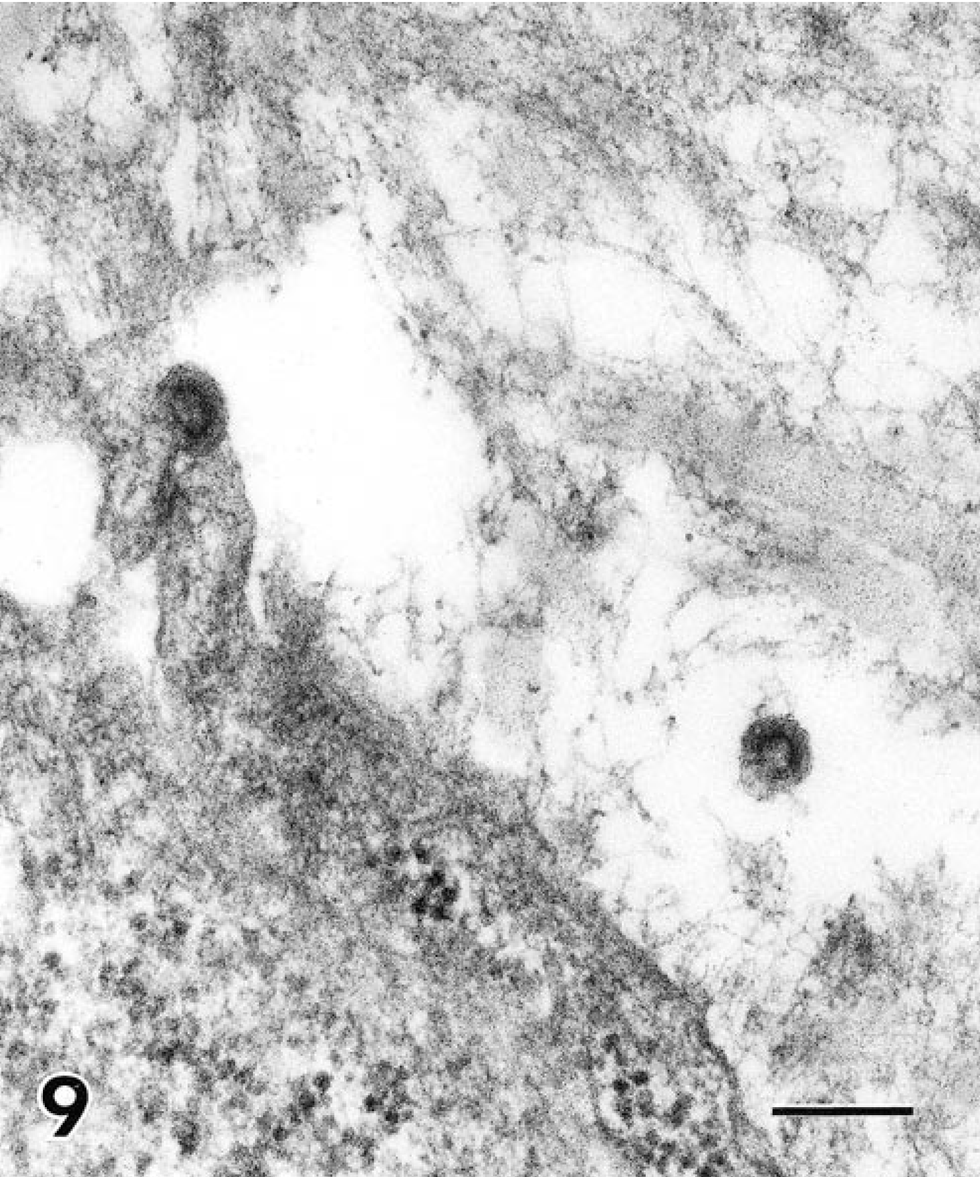
High magnification of cell membrane and extracellular space showing extracellular immature retrovirus and one particle with apparent attachment stalk to fibroblastic tumor cell. Bar = 200 nm.
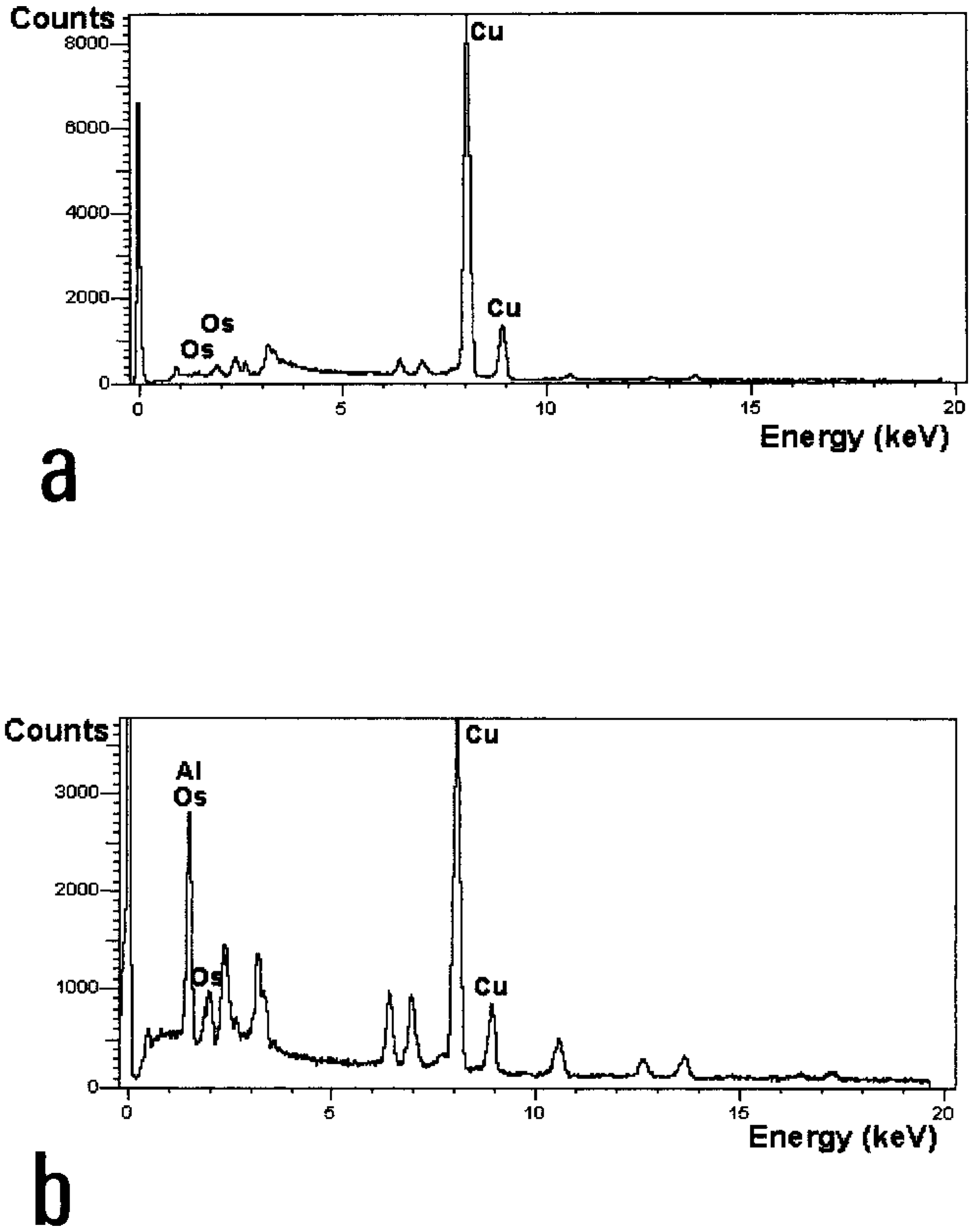
A tumor from one cat with intracytoplasmic crystalline material was examined by EDXS. In Fig. 10, the X-ray spectrum for the sarcoma that has undergone uranyl acetate/lead citrate stain and postfixation with osmium tetroxide is given alongside the spectrum of adjacent tissue that did not contain the crystalline material. The high intensity of the peak at approximately 1.5 keV shows the presence of aluminum, but additional study would be required to identify the exact aluminum compound present.
All tumors examined by immunohistochemistry expressed vimentin, whereas only one of seven showed scattered tumor cells immunolabeled for desmin intermediate filaments. Three of seven tumors examined expressed alpha smooth muscle actin; in these tumors, more than 50% of tumor cells were immunolabeled.
Discussion
The histologic diversity of feline vaccine-associated sarcomas has been defined by light microscopy, 7 10 and part of that heterogeneity was reflected in the tumors studied herein. The tumors studied were characterized by light microscopy as fibrosarcoma or malignant fibrous histiocytoma.
Within most tumors, there were fibroblasts and myofibroblasts. Myofibroblasts were characterized by bundles of actin microfilaments and intercellular attachments. Like fibroblasts, in the myofibroblasts, there were subplasmalemmal plaques, abundant, well-developed endoplasmic reticulum, and irregularly distributed intermediate filaments. Those tumors with smooth muscle differentiation often showed pinocytotic vesicles on the cell surface. They differed from typical smooth muscle cells, however, by having an incomplete external lamina, abundant rough endoplasmic reticulum, and apparent collagen production. Further, few or no individual cells showed all of the features characteristic of smooth muscle—dense bodies, subplasmalemmal plaques, pinocytotic vesicles, and complete external lamina. Myofibroblastic differentiation of a tumor arising at the site of an intramuscular rabies vaccine was previously reported, and in that study, the smooth muscle features noted by ultrastructure were corroborated by immunohistochemical reactivity of tumor cells for smooth muscle actin, 2 and similar immunoreactivity was detected herein in three of seven tumors studied.
Although cells with features of myofibroblasts were frequently encountered within tumors presumed related to vaccination, the origin of these cells, or the role played by these cells in the neoplastic process, remains unanswered. Myofibroblasts are implicated in a wide variety of disorders, including inflammation and wound healing, repair processes, proliferative disorders, and the desmoplastic response to tumors. 14 A limited number of sarcomas showing myofibroblastic differentiation (myofibrosarcomas) have also been described in human patients 11 and a myofibroblast-like sarcoma was described in the mammary gland of a cat. 6 The relationship between myofibroblasts, fibroblasts, and smooth muscle cells remains uncertain. Morphologically, myofibroblasts have features intermediate between those of the fibroblast and smooth muscle cell. 16 It is conceivable that undifferentiated fibroblasts differentiate into myofibroblasts according to microenvironmental stimuli or smooth muscle cells may acquire myofibroblastic features. 13 15
Spindle cells, giant cells, and histiocytoid cells were consistent features of the vaccine-associated sarcomas studied, suggesting some overlap in the categorization of these tumors. 7 In some studies, cells with two or more nuclei are considered giant cells. 12 Whether the giant cells observed in the vaccine-associated sarcomas examined were neoplastic giant cells or reactive (nonneoplastic) giant cells could not be determined on the basis of ultrastructural characteristics. The pleomorphic appearance of these giant cells within some tumors, however, suggested that these cells were neoplastic. In human patients with soft tissue sarcomas, the presence of neoplastic giant cells suggests an aggressive disease course linked to poor prognosis. 12 We are unaware of reports linking intratumoral giant cells in cats with tumor biological behavior.
Retrovirus particles were present in one tumor and appeared to be budding from tumor cells. The tumor had been collected from a 7-year-old domestic shorthaired cat that was serum enzyme-linked immunosorbent assay positive for feline leukemia virus (FeLV). The tumor was characterized histologically as a fibrosarcoma. The virus ultrastructure was consistent with FeLV, an oncovirus now classified as a γ retrovirus. 11 FeLV has been shown not to be causally associated with feline vaccine-associated sarcomas. 4 It is unclear from the literature, however, whether FeLV is expressed in tumor cells other than those derived from the hemopoietic system. When susceptible cats are deliberately infected with FeLV, it proliferates in bone marrow, lymphoid tissues, and intestine, 15 but it seems conceivable that the virus could also replicate in the rapidly dividing tissues of a sarcoma. Another possibility is that the cat was infected with a feline sarcoma virus—a virus that does bud from transformed fibroblasts, 5 although this would seem unlikely in a 7-year-old cat.
Intracellular crystalline particulate material was found in 5 of 20 tumors examined. Histiocytoid cells were identified in all tumors that contained particulate material, but there was no correlation between the presence of particulate material and giant cells. The nature of the particulate material observed within one of these tumors was determined to be aluminum-based by energy-dispersive X-ray spectroscopy. In one of the first reports of feline vaccine-associated sarcomas, dense crystalline material was recognized by electron microscopy within macrophages surrounding tumor cells. 9 Subsequent electron-probe microanalytical studies demonstrated aluminum in the cytoplasm of the macrophages within these sarcomas, suggesting the role of aluminum-containing adjuvant as irritant in the pathogenesis of vaccine-associated sarcomas. 9 The role of vaccine adjuvant in the etiopathogenesis of these tumors remains unclear. The most prevalent adjuvants used in licensed veterinary vaccines are aluminum salts and oil emulsions; all of these formulations are considered to act as depots for injected vaccines. 1 Aluminum hydroxide adjuvants are used in many human and veterinary vaccines, presumably because of their safety and low cost. Aluminum has been detected at the site of subcutaneous injections for up to 1 year in animals. 3 That the tissues collected in this study represented tumors that developed months to years after vaccination when the precise site of vaccination was unknown and that most tumors were several centimeters in diameter or greater at the time of excision suggest that a large amount of aluminum, indeed, was contained within these adjuvanted vaccines to allow its detection in randomly selected ultrathin (60–90 nm thick) tissue sections examined in the electron microscope.
The results of this study support previous morphologic observations of feline vaccine-associated sarcomas. The role of the myofibroblast in vaccine-associated sarcomas is unclear but perhaps reflects a continuum of the inflammatory response that characterizes, in part, these unique neoplasms. The role of adjuvant, similarly, in these tumors is unknown.
Footnotes
Acknowledgements
This work was supported in part by a grant from the Vaccine-Associated Feline Sarcoma Task Force. We appreciate the assistance of Drs. Tracy Gieger, Michael Kent, and Michael Lucroy for providing biopsy specimens.