
Abstract
Twenty-eight histologically confirmed cases of porcine leptomeningitis were examined retrospectively, with focus on the pathology of the inner and middle ear, brain, and vestibulocochlear nerve. Tissues were evaluated by histology and immunohistochemistry for Streptococcus suis serotype 2 antigen, and the bacteriologic results were recorded. Exudative otitis interna was diagnosed in 20/28 pigs (71%). The lesions primarily affected the perilymphatic ducts, with consistent involvement of the scala tympani. Perineuritis of the vestibulocochlear nerve was seen in all but four of the ears affected with otitis interna. Immunohistochemically, S. suis serotype 2 antigen was demonstrated in the leptomeningeal, perineural, and labyrinthine exudates in 11 cases. Otitis media was diagnosed in 10/28 pigs (34%), but evidence of extension to the inner ear was not observed. The findings were highly similar to descriptions of meningogenic labyrinthitis in humans and in laboratory animal models. Otitis interna in pigs can also develop via the meningogenic route and is not always, as generally stated, tympanogenic.
Otitis interna in swine and other animals is generally believed to be a complication of otitis media. 20 24 26 By contrast, the route of infection to the ear in humans is often from the brain in the presence of meningitis. 9 18 21 Hearing loss in humans due to meningogenic labyrinthitis is a common sequela to bacterial meningitis. 19
Meningitis is an important condition in swine, and Streptococcus suis is a primary causative agent. 8 16 Among the 35 serotypes of S. suis, type 2 is the most prevalent in porcine cases of meningitis. 22 Apart from meningitis, S. suis is also found in association with septicemia, polyserositis, and arthritis and has been implicated as a possible pulmonary pathogen. 22 Reports connecting S. suis to lesions of the ear in spontaneously infected pigs seem limited to a single study, in which the organism was isolated from the bulla tympani of a few animals with otitis media. 11 This paucity of information is remarkable, given the situation in humans, where S. suis serotype 2 is also a well-recognized cause of meningitis. Inner ear involvement leading to sensorineural hearing loss affects a majority of patients surviving S. suis meningitis. 1 The infection is prevalent in commercial swine operations and is a common cause of disease. 5 8 The lack of reports of S. suis involvement in disease of the porcine inner ear is likely due to a lack of routine examination of this organ.
Development of meningitis and suppurative labyrinthitis was discovered recently in a pig experimentally infected with S. suis serotype 2 in an aerogenous challenge model (L. W. Madsen, personal observations). These findings prompted a review of material from cases of meningitis collected at the Veterinary Diagnostic Laboratory, Kjellerup, Denmark. The aim of the present study was to elucidate the frequency and nature of lesions of the inner and middle ear of pigs with spontaneous meningitis. Here, we report the pathologic, immunohistochemical, and bacteriologic findings from this investigation.
Materials and Methods
The study included 28 pigs (mean age = 10.3 weeks [range, 2–19 weeks]; 13 females, 13 males, and two pigs of unrecorded sex) with histopathologically confirmed meningitis and from which at least one inner and one middle ear were available for histologic examination. Macroscopic lesions had been recorded (see Table 1). The animals had initially been selected on the basis of a clinical history or necropsy findings suggestive of meningitis or septicemia; all selected animals had a history of clinical neurologic signs or sudden death and/or necropsy findings suggestive of meningitis or polyserositis. Tissue collection was done at the Veterinary Diagnostic Laboratory, Kjellerup, Denmark, over a period of 3 months (November 1998–March 1999), during which time, a total of 742 complete porcine necropsies were performed.
Pathologic, immunohistochemical (IHC), and bacteriologic (Bact)∗ findings in 28 pigs with histologically confirmed leptomeningitis.
S. suis 2 and 7 = Streptococcus suis serotypes 2 and 7; P. multocida = Pasteurella multocida; A. pyogenes = Archanobacterium pyogenes; A. pleuropneumoniae = Actinobacillus pleuropneumoniae.
ND = not done.
The following tissues were evaluated by light microscopy: brain (cerebral hemisphere, hippocampus, cerebellum, pons, and medulla oblongata), inner ear (cochlea [n = 44] and vestibulocochlear nerve [n = 45]), and middle ear (cavum and bulla tympani, n = 47). The tissues were fixed in 10% neutral buffered formalin, processed through graded alcohols and xylene, and embedded in paraffin. Hematoxylin and eosin (HE) staining was performed on 4–5-µm tissue sections. Formic acid decalcification of inner and middle ears after initial formalin fixation was done as described previously. 17
S. suis type 2 antigen was detected in 4–5-µm tissue sections by immunohistochemistry using a 1:8,000 dilution of a commercially available polyclonal serum (article 22282, lot 2A, Statens Seruminstitute, Copenhagen, Denmark). The specificity of this serum for use in immunohistochemistry has previously been described. 4 The immunoreaction was demonstrated by the application of EnVision™ labeled polymer, alkaline phosphatase (K4018, DAKO, Glostrup, Denmark), and the substrate fast red (CatII 4210, Kem-En-Tec, Copenhagen, Denmark) according to the instructions of the manufacturers. The sections were counterstained with Harris hematoxylin.
Bacteriologic culturing from the brain, tympanic bullae, and tissues with macroscopic lesions was done aerobically at 37 C on 5% calf blood agar. Causative organisms were identified on the basis of morphologic and biochemical characteristics according to standard laboratory procedures. 15 For S. suis, species identification was based on a commercial system (API Rapid ID 32 STREP, BioMérieux sa, Marcy L'Etoile, France), and serotyping was done by the slide agglutination method using commercially available antisera (Statens Seruminstitute). 6
Results
The results from the histopathologic, immunohistochemical, and bacteriologic examinations are detailed in Table 1. By culture, 16 animals were infected with S. suis serotype 2 and five were infected with serotype 7. Other pathogens were cultured from two pigs only, and five animals were bacteriologically negative. Necropsy findings apart from meningitis included serositis (five pigs), polyarthritis (one pig), endocarditis (one pig), and necrotizing pleuropneumonia (one pig).
Meningitis, i.e., leptomeningitis (Fig. 1), in these animals was characterized by infiltrates of neutrophils with varying amounts of fibrin and/or mononuclear cells. In all but three pigs, the meningitis was diffuse, and often submeningeal encephalitis was found locally. The meningitis in pig Nos. 22 and 23 primarily affected the basal parts of the brain in the region of the internal auditory canal, whereas in pig No. 26, a single abscess was found in the right side of the cerebellum.
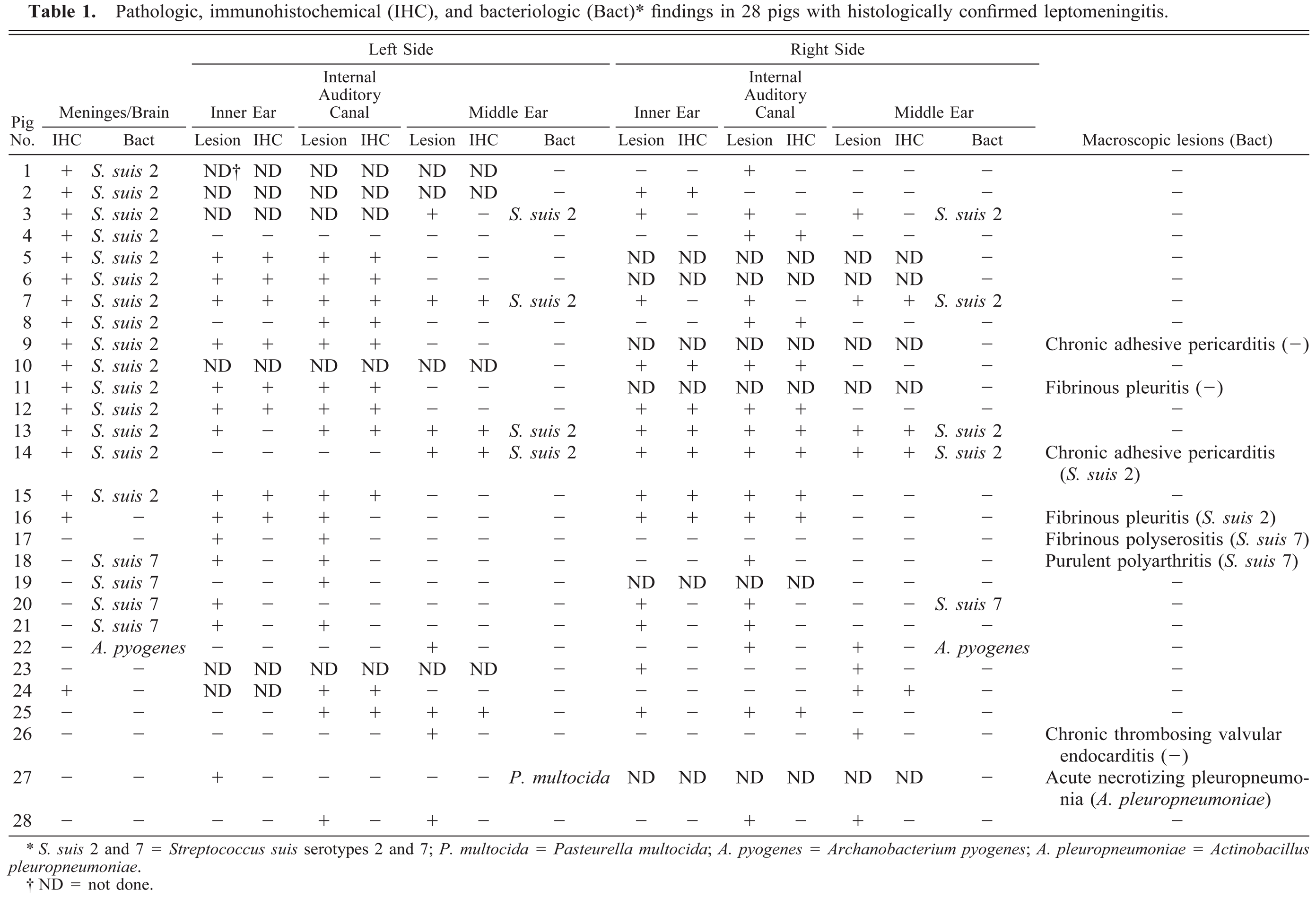
Cerebrum; pig No. 12. Leptomeningitis with mixed inflammatory cells and Streptococcus suis serotype 2 antigen (arrow) in the meningeal exudate. Immunohistochemistry, Envision method, Harris hematoxylin counterstain. Bar = 75 µm.
By immunohistochemistry, S. suis type 2 antigen was detected in the leptomeninges (Fig. 1) of 17/28 pigs (61%), including all the pigs from which S. suis type 2 was cultured and one microbiologically negative pig (No. 24). The immunohistochemical signal was generally associated with 1–3-µm round to oval structures resembling streptococci that were located intracellularly within macrophages or freely in the exudate.
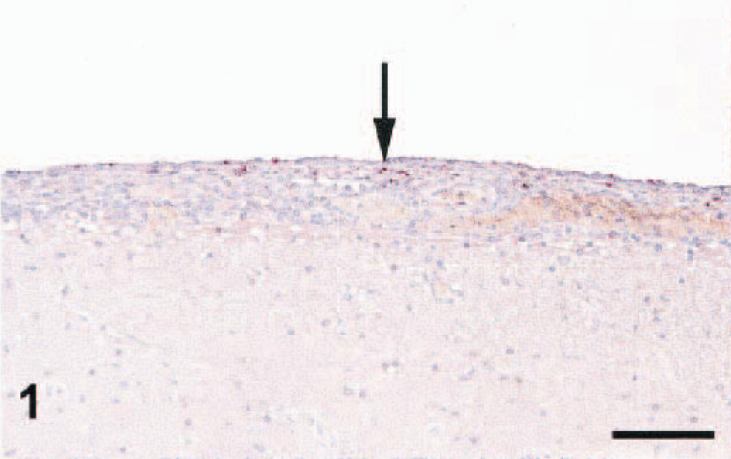
Otitis interna was present in 20/28 pigs (71%), and of the 16 animals from which both inner ears were available, seven were bilaterally affected. The otitis interna was characterized by a mixed inflammatory exudate comprising neutrophils, lymphocytes, and less often macrophages in the perilymphatic ducts of the cochlea (Figs. 2, 3). Exudation in the scala tympani was seen in all pigs, whereas the scala vestibularis was only found to be involved in three pigs (Nos. 11, 20, and 23). In two pigs (Nos. 20 and 23), suppurative inflammation with fibroplasia and apparent destruction of the organ of Corti were seen in the scala media. The vascular changes in the inner ear consisted of hyperemia and exudation of inflammatory cells, but thrombosis and vasculitis were not observed.
Cochlea; pig No. 12. Labyrinthitis with mixed inflammatory cells and Streptococcus suis serotype 2 antigen (arrow) in the exudate (E) in the scala tympani (ST). SV = scala vestibularis; SM = scala media; OSC = organum spirale Corti; GSC = ganglion spirale cochlea. Immunohistochemistry, Envision method, Harris hematoxylin counterstain. Bar = 75 µm.
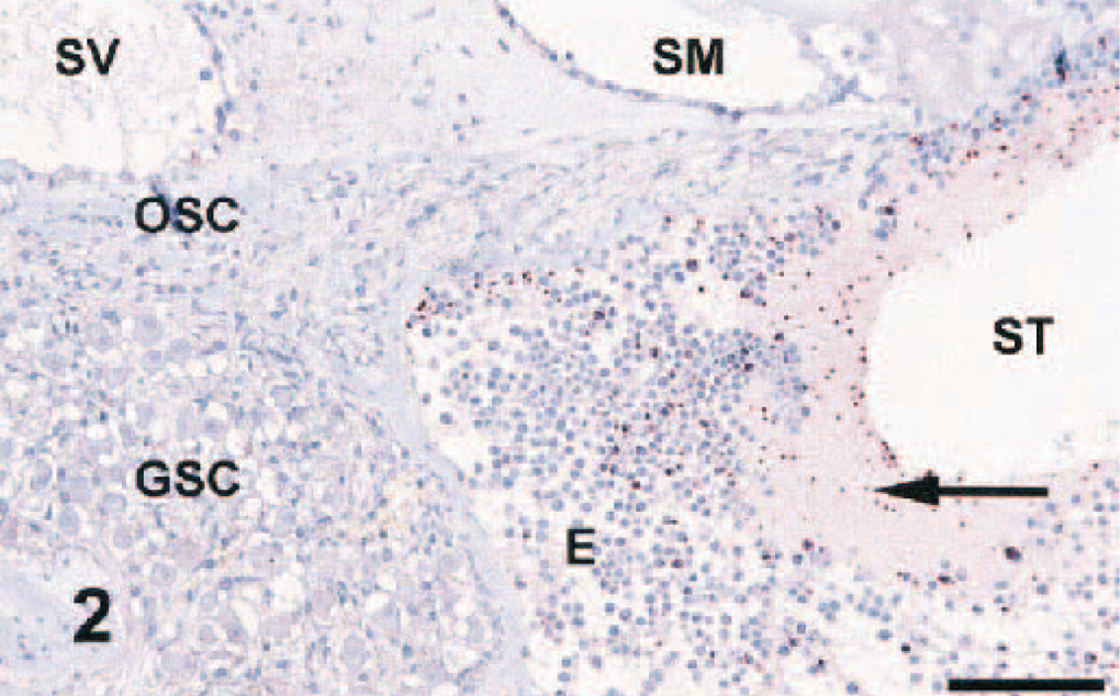
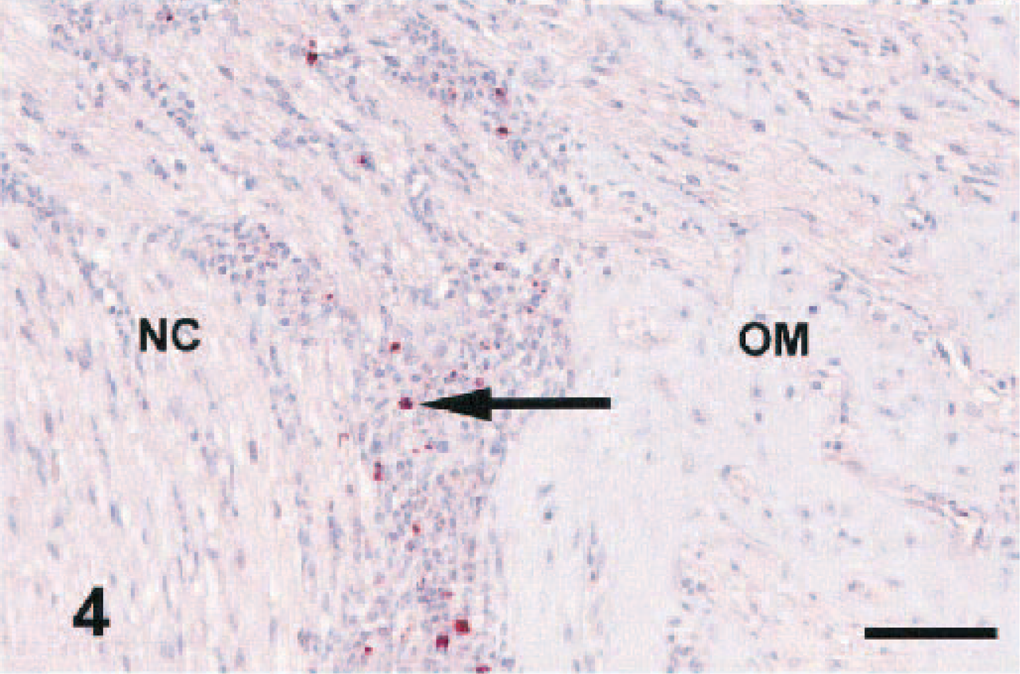
Cochlea; pig No. 10. Perineuritis and labyrinthitis. Exudate along the nervus cochlearis (←NC), where it enters the osseous modiolus (OM). Exudate is evident in the scala tympani (ST) but not in the scala media (SM). HE. Bar = 250 µm.
By immunohistochemistry, S. suis type 2 antigen was demonstrated (Fig. 2) in 15/44 (34%) of the inner ears. Thus, 11/16 pigs (69%) from which the organism had been cultured had demonstrable S. suis type 2 antigen in the inner ear.
Perineuritis (Figs. 3, 4) was found along the vestibulocochlear nerve in 34/45 (76%) of the examined inner ears, comprising all but four of the ears (23/27) in which otitis interna occurred. The type of perineural inflammation was generally similar to the type of meningitis present in the animal. Generally, the inflammatory changes were most profound along the proximal parts of the nerve, and in only 10/27 inflamed inner ears they extended into the cochlear modiolus (Fig. 4). Immunohistochemically, S. suis type 2 antigen was detected perineurally (Fig. 4) in 20/34 (59%) of the affected nerves, including two pigs (Nos. 24 and 25) from which the organism had not been cultured.
Cochlea; pig No. 10. Perineuritis with mixed inflammatory cells and Streptococcus suis serotype 2 antigen (arrow) in the exudate along the nervus cochlearis (NC), where it enters the osseous modiolus (OM). Immunohistochemistry, Envision method, Harris hematoxylin counterstain. Bar = 50 µm.
Otitis media was diagnosed in 10/28 pigs (36%), and of the 19 animals from which both middle ears were available, seven were bilaterally affected. In all cases, the inflammatory reaction was chronic and suppurative, and it generally affected both the cavum and bulla tympani. Rupture of the tympanic membrane was noted in one pig (No. 23). Otitis media was found in 7/27 ears (26%) in which there was otitis interna. Antigen from S. suis type 2 was detected in 8/17 (47%) of the inflamed middle ears, again including two pigs (Nos. 24 and 25) that were microbiologically negative.
Discussion
Exudative labyrinthitis was detected in a majority (20/28) of the pigs with meningitis. The inflammatory changes in the central nervous system and meninges were as previously described for bacterial meningitis due to pyogenic bacteria. 22 23 25 The character of the inner ear lesions was compatible with that reported in previous studies of pigs 20 24 and with descriptions from the human literature. 10 12 18 The high frequency (71%) of labyrinthitis is also comparable to human figures, where it was diagnosed in 49% of 41 temporal bones from patients with meningitis. 18 In humans, two main hypotheses on the pathogenesis of meningogenic labyrinthitis have been proposed. On the basis of the consistent involvement of the vestibulocochlear nerve, with perineural exudates extending into the cochlear modiolus, this nerve has been suggested as a common portal of entry. 18 Also, the cochlear aqueduct connecting the subarachnoidal space to the perilymphatic system has been incriminated as a possible route of spread. 7 18 In the present study, evaluation of the cochlear aqueduct was not routinely possible. However, the absence of vasculitis and thrombosis in the inner ear, the general perilymphatic distribution with the scala tympani most often affected, and the rare morphologic evidence of involvement of the neurosensory structures are similar to findings reported in humans with meningogenic labyrinthitis. 18 The parallel detection of S. suis type 2 antigen in meningeal, perineural, and labyrinthine exudates in 11 of the pigs is compatible with spread from the meninges to the cochlea. Moreover, similarities of the cochlear lesions in the pigs and in laboratory animal models of meningitis were evident. 2 3 13 14
These cases of meningitis had not been selected on the basis of clinical or gross otologic findings, and yet labyrinthitis was found in a majority of them. Although otitis media was present in some pigs (10/28), there was no evidence of tympanogenic spread to the inner ear. Therefore, the development of otitis interna was most likely a sequela to meningitis and not, as generally believed, a complication of otitis media. In pig Nos. 22 and 23, the focal meningitis in the region of the internal auditory canal was suggestive of spread from the affected ear. This otogenic route of meningeal infection has previously been suggested in pigs. 24 25 In pig No. 26, a cerebellar abscess was found with no corresponding ear lesions. Given the valvular endocarditis also present in this pig, this brain lesion was probably hematogenous. 25
S. suis was the dominating etiology of meningitis in the present study. However, it remains unclear whether the high correlation of meningitis and otitis interna is unique to this organism or whether it applies to porcine meningitis in general.
Coexistence of otitis interna should be considered when examining pigs with meningitis, and animals recovering from meningitis could suffer from hearing loss and vestibular dysfunction, as is the case in humans. Thus, pigs should be evaluated as a possible animal model for meningogenic labyrinthitis.
Footnotes
Acknowledgements
We thank the technical staff at our laboratories for skillful assistance. This work was financially supported by the Danish Bacon and Meat Council and by an R. N. Thomsen grant.