
Abstract
Quinolones and magnesium deficiency cause similar lesions in joint cartilage of young animals. Chondrocytes cultivated in the presence of quinolones and in Mg-free medium show severe alterations in cytoskeleton and decreased ability to adhere to the culture dish. We investigated whether Mg2+ supplementation can prevent quinolone-mediated effects on chondrocytes in vitro. Chondrocytes cultivated in Dulbecco's modified Eagle's medium/HAM's F-12 medium were treated with ciprofloxacin (80 and 160 μg/ml) and enrofloxacin (100 and 150 μg/ml). Mg2+ was added at a concentration of 0.0612 mg/ml (MgCl) and 0.0488 mg/ml (MgSO4) or a triple dose. In addition, cells were cultivated in Mg-free medium and accordingly treated with Mg2+ supplementation. After 5 days in culture, the number of adherent cells per milliliter was determined. The number of chondrocytes in quinolone-treated groups decreased to 12-36% that of the control group within the culture period. With Mg2+ supplementation, the number of attached cells increased to 40-70% that of control cells. The threefold dose of Mg2+ led to better results than did the single dose. Cell proliferation tested by immunohistochemical staining with Ki67 (clone MIB5) decreased from 70% in control groups to 55%, 48%, and 30% in enrofloxacintreated groups in a concentration dependent manner (50, 100, and 150 μg/ml). Addition of Mg2+ did not increase the rate of cell proliferation. These results suggest that a great part of quinolone-induced damage is due to magnesium complex formation, as Mg2+ supplementation is able to reduce the effects in vitro. However, quinolone effects on cell proliferation seem to be an independent process that is not influenced by magnesium supplementation.
The chondrotoxic effect of quinolones, a group of very important antibacterial drugs in human and veterinary medicine, has been traced back to their ability to form chelate complexes with divalent cations, especially magnesium. 18 22 In vivo studies have shown that feeding a Mg-deficient diet to juvenile rats led to lesions in articular cartilage identical to those observed after quinolone treatment. 23 26 26 This finding was confirmed by in vitro studies, in which detachment of chondrocytes of different species and severe alteration of the cytoskeleton were observed after quinolone treatment; these changes were similar to those observed with cultivation of cells in Mg-free medium. 9–11, 29 Lack of extracellular Mg2+ impairs the function of integrins. 7 20 21 These transmembrane proteins connect the cells to extracellular matrix (ECM) proteins, and their intracellular segment is linked to the cytoskeleton. 1 8 15 Integrins generate a proliferative response when stimulated by binding to ECM proteins such as fibronectin and regulate matrix protein synthesis and matrix-degrading enzyme production. 2 6 Results of in vitro studies of canine and rabbit articular cartilage (explants and monolayer culture) have suggested that quinolones inhibit glycosaminoglycan production, mitochondrial function, and DNA synthesis. It is not clear from these experiments whether observed changes are directly caused by quinolones or are a consequence of transmembrane signalling. 5 14 15 17
To explore whether or not Mg2+ supplementation could prevent quinolone-induced arthropathy, Stahlmann and coworkers found that a Mg2+ supplemented diet reduced the number of lesions in rats after quinolone treatment. 24 Addition of tocopherol to the diet improved the results significantly. The authors concluded that the lack of magnesium induces O2 radical formation, which can be prevented by supplementation of vitamin E. Based on our previous studies, we tested the hypothesis that quinolones exert their effects by creating a Mg2+ deficiency. The prediction was that there would be a reversal of the negative effects by addition of magnesium to the cultures along with the drug. For that purpose, chondrocytes were cultivated in the presence of ciprofloxacin and enrofloxacin in Mg-free medium. Magnesium was added to test groups in two different concentrations. The number of cells and the cell proliferation rate after 5 days of culture was determined.
Materials and Methods
Culture of chondrocytes
Articular cartilage of one horse (2 years old, female) and one dog (17 weeks old, female) euthanized for reasons other than musculosceletal disease was obtained by dissection and cut into small pieces (2 × 2 mm). Cartilage pieces were trypsinized (0.05% trypsin, 0.2 g ethylenediaminetetraacetic acid) [EDTA]; (Sigma, Austria) for 30 minutes at room temperature, treated with 0.2% collagenase for 30 minutes at room temperature, and subsequently incubated in 0.04% collagenase (449 U/mg) at 37 C overnight. Cells were filtered through a nylon mesh (50 µm pore size) to remove matrix residues, centrifuged, and seeded (106 cells/flask) in the respective culture medium.
Cell culture medium of control groups was a mixture of Dulbecco's Modified Eagles Medium (DMEM) and HAM's F-12 medium (1:1) (Sigma) supplemented with 10% fetal calf serum (FCS),
Magnesium supplementation
In Mg2+-supplemented groups, MgCl and MgSO4 were added at the time of seeding in concentrations specified by the manufacturer for normal medium content (DMEM/Ham's F-12, Sigma; 0.0612 mg/ml MgCl and 0.0488 mg/ml MgSO4 respectively) as a single (Mg1) and a triple (Mg3) dosage. Mg2+ was added to control groups as previously described to evaluate the basic effect of Mg2+ supplementation. Mg2+ concentrations were determined by results of previous experiments (data not shown).
Evaluation
Cells were cultivated for 5 days (38 C, 5% CO2) without changing the medium. Cells were evaluated microscopically, removed from the culture dish by trypsinization (0.05% trypsin, 0.2 g EDTA) at 37 C for 5 min, and resuspended in control medium. Two lots of each test group were counted using a hemacytometer (Bright-line, Sigma). Experiments with horse chondrocytes were repeated 12 times, those with dog chondrocytes were repeated four times, and mean values were determined. Cells from the same donor and the same passage were used for a whole set of study groups.
Statistics
Normally of the distribution of values was tested evaluated with the Kolmogorov-Smirnov test. The general linear model for repeated measurements was used. The individual differences were tested with simple contrasts. P values of ≤0.05 were considered significant.
Cell proliferation
Immunohistochemical staining with Ki67 (clone MIB5, dilution 1:50; Immunotech, France) was used to determine chondrocyte proliferation rate in all test groups. For this purpose, cells were grown on eight-well chamber slides (Lab-Tek, USA) at a density of 25,000 cells/chamber under identical culture conditions for 3 days (37 C, 5% CO2). Slides were washed in phosphate-buffered saline and fixed in ice-cold methanol for 5 min. Antigen retrieval was performed by microwave heating in citrate buffer (pH 6.0) three times at 5-minute intervals. Binding of primary antibody was demonstrated using a Dako (USA) envision system, followed by diaminobenzidine reaction and counterstaining with hematoxylin. In 10 randomly chosen fields (average 300 cells in every test group), Ki67-positive and negative adherent cells were counted using Metreo KAPPA ImageBase (USA) image processor software.
Results
As in previous studies, the most obvious effect of quinolone treatment of chondrocytes in vitro was that cells detach from the culture dish and change their cell shape. The extent of damage was strictly concentration dependent. There were no differences between the two species and among passage numbers. After 5 days of culture, the number of chondrocytes still attached to the culture dish was reduced to 10% and 20%, respectively, that of controls in the ciprofloxacin (CFX) 160 µg/ml and enrofloxacin (ENR) 150 µg/ml groups and to 34% and 36% that of controls in the CFX 80 µg/ml and ENR 100 µg/ml groups. Cultivation of chondrocytes in Mg-free medium led to a 35% decrease in number of adherent cells. All test groups differed significantly from control groups.
Addition of MgSO4 and MgCl led to a significant increase (P ≤ 0.05) in number of adherent cells as compared with unsupplemented medium in all test groups except the Mg-free + Mg3 group. The number of cells in the CFX 80 group increased by 12% (P ≤ 0.001) with the addition of a single dose of Mg2+ and by 20% (P = 0.001) with a triple dose. In the CFX 160 group, cell numbers increased by 18% with a single dose and by 26% (P ≤ 0.001) with a triple dose. In test groups treated with enrofloxacin, the increase was 24% and 34% (single dose and triple dose; P ≤ 0.001) in the ENR 100 group, and 15% and 34% (single dose and triple dose; P ≤ 0.001) in the ENR 150 group. Test groups with Mg-free medium showed a significant increase in cell number (13%; P = 0.045) with a single dose of Mg2+. A threefold Mg2+ supplementation increased cell number by 11% (not significantly different from Mg-free group). Results are summarized in Fig. 1.
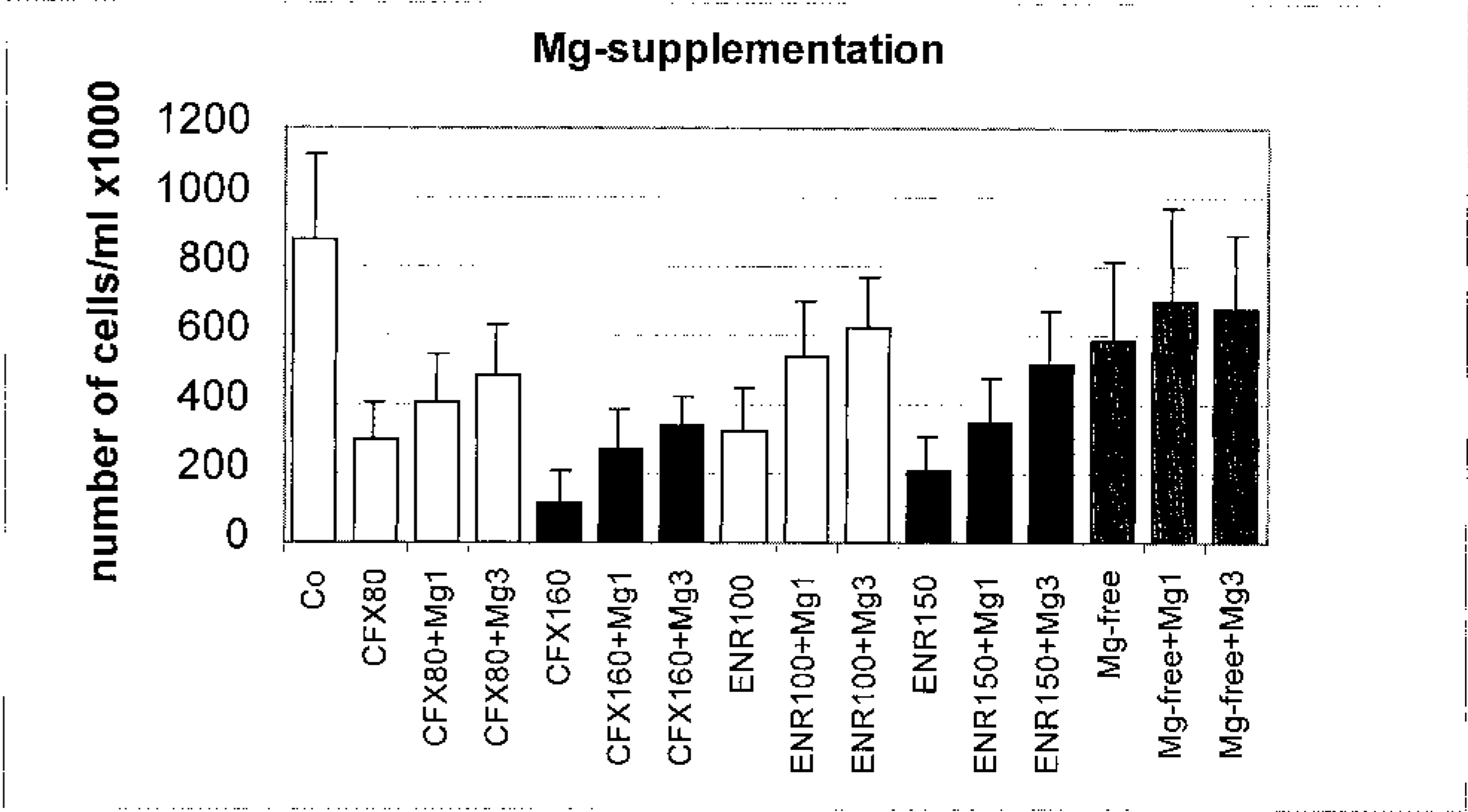
Chondrocytes. Number of chondrocytes (×1,000) after 5 days in culture in control and test groups. Magnesium was added to ciprofloxacin-and enrofloxacin-treated groups and to Mg2+-free groups in single (Mg1) and triple (Mg3) doses. Note the significant increase in cell number in all test groups except the Mg-free + Mg3 group.
Addition of a single dose of Mg2+ to control groups had no effect at all, whereas addition of a triple dose resulted in a slight decrease in cell number. We found a remarkable change in chondrocyte morphology in Mg2+ treated groups in that cells resembled more outspread, stellate chondrocytes of control groups rather than the more spindle-shaped or spherical cells of quinolone-treated and Mg2+-free test groups. Effects of Mg2+ supplementation, which include decreased cell loss and morphologic changes, were readily apparent in the different study groups (Figs. 2–5).
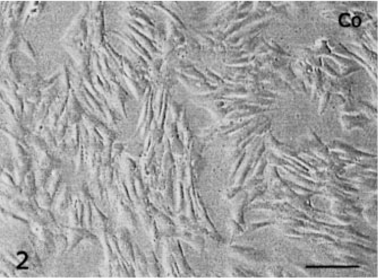
Chondrocytes; Horse, Passage 4. Bar = 100 µm. Fig. 2. Control group after 5 days of culture.
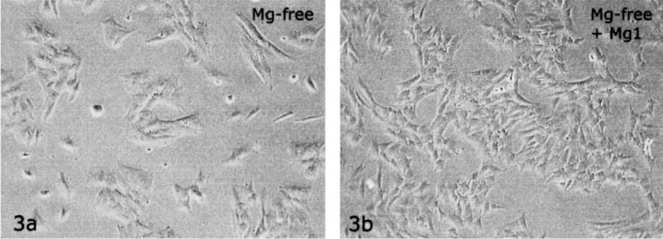
Chondrocytes cultivated in Mg-free medium for 5 days.
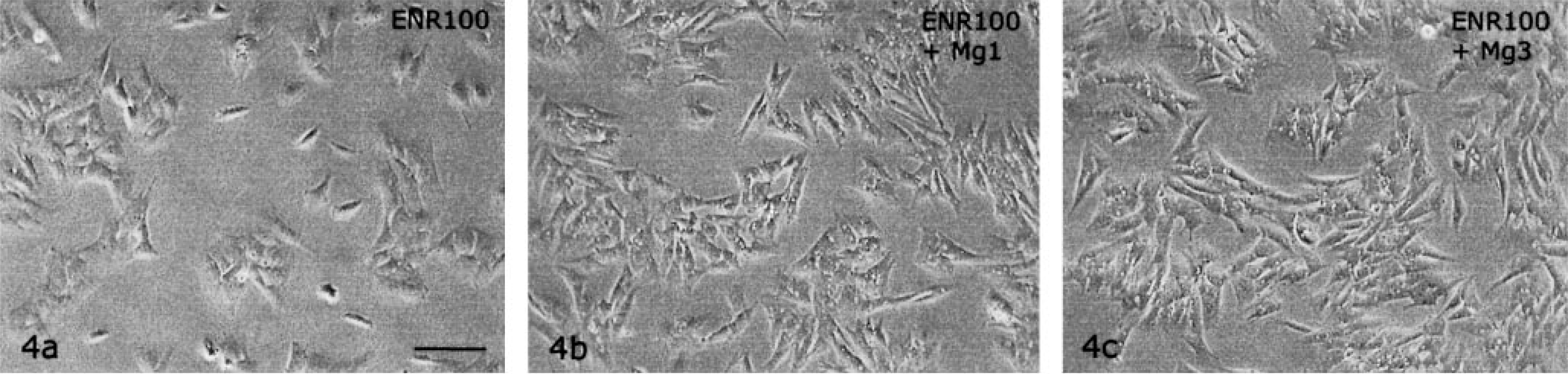
Chondrocytes cultivated in the presence of 100 µg/ml enrofloxacin (ENR 100) for 5 days.
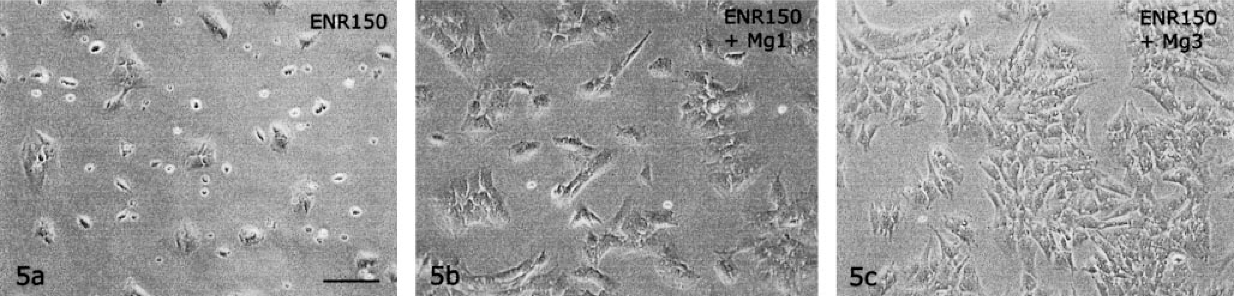
Chondrocytes cultivated in the presence of 150 µg/ml enrofloxacin (ENR 150) for 5 days.
Immunohistochemical staining for Ki67 proliferation marker revealed a dose-dependent decrease of cell proliferation. Whereas 70% of chondrocytes in control groups were Ki67 positive, proliferation rates were reduced to 55% (ENR 50), 48% (ENR 100), and 30% (ENR 150), similar to reductions in CFX-treated groups (53% for CFX 40 and 49% for CFX 80). It was not possible to evaluate the CFX 160 group because of severe cell loss.
Addition of Mg2+ did not increase the rate of cell proliferation (e.g., in test group CFX 80 + Mg1, cell proliferation rate was 49.2% and in CFX 80 + Mg3 it was 49.1%). In groups cultivated in Mg2+-free medium, we found 47% Ki67-positive cells, and addition of Mg2+ only slightly increased cell proliferation (53% for Mg1 and 55% for Mg3). Results of determination of the proliferation rate are depicted in Fig. 6.
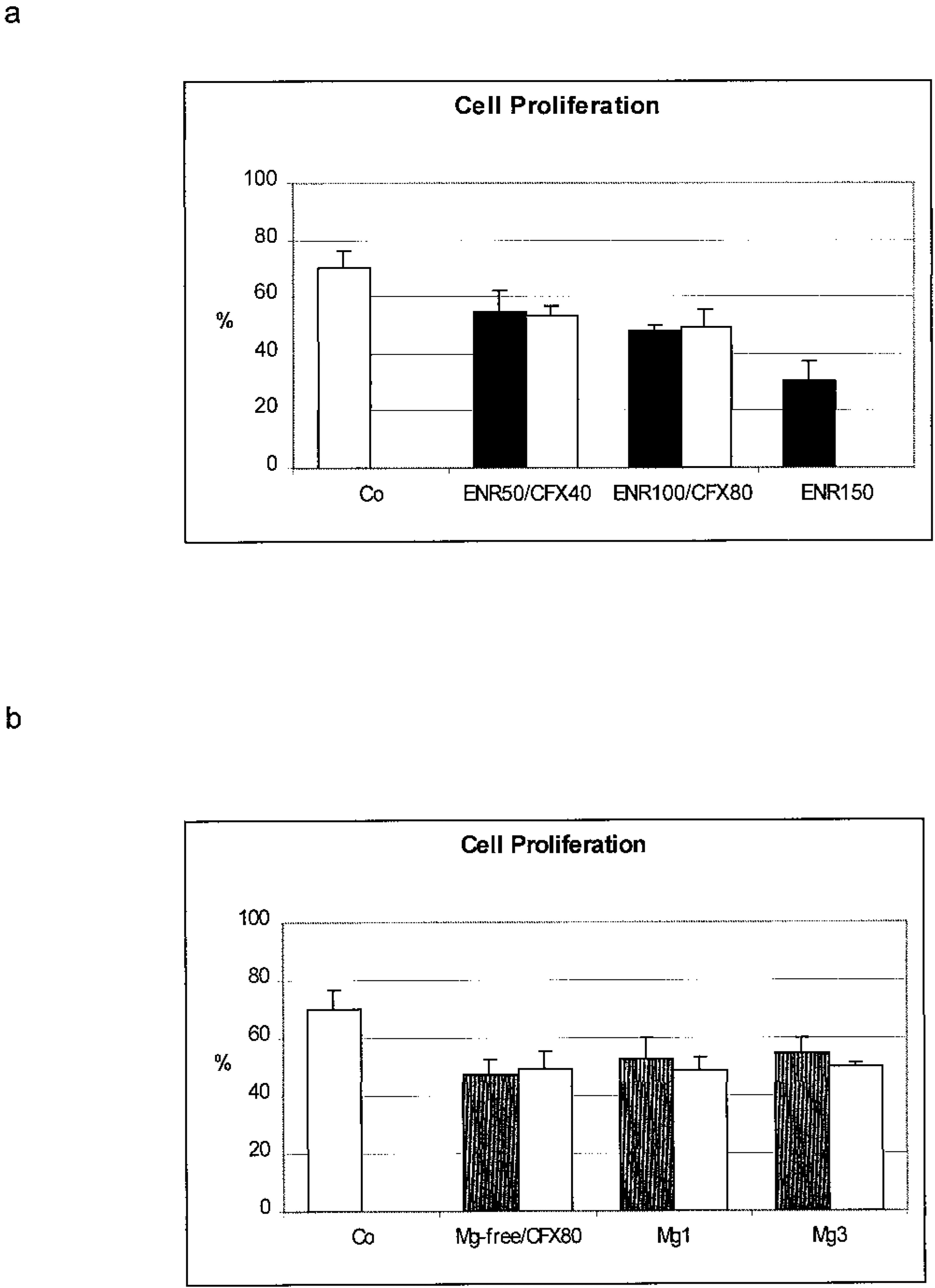
Cell proliferation (% of Ki67-positive Chondrocytes ENC = enrofloxacin; CFX = ciprofloxacin).
Discussion
All members of the fluoroquinolone family have the potential to damage articular cartilage in juvenile mammals. 3 13 19 All fluoroquinolones form stable chelate complexes with divalent cations, preferably Mg2+, situated between the ketone and the carboxylate groups. 18 22 This common property has led to the speculation that Mg2+ deficiency is a primary cause of cartilage damage. Studies on juvenile rats showed that feeding a Mg2+-deficient diet led to lesions identical to those found with quinolone treatment. 23 26 28 This finding was confirmed by our in vitro studies cultivating chondrocytes in Mg2+-free medium. In previous studies, quinolone treatment plus Mg2+-free medium increased the negative effects on cell morphology and adhesion seen in the quinolone or Mg2+-free groups. 9 10 11 In the present experiments, we examined whether Mg2+supplementation could compensate for quinolone-induced effects in chondrocyte culture. Significant positive effects were observed in all Mg2+-treated groups, especially in the triple-dose groups. These results are in agreement with in vivo experiments with immature rats. 24 This positive effect was specific for ciprofloxacin, enrofloxacin, and Mg2+-deficient groups, whereas addition of Mg2+ had no influence on control cells.
Mg2+ supplementation had a twofold effect on quinolone-treated cultured chondrocytes: the number of cells adhered to the culture support was increased, and cell morphology was comparable to that of control cells. These results suggest that addition of magnesium results in restoration of extracellular Mg2+-dependent interactions, such as integrin function. Integrins bind Mg2+ ions mainly at the EF hand-like motif and the I-domain of the alpha subunit and mediate a number of processes such as cell adhesion, migration, and growth. 7 8 21
Mg2+ supplementation never led to a full recovery of treated chondrocytes to the level of control cells. Therefore the proliferation rate of cells in quinolone-treated and Mg2+ supplemented groups was tested. As in previous studies, cell proliferation was impaired after quinolone treatment. 10 11 Cell proliferation decreased in a dose-dependent manner. This finding is in accordance with those of other authors, who observed reduced DNA synthesis in cultured rabbit chondrocytes after levofloxacin treatment. 17
The rate of cellular proliferation in both quinolone-treated and Mg2+ free medium was not responsive to Mg2+ supplementation. Addition of Mg to Mg2+-free medium was expected to result in cell growth comparable to that of the control group. However, this was not the case. The proliferation rate in Mg2+ free groups was as low as that of controls and increased only slightly with Mg2+ supplementation (Fig. 6). This slow rate of growth may explain why the number of cells never reached the amount for the control groups. This effect could be due to the influence on different Mg compartments (i.e., drainage of preexisting intracellular Mg2+ stores induced by the initial lack of available Mg2+ ions), the timing of supplementation, or the proportion of ions in the culture medium. Possible other reasons for these observed effects could be the production of oxygen-derived species, as described by other authors. 14 24 27 The number of lesions in articular cartilage was significantly reduced with magnesium plus vitamin E supplementation. 24 Also, quinolones form chelate complexes with other divalent cations, such as Ca, Mn, and Zn, whose influence in connection with quinolones has not been investigated.
Our results confirm that Mg2+ supplementation had a significantly positive effect on quinolone-treated chondrocytes, as shown by in vivo experiments. 24 As a practical consequence, before treating young animals with quinolone antibiotics, the overall magnesium intake should be checked, and oral Mg2+ supplementation might be considered. To avoid pharmacokinetics interactions, Mg2+ and quinolones must be administered by different routes (orally versus intravenous or subcutaneous). Despite these findings, caution should be taken when treating juvenile animals with quinolones because Mg2+ supplementation may not fully prevent damage but may only improve observed effects 24 and because high levels of magnesium antagonize the penetration of fluoroquinolones into the bacterial cell and therefore increase the minimum inhibitory concentration. 18
Footnotes
Acknowledgements
We thank Magdalena Helmreich for excellent technical assistance.