
Abstract
Separation of the femoral capital epiphysis is associated with severe trauma in most species. This report describes 13 cats with slipped capital femoral epiphysis characterized by a distinctive lesion in the physeal cartilage. The lesion consists of irregular clusters of chondrocytes separated by abundant matrix on both the epiphyseal and metaphyseal side of the cleavage site. The affected population in this study is 85% male, 90% overweight, 23% Siamese, and 4.5–24 months old. The histopathology and demographics are similar to slipped capital femoral epiphysis in humans, which most often affects overweight adolescent boys.
Keywords
Separation of the femoral capital epiphysis is usually associated with severe trauma in dogs, 11 19 28 calves, 20 and foals. 6 15 26 The condition in pigs is termed epiphysiolysis and is considered a manifestation of osteochondrosis, with only minimal trauma required. 48 Atraumatic slipped capital femoral epiphysis has also been described in the coypu (Myocastor coypus). 25 In humans, the condition is well described as an atraumatic separation that occurs most often in obese adolescent males. 7 This report describes a unique histopathologic lesion associated with physeal separation in 13 cats that have similar sex distribution and weight abnormalities as the human cases.
In small animals, the physes of the femoral head and greater trochanter contribute approximately 30–40% of the longitudinal growth of the femur. 36 The physis is made up of five zones: 1) reserve chondrocytes, 2) proliferating chondrocytes, 3) mature chondrocytes, 4) hypertrophic chondrocytes, and 5) calcified cartilage. 8 Chondrocytes in the reserve zone are scattered singly or in couples (mitotic pairs) within relatively abundant cartilage matrix. Cell division and matrix production occur at a slow rate within this population. In the proliferating, mature, and hypertrophic chondrocyte zones, the cells are arranged in rows that are parallel to the long axis of the bone. The cells are round in the reserve zone, flattened with their long axis transverse to the long axis of the bone in the proliferating zone, and polygonal with abundant cytoplasmic glycogen in the hypertrophic zone. The cells within the hypertrophic zone undergo an eightfold increase in cell volume without any increase in bone width. Therefore, the amount of matrix between the cells is markedly decreased. 8 It is this zone of hypertrophied cartilage cells separated by thin strands of matrix that is the weakest and experimentally the most likely to fracture following trauma. 28
Traumatic fractures of the growth plate have been classified by Salter and Harris into five types, with decreasingly favorable prognoses for normal growth plate function with each succeeding type. 43 The true slipped epiphysis is classified as type I, in which only the physis is involved. In small animals, this is the most common type of physeal fracture and the proximal femur is the most common site. 36 The histopathology of this type of fracture has been described in dogs euthanized following motor vehicle trauma. The findings in these dogs differed from experimental animals in that the fracture lines often crossed through multiple zones of the physis resulting in separation of the proliferative cartilage from the epiphysis. However, the chondrocytes retained their linear arrangement on both sides of the fracture site for 1–4 days following the injury. 28
Slipped capital femoral epiphysis (SCFE) is a well-described entity in humans, which primarily affects overweight adolescent boys. 7 30 33 The male-to-female ratio ranges from 2:1 to 4:1. 30 37 The average age at diagnosis is slightly younger for girls (12 years) than boys (13 years). 30 33 The onset is atraumatic and often insidious. 7 Twenty-one to 80% of reported cases are bilateral. 33 Undiagnosed cases are thought to be a frequent cause of degenerative joint disease of the hip in later life. 7 44 Blacks are more often affected than whites, and the familial incidence ranges from 3 to 35%. 5 37 Males and females are equally affected within family groups, and an autosomal dominant inheritance with variable penetrance is suggested. 5 37 This report describes the histopathology of SCFE in cats with similar weight, sex, age, and clinical characteristics as SCFE in humans.
Materials and Methods
Sixteen femoral heads and five femoral necks from 13 cats submitted to the University of Pennsylvania School of Veterinary Medicine biopsy service between 1987 and 2000 were reviewed. The tissues were emersion fixed in 10% formalin, decalcified in 15% formic acid, sectioned along a frontal plane to include the ligamentum teres, rinsed and dehydrated in a graded series of ethanol, and embedded in paraffin. Five-micrometer sections were stained with hematoxylin and eosin (H&E), periodic acid-Schiff (PAS), PAS following digestion with amylase (PAS-diastase), and Alcian blue (1% Alcian blue in 3% acetic acid, pH 2.5). Control tissues consisted of proximal femurs from cats with open growth plates (3–8 months of age).
The medical records of the 13 affected cats were reviewed for age, weight, breed, sex, history of trauma, duration of symptoms, and bilaterality of limb involvement. The comparison of weights of the affected cats to unaffected age-matched controls and the comparison ages of the affected males to affected females were compared by t-test. The sex and breed distributions were compared to the biopsy service population (13,250 cats) and measured by Fisher's exact test. P values less than 0.05 were considered significant.
Results
Of the 30 sections of femur submitted from cats between 1987 and 2000, 13 consisted of femoral heads with very similar lesions. The characteristic findings were an intact epiphysis consisting of viable bone (Fig. 1) with an unusually wide physis containing irregular clusters of chondrocytes in an abundant extracellular matrix and necrotic cartilage at the cleavage site (Fig. 3). Five of those 13 submissions also included the femoral neck (Fig. 2). The histopathology of the femoral necks was very similar, with necrotic cartilage at the cleavage site, chondrocyte clusters, and viable metaphyseal bone (Fig. 4). The chondrocytes within these abnormal clusters were round to polygonal, with scant cytoplasm and minimal cytoplasmic glycogen (Figs. 3, 4), most closely resembling the mitotic pairs of the reserve zone. The thickness of the physis in the affected cats (estimated by combining the remaining physis attached to the epiphysis and metaphysis) was over twice that of the normal physis of an 8-month-old cat (Fig. 5).
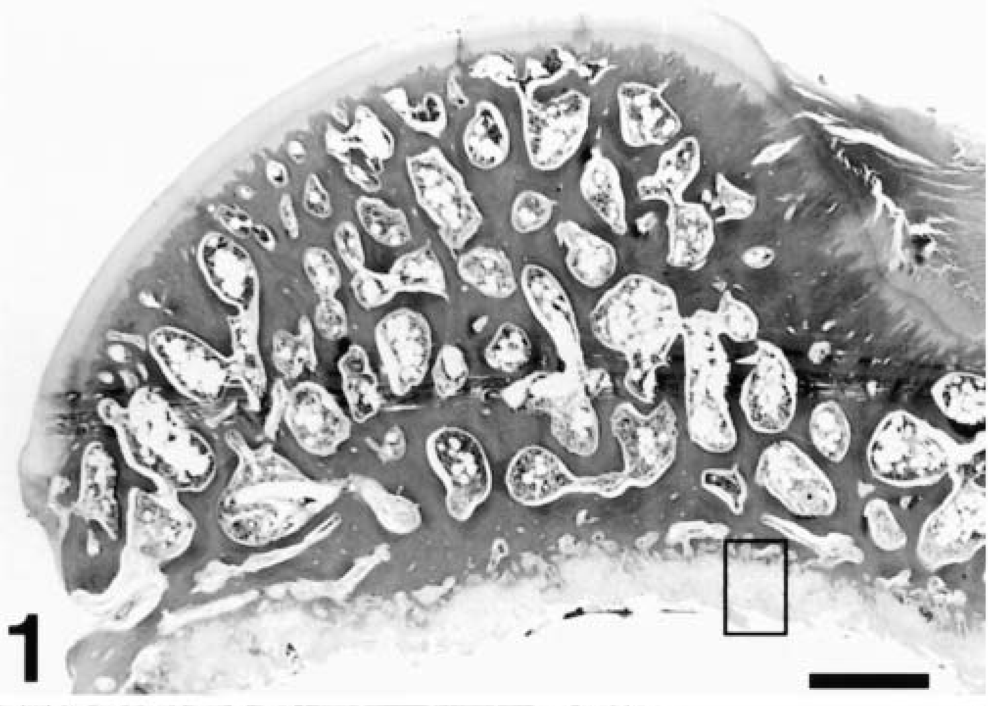
Femoral capital epiphysis; cat 2. The entire slipped capital epiphysis with viable bone and marrow. Notice the abnormally thick physeal cartilage along the cleavage site. H&E. Bar = 1,111 µm.
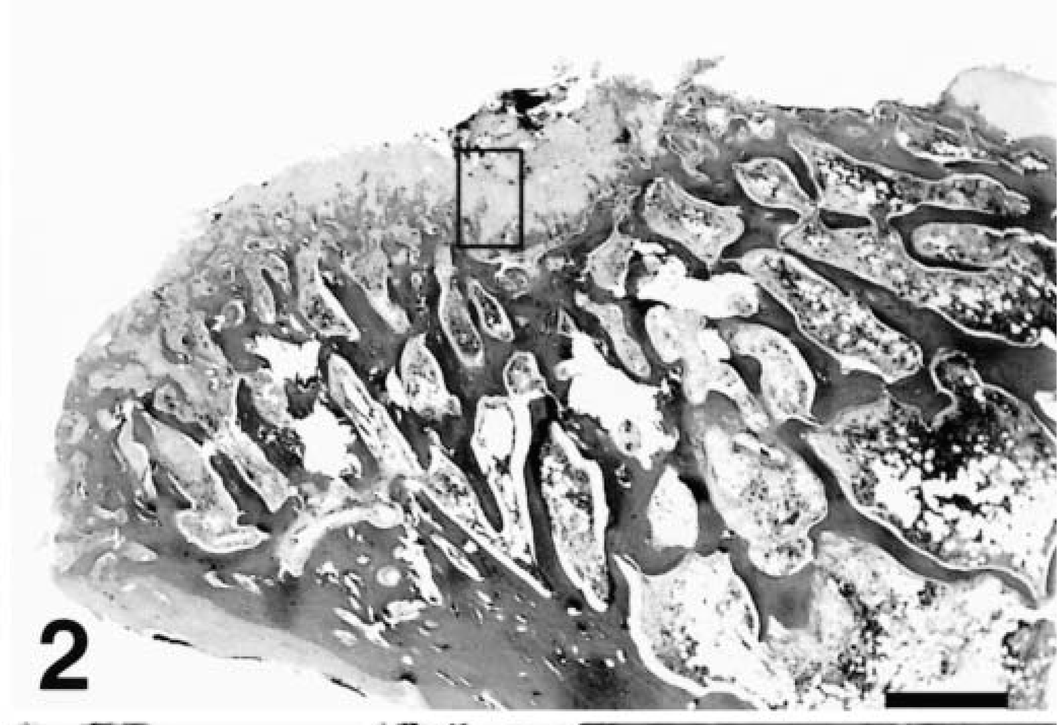
Femoral metaphysis; cat 2. The femoral neck metaphysis with abnormally thick physeal cartilage along the cleavage site. H&E. Bar = 1,111 µm.
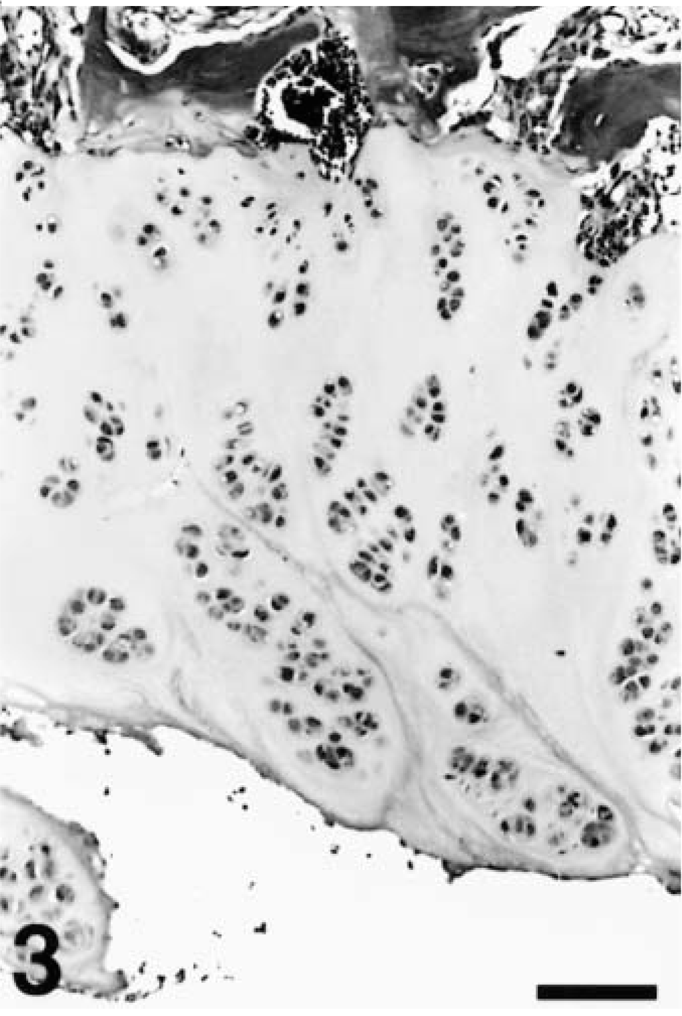
Epiphyseal side of physeal separation; cat 2. Higher magnification of rectangular area in Fig. 1 illustrating irregular chondrocyte clusters in an abundant extracellular matrix attached to the epiphysis. H&E. Bar = 84 µm.
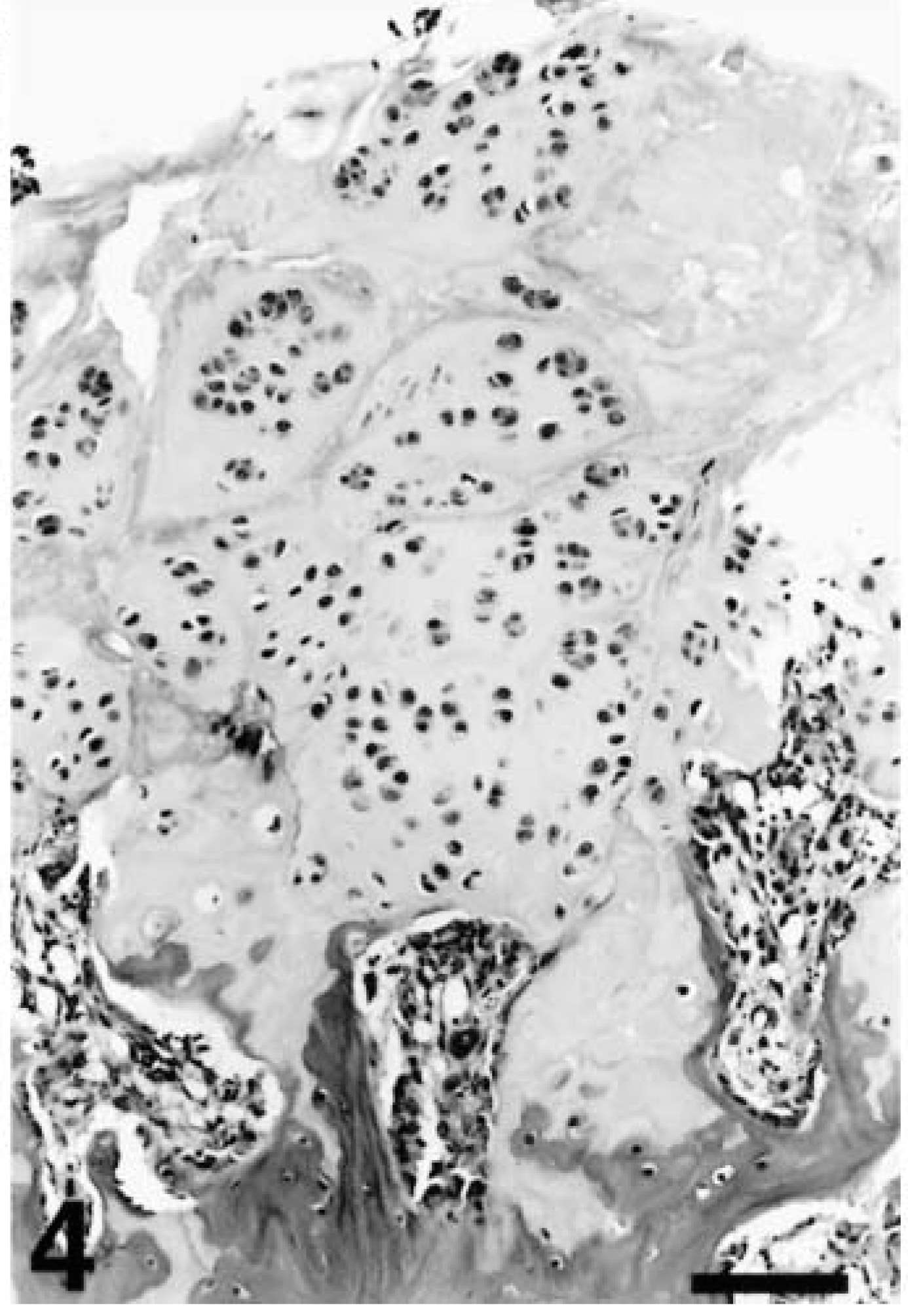
Metaphyseal side of physeal separation; cat 2. Higher magnification of rectangular area in Fig. 2 illustrating irregular chondrocyte clusters attached to the metaphysis. H&E. Bar = 84 µm.
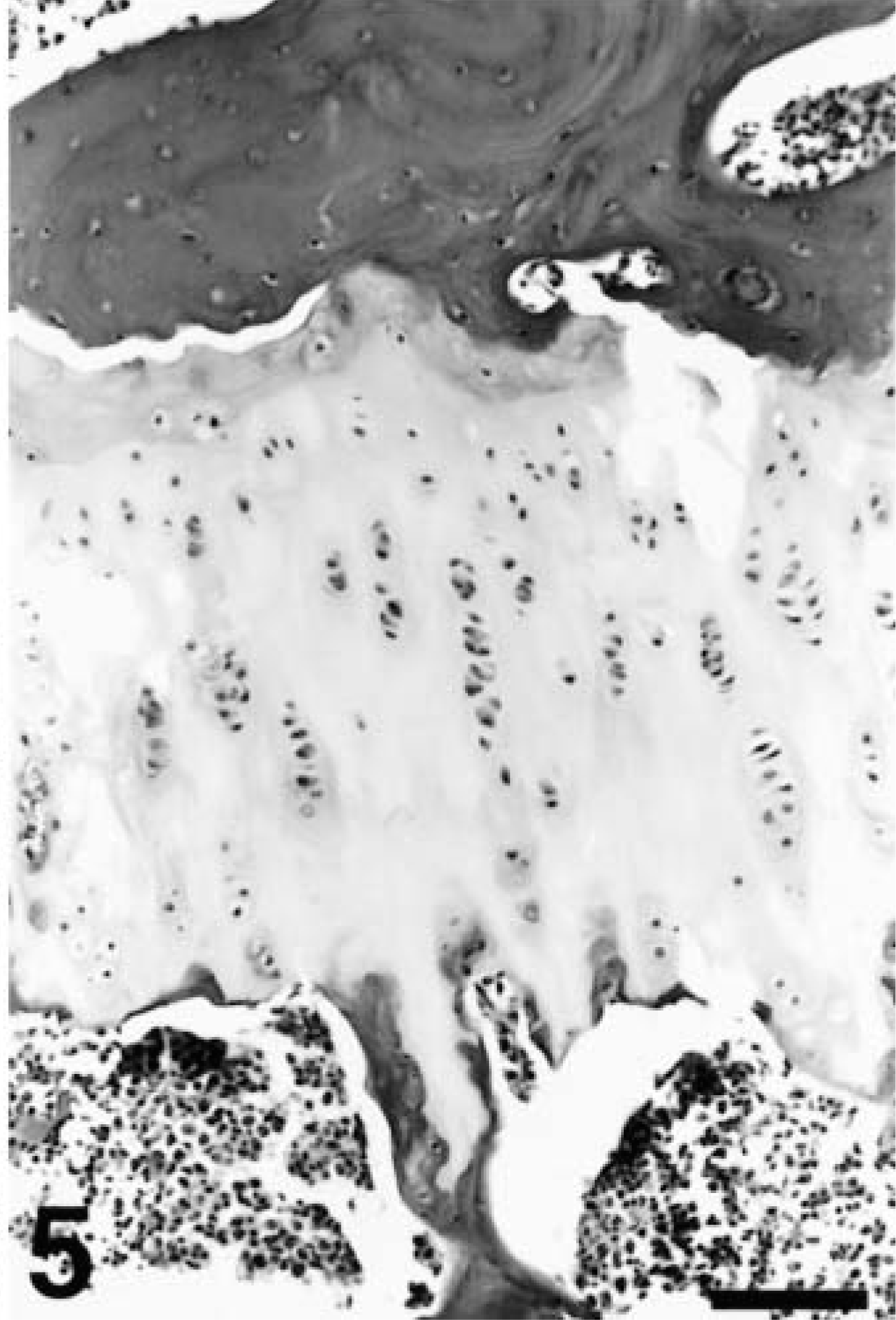
Normal physis; 8-month-old cat. Notice the regular linear arrangement of chondrocytes and the thickness of the physis compared with Figs. 3 and 4. H&E. Bar = 84 µm.
In six cats, there were also multifocal areas of granulation tissue at the cleavage site consistent with callus formation. These cats also had fibrillation of the superficial articular cartilage, presumably a secondary change indicating early degenerative joint disease. The duration of symptoms in those cases ranged from 2 to 8 weeks. The articular cartilage of the remaining cats was normal. In one case, the epiphyseal bone and marrow was necrotic, but the characteristic chondrocyte clusters were present and there was no evidence of collapse.
The Alcian blue and PAS stains were the most useful for staining the extracellular matrix of the affected and unaffected growth plates. The Alcian blue staining pattern of the normal growth plates (from a 3-month-old cat) had irregular clumps of densely stained matrix in a lightly stained background in the resting zone, which gave way to intense staining of the territorial matrix (perilacunar rims and transverse septa) and light staining of the extraterritorial matrix (all matrix away from the chondrocytes) in the proliferative zone. The affected growth plates had irregular clumps of densely stained matrix, similar to that in the resting zone of the normal growth plate, which extended throughout the remaining physis to the cleavage site. The PAS staining was diffusely more intense in the extracellular matrix of the resting zone of the normal growth plates than in the other zones. The PAS staining of the affected growth plates showed no such stratification of PAS staining. PAS staining was not affected by amylase treatment.
The sex, age, weight, and breed of the affected cats are shown in Table 1. Eighty-five percent (11/13) of the affected animals were male. This is significantly greater (p = 0.009) than the percentage of males in the biopsy population (45.5%). The average age of the affected cats was 16.3 months (range 4.5–24 months). The males (n = 11) were significantly older (average 17.8 months) than the females (n = 2; average 8.4 months; p = 0.04).
The breed, age, sex, weight, and bilaterality of the 13 cases of feline physeal dysplasia. Asterisks indicate animals described as overweight or obese. DSH = domestic short hair; SIAM = Siamese; M = male; F = female; MC = male casterated; FS = female spayed; NA = information not available.
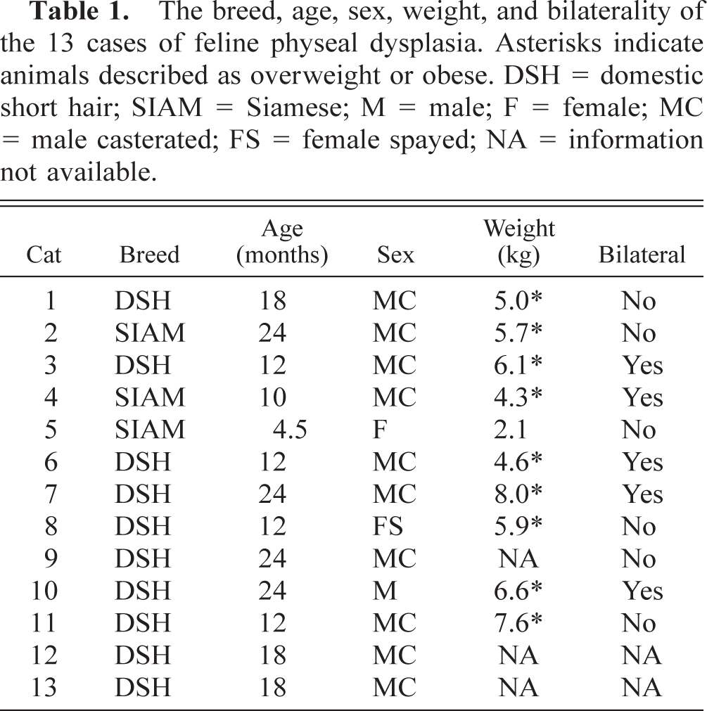
Nine of the ten cats for which weights were available were considered overweight or obese. The average weight of the affected cats (5.6 kg) was significantly higher than that of the control population (4.4 kg; p = 0.015). Twenty-three percent (3/13) of the affected cats were Siamese. This is significantly greater (p = 0.025) than the percentage of Siamese in the control population (5%; 662/13,250). There was no difference between the average weights of the affected Siamese and domestic short hair cats.
Five of the 13 cats had bilateral SCFE. Most presented with simultaneous bilateral lameness. However, cat 6 had two separate surgeries 1 month apart and cat 4 had surgeries 2 months apart, with only the first (left) femoral head submitted for examination.
None of the cats had a history of trauma prior to the onset of lameness. All 11 of the 13 cats for which information was available were indoor-only cats. Cat 8 was reported to have a male littermate with a very similar lameness, but additional information regarding that cat was not available. The 11 cats for which follow-up information was available had uneventful recoveries and good limb function following femoral head ± neck excision.
Discussion
The radiographic features of slipped capital femoral epiphysis (SCFE) in 10 cats have recently been reported by Forrest et al. 18 The age range (12–21 months) and male predisposition (90%) of the affected cats in that study were similar to the findings here. However, the weights of the cats and histopathologic findings were not reported. A single case of bilateral SCFE has also been reported in a cat in Switzerland. 46
Plain radiography is often diagnostic of SCFE, and in humans, the femoral heads are rarely excised. 7 Therefore, there are few reports of the histopathology of this condition in humans. One surgical correction method involves removal of a cylindrical core of growth plate from the femoral neck and replacement with a peglike graft of iliac bone. The light microscopic and histochemical studies of these small samples showed changes identical to those described here in cats. 1 The ultrastructural study demonstrated haphazardly arranged collagen, decreased amounts of collagen, and chondrocyte degeneration and death. 2
The age of the cats in this study ranged from 4.5 to 24 months. Since this lesion requires an open physis, all but one (cat 5) of the cats in this study are older than expected. The growth plates close between 7 and 9 months in normal, healthy, nonneutered cats. 38 Although gonadectomy is known to delay physeal closure, 42 one of the affected cats in this study was a 24-month-old intact male that had bilateral SCFE (cat 10). Therefore, the prolonged growth phase caused by early neutering cannot explain this lesion in all cases. The histopathology of this lesion in cats is suggestive of a physeal dysplasia that results in persistence of an open, disorganized growth plate that cannot resist the shear forces associated with normal activity. It is unclear whether obesity contributes to the slippage by the increased trauma of additional weight or whether it is another manifestation of an underlying metabolic disorder that results in both physeal dysplasia and obesity.
Epiphysiolysis in pigs is considered to be a manifestation of osteochondrosis in which a defect in endochondral ossification results in a weakened physis. It may be unilateral or bilateral and occurs with equal frequency in sows and boars between 5 months and 3 years of age. 32 Although nutritional, genetic, and toxic etiologies have been investigated, only rapid growth and excellent body condition have been consistently associated with the syndrome. 13 44 Vascularization disorders of the metaphysis 48 and physis 9 have been suggested, but the histopathology of the lesion closely resembles that of humans and cats, i.e., clusters of chondrocytes on both sides of the cleavage site. 24 Irregular chondrocyte clusters have also been described in a small percentage of clinically normal pigs, 47 suggesting that the chondrocyte lesion precedes the separation.
Osteochondrosis is a multifactorial disorder that has many clinical manifestations in a variety of species. The typical growth plate lesion is characterized by a focal failure of endochondral ossification that results in retained cartilage that extends into the metaphysis. The chondrocytes within this metaphyseal cartilage core maintain their alignment but persist because they are not replaced by bone. 14 Feline physeal dysplasia differs from osteochondrosis in two ways: 1) it is more diffuse, affecting the entire physis, and 2) the chondrocytes are arranged in disorganized clusters rather than parallel rows. In addition, domestic short hair and Siamese cats have not been selected for rapid growth, a factor closely correlated to the incidence of osteochondrosis in dogs, pigs, poultry, and horses. 14 23
Femoral capital physeal fractures in foals are typically associated with severe trauma. 15 45 In calves, SCFE is more common in the heavily muscled breeds and is often related to dystocia and forced traction delivery. 20 In both foals and calves, there is a slight female predominance and bilateral cases are rare. 6 15 20 26
Proximal femoral epiphyseal separation in the dog is usually associated with severe trauma, such as being hit by a car. 19 28 These are most often classified as Salter–Harris type I fractures. 11 43 In some reports, there is no sex or breed predisposition, 10 11 while others report large breed male dogs to be overrepresented, as they often are in reports of other traumatic injuries. 19 28 31 Histopathologic studies of these cases report that the fracture line crosses through multiple zones of chondrocytes but without loss of the linear orientation of those cells. 28
In one recent report of bilateral slipped capital epiphysis in two overweight male Shetland sheepdogs, the separation occurred without a history of trauma and the histologic appearance was similar to the cats reported here. 12 The term epiphysiolysis was used in that report as it is in pigs. However, since this is a disease of the physis (formerly termed the epiphyseal plate), the term physeal dysplasia (first used by Forrest et al. 18 ) is more accurate.
Experimental separation of the femoral head in rabbits results in consistent cleavage through the zone of hypertrophied chondrocytes and rapid avascular necrosis of the epiphysis without the formation of chondrocyte clusters. 22 These are similar to findings in the Salter–Harris type I fracture in humans 43 and dogs. 11 31 It is surprising that so few cases of SCFE in cats and humans result in avascular necrosis since the entire femoral capital epiphysis is intra-articular and therefore dependent on the ascending arteries of the femoral neck. 4 43 However, surgeons removing the femoral heads of dogs with atraumatic SCFE 12 and the surgeons who removed the femoral heads of the cats in this study report that the physis is not completely separated but attached at the periphery along the perichondral fibrocartilaginous complex. Presumably the slower separation in SCFE allows the arteries of the synovial membrane plexus to remain attached and supply the separated epiphysis. 4 The ligamentum teres does not contribute to the blood supply of the femoral head. 4 17 29
Endocrine disorders that affect the life cycle of physeal chondrocytes are diagnosed in 5–30% of humans with SCFE. 34 39 Hypothyroidism and growth hormone deficiency are the most commonly diagnosed disorders. 34 Thyroxine (T4) is necessary for proliferation and maturation of chondrocytes, and growth hormone stimulates mitosis of the proliferating chondrocytes via somatomedins such as insulin-like growth factor 1. 35 Interestingly, experimental studies in rats have shown that exogenous growth hormone results in a thicker, more fragile physis. 21 41 The role of these hormones in the pathogenesis of SCFE is still poorly understood. Hypothyroidism and growth hormone deficiency have rarely been reported in cats. 16 None of the cats in this study showed any signs of endocrine imbalance other than obesity.
One hypothesis to explain the relationship between obesity and the physeal lesion is an abnormality in insulin metabolism. Insulin acts as a survival factor preventing chondrocyte death, 27 and insulin-like growth factor 1 is a chondrocyte and adipocyte mitogen. 3 40 In horses, high circulating levels of insulin from high energy feeding has been hypothesized to affect chondrocyte maturation and lead to osteochondrosis. 27 A similar mechanism could explain the obesity and physeal dysplasia in these cats. Alternately, there could be an underlying abnormality in insulin metabolism, such as a receptor defect, that could result in both adipocyte and chondrocyte survival. The prolonged chondrocyte survival in the absence of other appropriate growth factors to direct differentiation would result in the persistence of an open, disorganized physis. This hypothesis is supported by the similarities between the morphology of the chrondrocytes forming the abnormal clusters and those within the reserve zone of normal growth cartilage.
The higher than expected number of Siamese cats affected, the male predominance, and the suspected occurrence of SCFE in sibling cats (cat 8 had a male littermate that was also lame) are all supportive of a genetic etiology. The pathogenesis of SCFE in humans is poorly understood, 44 and pathological materials are rarely available. 1 2 The similarity of the lesion in humans, pigs, Shetland sheepdogs, and cats suggests that these species have an analogous physeal dysplasia that results in persistence of an open, disorganized growth plate that cannot resist the shear forces associated with normal activity. This lesion is responsible for a small minority of SCFE in dogs, and the condition in pigs does not have the same male predominance as in cats and humans. Therefore, the cat may serve as an animal model in which to study the role of genetics, nutrition, obesity, endocrine imbalances, and other factors in the development of this lesion.
Footnotes
Acknowledgements
I thank Dr. Frances S. Shofer for assistance with statistical analyses.