
Abstract
A 7-year-old spayed female domestic shorthair feline presented with tachycardia and was later euthanized due to a declining condition. On gross examination, the thoracic cavity contained an expansile, multiloculated mass that displaced the lungs dorsocaudally. The mass, within the pericardial sac, compressed adjacent myocardium. Cut surface revealed variably sized, fluid-filled spaces with multiple foci of hemorrhage and necrosis. Histologically, the mass was composed of solid foci of polygonal cells admixed with colloid-containing follicles. Immunohistochemical staining for thyroglobulin was positive, and staining for calcitonin was negative. Grossly, thyroid glands were normal, and serum thyroxine was within reference intervals.
Thyroid neoplasia occurs most commonly in aged dogs, cats, and horses. 3 Thyroid nodules in older cats typically represent functional multinodular (adenomatous) hyperplasia associated with hyperthyroidism. 1 Malignant tumors in cats are highly metastatic and also produce excess thyroid hormone. 1 Although thyroid masses in the dog are less common than in the cat, they are usually malignant. 1 Hyperthyroidism usually does not occur, and clinical signs tend to result from the mass impinging on surrounding tissues. 1 In horses, thyroid adenomas are more common than carcinomas. 3 Thyroid nodules in human beings are usually benign. 1 In dogs, cats, and human beings, tumors usually arise from epithelial follicular cells, but tumors can also arise from parafollicular C cells, especially in the horse. 1,3 In addition, thyroid lymphoma and sarcoma can arise from lymphocytes and stromal cells, respectively. 1
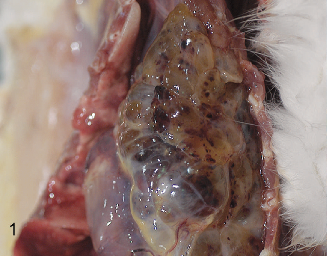
Thorax; cat. Expansile, multiloculated, fluid-filled mass within the thoracic cavity.

Heart; cat. Thoracic mass contained within the pericardial sac and compressing adjacent myocardium.

Intrapericardial ectopic thyroid carcinoma; cat. Neoplastic tissue composed of solid foci of polygonal cells admixed with colloid-containing follicles. Hematoxylin and eosin. Bar = 1.0 mm.

Intrapericardial ectopic thyroid carcinoma; cat. Polygonal neoplastic cells lining colloid-containing follicles. Hematoxylin and eosin. Bar = 100 μm.

Intrapericardial ectopic thyroid carcinoma; cat. Polygonal neoplastic cells with distinct borders, moderately abundant eosinophilic cytoplasm, and round to oval nuclei with stippled chromatin and 1 magenta nucleolus. Hematoxylin and eosin. Bar = 30 μm.

Intrapericardial ectopic thyroid carcinoma; cat. Colloid, neoplastic cells lining follicles and neoplastic cells within solid areas showing positive staining thyroglobulin. 3,3′-diaminobenzidine chromogen, Hematoxylin counterstain. Bar = 60 μm.
Clinical signs in cats with both benign and malignant thyroid neoplasms include anorexia, weight loss, hyperactivity, polydipsia, polyuria, and increased defecation and stool volume. 1,4 There may be an abnormal coat, tachycardia, or a heart murmur. 1 Concentrations of triiodothyronine and thyroxine are elevated along with liver enzyme activities. 1 Cardiomyopathy may occur with benign and malignant tumors. 1 The current report describes the clinical history and gross, microscopic, and immunohistochemical features of an intrapericardial ectopic thyroid carcinoma in a cat.
A 7-year-old spayed female domestic shorthair feline presented with a 1-month history of vomiting, lethargy, anorexia, and weight loss. On physical examination, the cat had an unkempt hair coat, a tooth root abscess, submandibular lymphadenopathy, and tachycardia. Hematology findings included a macrocytic, hypochromic, nonregenerative anemia. The cat was euthyroid, and tests for Feline leukemia virus and Feline immunodeficiency virus were negative. Over the next month, the anemia worsened, and the cat developed a leukocytosis, characterized by a left shift with lymphocytosis, and a monocytosis. The leukocytosis, left shift, and monocytosis were attributed to the tooth root abscess and the lymphocytosis from chronic antigenic stimulation. Due to the declining condition, euthanasia was elected, and a postmortem examination was performed.
Gross examination revealed a thin cat with scant adipose tissue. A tooth root abscess of the right maxillary canine extended dorsally through the hard palate and maxillary sinus and emerged through a focus of ulcerated skin lateral to the right nares. Likely associated with oral findings, submandibular lymph nodes were enlarged twice their normal size. A 9 cm × 4 cm × 4 cm, expansile, multiloculated mass occupied the thoracic cavity and displaced the lungs dorsocaudally (Fig. 1). Cranial lung lobes were pale and collapsed, and other lobes were hyperemic and edematous. The mass was entirely contained within the pericardial sac and compressed the adjacent myocardium (Fig. 2). On cut surface, the mass was composed of variably sized, fluid-filled spaces with multiple foci of hemorrhage and necrosis. Differential diagnoses included neoplasms of ectopic thyroid or parathyroid tissue, chemodectoma, rhabdomyosarcoma, mesothelioma, hemangiosarcoma, thymoma, lymphoma, and metastatic neoplasms.
Tissues were fixed in 10% neutral buffered formalin, processed routinely, embedded in paraffin, sectioned at approximately 4 μm, and stained with hematoxylin and eosin. Immunohistochemical staining was performed for thyroglobulin and calcitonin using normal feline thyroid glands as a positive control. Antibodies used included anti-human thyroglobulin (rabbit polyclonal, 1:1,500) a and anti-human calcitonin (rabbit polyclonal, 1:1,000). a The substrate-chromogen system 3,3′-diaminobenzidine a was used to visualize the immune reactions.
Histologically, the mass was moderately cellular, well demarcated, and multilocular and was composed of polygonal cells arranged into solid areas and follicles of varying sizes and shape filled with a pale, eosinophilic material (colloid; Figs. 3, 4). The polygonal cells had distinct cell borders and moderately abundant eosinophilic cytoplasm. Nuclei varied from round to oval to fusiform and had finely stippled chromatin and 1 magenta nucleolus (Fig. 5). There was moderate anisocytosis and anisokaryosis. Mitoses were 3 per 10 high-power fields at 400×. There was no evidence of vascular invasion. Approximately 20% of the tissue was necrotic. Throughout the fibrovascular stroma were small collections of lymphocytes, plasma cells, and scattered neutrophils. Occasional follicles contained sloughed cells and small collections of free red blood cells. There was no evidence of metastasis to the submandibular lymph nodes, although they were grossly enlarged.
Staining for thyroglobulin was positive within colloid, neoplastic cells lining follicles and within neoplastic cells in solid areas (Fig. 6). Staining for calcitonin was negative (data not shown). Results indicate that the origin of the neoplastic cells was thyroid follicular epithelium. Therefore, other differential diagnoses were excluded. Based on the location, histologic features of malignancy, and immunohistochemistry, intrapericardial ectopic thyroid carcinoma was diagnosed.
Accessory thyroid parenchyma is relatively common in cats and dogs and may occur anywhere from the larynx to the diaphragm. 4,5 The aberrant tissue can result from failure to descend from the floor of the pharynx to the normal cervical location or from tissue descending beyond its normal adult location. 5 The thyroid is intimately related to the aortic sac in its development, and this explains the frequent finding of accessory thyroid tissue in mediastinal structures. 4 Approximately half of adult dogs have accessory thyroid tissue within the adipose tissue on the intrapericardial aorta, and tumors of this tissue should be considered in the differential diagnosis of heart-base tumors. 4
In cats, ectopic thyroid tissue has been reported in the tongue and in a mediastinal thyroglossal duct cyst. 8,10 A functional adenoma was reported from ectopic tissue within the thoracic inlet. 9 Intracardiac ectopic thyroid carcinomas have been reported in dogs. 2 Although ectopic thyroid masses have been reported within the pericardial sac of dogs and human beings, there are no reports in cats. 6,7,11 Therefore, it is important to consider these tumors as a differential diagnosis for cardiac-associated neoplasms in the cat.
Footnotes
a.
Dako North America Inc., Carpinteria, CA.