
Abstract
As the human and mouse genome projects approach their goals, initiatives in functional genomics are advancing. When the nucleotide sequences are available, identification of gene functions will assume even greater importance. Determination of gene products and their proximal biochemical functions provide a part of the picture, but determination of their functions in the context of the whole organism is the ultimate goal. The manipulated mouse genome has become accepted as a model for understanding the genetic basis of human conditions and diseases. Consequently, biomedical research institutions have seen significant increases in the use of mice since the early 1980s, and these increases are largely attributable to the use of genetically modified mice. The role of comparative pathology in research on mutant mouse models of disease is increasing in response to these trends. Evaluation and phenotypic characterization of mutant mice, via clinical and anatomic pathology techniques, will be an important component of functional genomics initiatives.
Reverse genetics and forward genetics are the two major approaches to functional genomics research (Table 1). 131 Reverse genetics or genotype-driven experiments typically involve directed mutagenesis. The gene, gene product, and often some of its proximal functions are known. An exogenous gene is inserted by insertional mutagenesis or transgenesis to create a transgenic mouse, or an endogenous gene is disrupted or removed by targeted mutagenesis to create a knockout mouse. Newer technologies, such as Cre-lox, generate conditional mutants that express the gene product only in specific tissues or at specific times in response to specific stimuli. The resultant genetically engineered mice (GEM) frequently are in short supply, and the desired phenotypic manifestation of the mutation, referred to simply as the phenotype, often is not immediately obvious or there may be an unexpected phenotype. Forward genetics or phenotype-driven experiments typically involve random mutagenesis. The G0 animals are exposed to a mutagen, and the offspring are evaluated for deviants or mutant phenotypes. N-Ethyl-N-nitros urea (ENU) is the most common mutagen used in mice today and produces point mutations scattered throughout the genome. 22 67 148 The male is exposed to ENU and then is bred to nonmutagenized females. In the first generation (G1), any deviants or mutant phenotypes should be the results of a dominantly expressed mutation that is expressed in the heterozygous state. The second or third generations (G2 or G3) are evaluated for recessively inherited mutations that are expressed in the hemizygous or homozygous state, respectively. Hundreds or thousands of G1, G2, or G3 mice may be evaluated by high-throughput, noninvasive methods to detect grossly visible or biochemical defects (phenotypes) of interest. 16 Then the genetic defect that caused the phenotype must be determined.
Research approaches and experimental techniques in functional genomics.
Genetic screening of offspring of mutagenized male mice or breeding strategies that expose mutations in specific genes or regions transform random mutagenesis into another genotype-driven technique. 95 DNA from G1–G3 mice can be screened for specific mutations or for mutations in specific genes or loci by sequencing techniques that have been facilitated greatly by progress toward genetic and physical maps of the mouse genome and creation of a database of expressed sequence tags and of cDNA libraries. 27 86 An extremely rapid, high-throughput technology that remains very expensive but holds great promise is microarrays that detect single nucleotide polymorphisms in DNA sequences. Microarrays also can be used to detect gene expression at various stages of development or disease progression. 154 The mutants of interest identified by these types of genetic techniques require phenotypic characterization to determine the functional effects of the mutations and ultimately to determine the biologic function of the mutated gene.
In this discussion, we emphasize anatomic pathology evaluations that are of primary utility in evaluating GEM produced by transgenesis or targeted mutagenesis and related technologies. There is no single ideal, affordable method to evaluate mutant animals, and this discussion is not intended to provide dogma regarding their evaluation. Methods will evolve based upon the objectives of the studies and the availability of emerging technologies, especially those that permit longitudinal studies of individual animals. Methods of evaluation must complement the logistic and economic resources, interests, and expertise of the institutions. Excellent texts are now available or forthcoming. 127 146
Some important considerations in the evaluation of mutant mice include 1) correct nomenclature for mice, genes, and mutations, 2) reporting methods and terminology, 3) reproducibility and consistency of techniques, 4) labeling and identification, 5) common lesions of background strains, 6) health and pathogen status of animals, 7) selection of animals, 8) comprehensive baseline characterization, 9) evaluation of early embryonic death and perinatal mortality, 10) stage 2 characterization (targeted evaluations), and 11) impact of new and emerging technologies.
Correct Nomenclature
Correct nomenclature for mice, genes, and mutations should be used instead of slang or jargon terms. Correct nomenclature provides important information regarding background strains, methods of generation of the mutant, and institutions of origin. 25 45 Although abbreviations may facilitate communication among people involved in a study, correct nomenclature is important to prevent confusion in communication with others or in publications and to prevent attempts to reproduce or continue studies on incorrect or inappropriate animals. A recent example of such confusion is “the 129 strain,” which recently was shown to comprise 16 substrains. 41 122 135 Appropriately and necessarily, reviewers of manuscripts are becoming more critical and demanding with regard to the use of correct nomenclature, and it is in the pathologists' best interest to require correct nomenclature from investigators and to use it in reports and publications. An apparently trivial but important point is that Roman-type (serif) fonts should be used instead of Arial-type (sans-serif) fonts (e.g., to distinguish 1, l, I). The most recent revisions of Nomenclature Guidelines and updates to gene names, mouse names, and registration codes are available via the World Wide Web. 25
Reporting Methods and Terminology
Reporting methods and terminology can vary markedly among laboratories. Compatibility of reporting methods with data entry, archiving, and information retrieval is becoming an increasingly important issue 3 as databases such as the International Mouse Strain Resource develop. 27 Use of widely accepted anatomy and pathology terminology is critical to effective communication, and “codeability” of the terms is an issue when database programs are in use or their use is anticipated. Coded terminology systems are used widely in industry, and it seems inevitable that their use will become more widespread. For those who are not using such systems already, it may be advantageous to prepare reports and documents in formats that are likely to be compatible with such systems. Advantages include simplified data entry, retrieval, and collation. A disadvantage of some available systems is that they tend not to be very plastic; they may not include lesions or changes peculiar to mice or to a specific mutant or accommodate new or different lesions easily, and clarifying comments in the report that are important to the study may not be retrieved easily. Comprehensive systems are being developed to facilitate data entry of experimental, genetic, and clinical information as well as clinical and anatomic pathology information, to coordinate quantitative information into spreadsheet and statistical programs, to archive images and verbal and quantitative data efficiently, and to permit easy retrieval of archived data in various formats via various selection criteria. In general, these systems remain cost prohibitive in academic settings, but impetus for functional genomics may help to improve the technology and accessibility of such systems.
A relatively common situation in studies involving mutant mice is that mice of different ages are examined months apart as they become available or even years apart in longitudinal aging studies. It may be important to reevaluate all specimens and reports when all specimens and data are finally available. Although the morphologic diagnoses should not change substantively, final evaluations should be based on all available data. Qualitative modifiers (e.g., mild, moderate, severe) seem to be especially prone to variation over time and among evaluators, and quantification should be used whenever possible.
Reproducibility and Consistency
Reproducibility and consistency are especially important with regard to specimen collection techniques and evaluation techniques. Often mice or specimens involved in a single study are submitted over long periods of time and by different graduate students or postdoctoral trainees. A clearly written outline of tests to be performed, equipment to be used (e.g., sizes of needles, syringes, tubes), and specimens to be collected and descriptions of any special techniques will provide continuity and prevent irreversible omissions that result in irreconcilable gaps in data. A detailed standard operating procedure (SOP) as is common practice in toxicology studies may be preferred, or a simple checklist format may suffice.
Labeling and Identification of Animals and Specimens
Labeling and identification of animals and specimens may seem to be simple procedures that are not the pathologists' responsibility. However, these procedures can become the pathologist's problem when, for example, the “wrong” mice have an important lesion. Mice of similar colors and sizes can be returned to the wrong cages, transponders can be lost, and other identification systems for live mice, such as ear punch, toe clip, and tattoos, can be difficult to implement clearly and to interpret subsequently. Redundant labeling and documentation by the pathology laboratory may prevent such problems and may provide information that can unravel confusion resulting from misidentified specimens or establish that the misidentification occurred before submission. Several precautionary practices can be applied in the pathology laboratory.
Attach the cage card to the submission/report form or save it with the specimens in fixative.
Include a drawing of ear punch or toe clip or other identifying features on the submission/report form, and save ears, toes, or other identifying features with specimens in fixative.
Save the pelt or representative specimens of it to document color and to demonstrate percentage of chimerism if applicable.
Label container bottoms as well as their lids. It is easy to apply the wrong lid when several containers are open. Saving labeled cassettes in the containers also facilitates identification of the correct container.
Select a cassette/slide numbering system that consistently places tissues/organs in the cassettes/slides of the same numbers, so that archived specimens can be retrieved easily.
Radiographs should include an internal label (i.e., not applied postprocessing) and a densitometry standard such as an aluminum step wedge that will facilitate subsequent densitometric and morphometric image analysis.
Photographs should include an internal label and a ruler to facilitate subsequent morphometric image analysis.
Common Lesions of Background Strains
Common lesions of background strains, also known by the oxymoron “normal pathology,” are becoming more widely recognized as an important issue in evaluation of mutant mice. Accurate assessment of the phenotypic result of a spontaneous or induced genetic mutation requires comprehensive knowledge of the phenotype of the background or control strain to recognize true diversions from “normal.” Among “normal” inbred strains of mice, there is significant genetic, morphologic, and physiologic variation and consequent variations in characteristics such as susceptibility to or manifestations of infectious diseases, tumor susceptibility, 32 83 84 93 129 141 longevity, 47 bone mass or density, 8 70 71 brain weight, 110 and brain anatomy. 147 Even closely related substrains can vary significantly with regard to immune status, fertility, behavior, and other phenotypic traits. 77 109 There is abundant clinical pathology and morphologic information on the B6C3F1 hybrid (offspring of C57BL/6 males mated to C3H females) that is used commonly in toxicology studies, 66 and the National Toxicology Program maintains a database of information representing more than 50,000 B6C3F1 mice. 85 Such information is widely dispersed in the literature regarding the inbred mouse strains that commonly are used in the development of mutant mice (e.g., C57BL/6, C3H, 129, FVB). The strains have changed over time and many generations, and specimen collection and evaluation techniques have changed as well. The Jackson Laboratory, with federal support, maintains information on inbred strains as well as several databases, including the Mouse Genome Database, Gene Expression Database, and Mouse Tumor Biology Database at Mouse Genome Informatics Web Site. 11 94 In addition, the Jackson Laboratory's product information resource provides additional data and references on strains currently available at the Jackson Laboratory, with links to their databases and references.
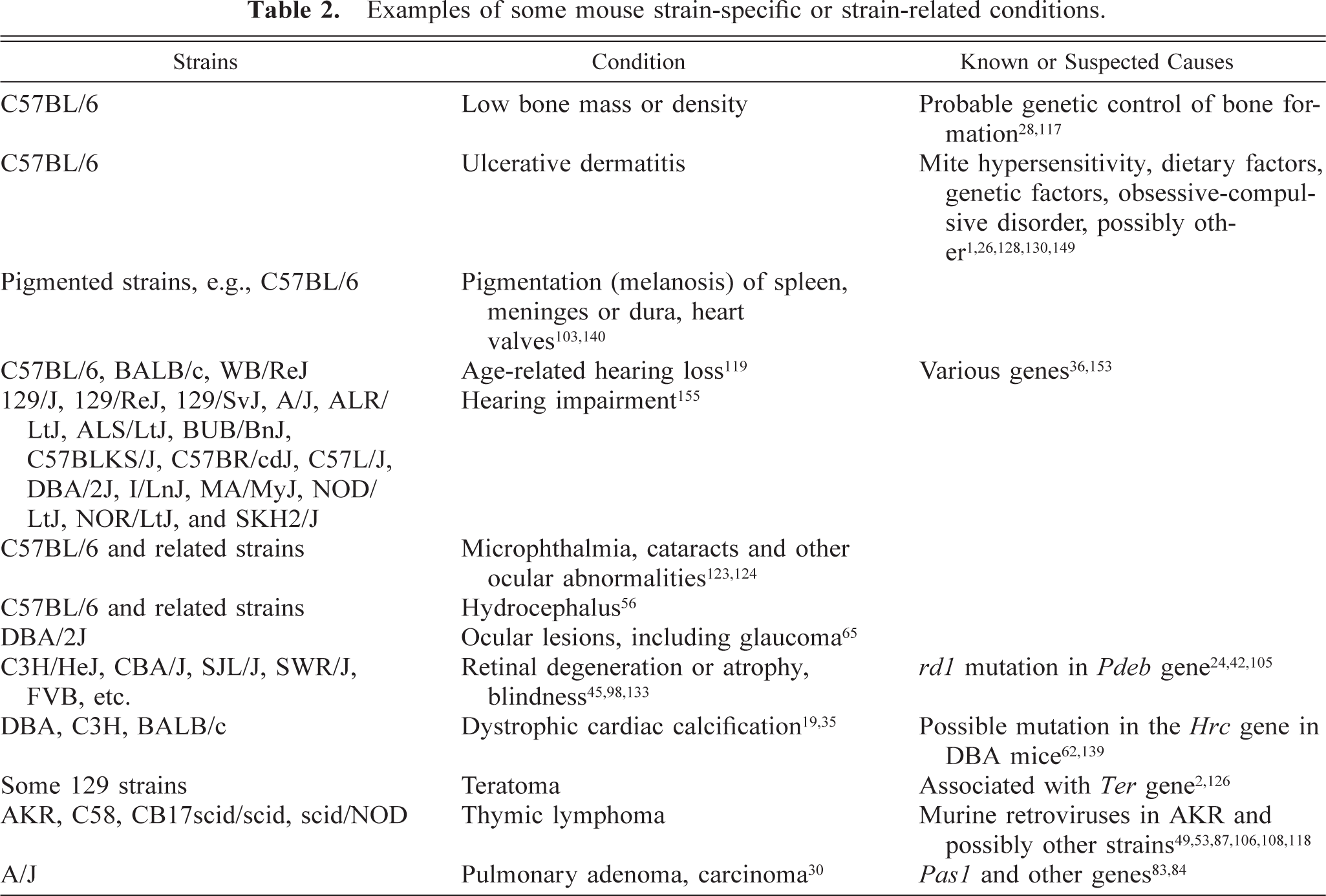
Several mouse-strain–specific or strain-related conditions can obfuscate evaluation of mutant animals (Table 2).
Examples of some mouse strain-specific or strain-related conditions.
Relatively low bone density (osteopenia, osteoporosis) of C57BL/6 mice contrasts with relatively high bone density of C3H mice and has been attributed to genetic control of bone formation and resorption. 28 71 117
Ulcerative dermatitis can occur with high frequency in some colonies of C57BL/6 mice and has been attributed to hypersensitivity to mites, dietary factors, geneitc factors, and obbsessive-compulsive disorders. 1 26 128 130 149
Pigmentation (melanosis) of various tissues, such as spleen, meninges or dura, and heart valves, occurs in C57B1/6 and other pigmented mouse strains. 103 140
Age-related hearing loss (presbyacusis) occurs in C57B1/6, BALB/c, and WB/ReJ mice, is associated with progressive loss of hair cells and spiral ganglion cells in the cochlea, and has been linked to several genes in these strains. 36 119 153
Elevated auditory-evoked brainstem response (hearing impairment) thresholds before 3 months of age have been demonstrated in 129/J, 129/ReJ, 129/SvJ, A/J, ALR/LtJ, ALS/LtJ, BUB/BnJ, C57BLKS/J, C57BR/cdJ, C57L/J, DBA/2J, I/LnJ, MA/MyJ, NOD/LtJ, NOR/LtJ, and SKH2/J mice. 155
Microphthalmia, up to 12% incidence, with highest frequency in female mice and in the right eye has been reported in C57BL/6 and related mouse strains. Cataracts and other ocular anomalies and hydrocephalus have also been reported. 56 123 124
A variety of ocular lesions, including glaucoma, have been reported in DBA/2J mice. 65
Retinal degeneration or atrophy, with blindness, occurs in C3H/HeJ, CBA/J, SJL/J, SWR/J, FVB, and other strains 45 98 133 because of homozygosity for the rd1 mutation in the Pdeb (phosphodiesterase, cGMP, rod receptor, beta polypeptide) gene, 24 formerly known as the r, rd, or rd1 gene, in these strains. Pdeb rd 1 is a nonsense mutation that results in a deficiency in rod photoreceptor cGMP phosphodiesterase and in degeneration and loss of photoreceptor cells. 42 105
Dystrophic cardiac calcification (cardiac calcinosis, myocardial mineralization, epicardial mineralization) occurs commonly in BALB/c, DBA, C3H (derived from DBA), and related strains. 19 35 In DBA and related strains, dystrophic calcification occurs in other soft tissues, especially kidney, tongue, and heart, and the trait maps to the region of the candidate gene Hrc (histidine-rich calcium-binding protein) on chromosome 7. 62 139
Teratomas in 129-related strains have been associated with the gene ter. 2 126 Extragonadal teratomas are reported in chimeric mice generated from 129 derived embryonic stem cells. 10
A high incidence of mammary gland tumors occurred in some C3H strains (e.g., C3H/HeJ and C3H/OuJ) that carried exogenous milk-transmitted murine mammary tumor virus (MMTV) or Bittner agent. 12 33 138 Except where it has been preserved intentionally for experimental purposes, exogenous MMTV probably has been eliminated from most laboratory colonies by rederivation.
T-cell lymphoblastic lymphoma (thymic lymphoma) is a common cause of death in AKR mice by 1 year of age, with a peak incidence at 3–6 months, and is attributed to complex interactions between the ecotropic murine retrovirus AKV and other murine retroviruses. 49 52 53 118 C58, CB17scid/scid, or scid/NOD mice also have high incidence of thymic lymphoma in which endogenous murine retroviruses have been implicated. 23 78 87 106 108
Pulmonary adenomas or carcinomas (alveolar bronchoalveolar adenomas or carcinomas) occur with high incidence at an early age in A/J mice 30 and are linked to the Pas1 (pulmonary adenoma susceptibility 1) gene. Susceptibility and resistance to development or induction of lung tumors in various mouse strains is attributed to complex relationships among resistance and susceptibility genes and among other genes. 83 84
Familiarity with inbred strains and their “normal pathology” will enable pathologists to select appropriate strains for their own research, to advise investigators regarding appropriate strains for various research purposes, and to identify probable strain-related phenomena. For example, C3H and FVB strains may not be ideal for evaluation of mutations that are anticipated to affect vision or behavior because these mice are blind by 4–5 weeks of age, with retinal atrophy or degeneration. A high incidence of hydrocephalus and ocular or craniofacial abnormalities reported in the mutant mice may be related to the gene or mutations of interest or may be related to the C57BL/6 background.
Health and Pathogen Status of the Colony
The health and pathogen status of the colony can impact phenotype significantly. Intercurrent diseases or conditions related to infectious agents or other environmental factors can interfere with research investigations, including attempts at phenotype characterization. Often, manifestations of the diseases or conditions are affected by strain background. An added concern is that mutant mice may respond to infectious agents, environmental factors, or drugs in unexpected ways, and agents that produce no apparent disease in mice that have been studied could be devastating to certain mutants. Several infectious agents were found in research colonies in the United States in 1998 64 and could have obfuscated mouse research.
Mouse hepatitis virus (MHV), a coronavirus, can affect immune function and gastrointestinal, hepatic, and central nervous system morphology. Different inbred strains and mutant mice may manifest specific and characteristic responses to MHV. 4 6 72 79 104
The mouse parvoviruses, minute virus of mice (MVM) and mouse parvovirus, have immunomodulatory effects in vivo 90 and adverse effects on contaminated cell cultures in vitro. 89 99 Although natural disease (i.e., not experimentally induced) in mice is inapparent, experimental infections with MVM result in strain dependent manifestations of disease, such as runting, cerebellar hypoplasia, and renal or intestinal hemorrhage. 18 63
Various murine Helicobacter species affect gastrointestinal and hepatic morphology. Different inbred strains and mutant mice manifest specific and characteristic responses to Helicobacter, including inflammatory and proliferative bowel diseases, hepatitis, and hepatocellular neoplasms. 20 29 43 44 48 59 142 143 145 150 Similarly, Citrobacter rodentium (C. freundii 4280) is the agent of transmissible murine colonic hyperplasia 7 112 and could complicate studies of inflammatory bowel disease or other conditions. 5 54 82
Mycoplasma pulmonis and Sendai virus affect immune function 39 75 76 92 and pulmonary morphology. Infection with these agents can affect mouse production and complicate research on other respiratory tract diseases, immunology, and other conditions. 57 80 101 Different inbred strains or mutant mice can manifest specific and characteristic responses to these agents. 17 38 74 100 102 111
Examples of environmental factors that could or have influenced experimental parameters in rodent studies include light and humidity, 60 phytoestrogens or other endocrine disrupters in rodent feed or bedding, 91 134 feed restriction, 15 116 118 acid, chlorine or antibiotics in drinking water, 51 and various other husbandry factors, 81 including restraint. 50
Selection of Appropriate Mutant and Control Animals
Selection of appropriate mutant and control animals may seem to be another issue that is and should remain outside of the pathologists' realm. Issues include age, strain, and generation (i.e., backcross generation N(1 − x) in development of congenic strains or generation postmutagenesis, G(1 − x) after ENU exposure of the G0 male).
There is no single best age for evaluation of mutant mice. Ideally, simple, inexpensive, and noninvasive technologies could be used throughout the animals' lives to determine the optimal window for terminal, comprehensive evaluations, and adequate and appropriate control animals would be available concurrently. Frequently, such technologies are not available. Per diem rates and other expenses exert strong pressure to develop mutant animal models in which the phenotype is expressed early and obviously. A practice of performing baseline evaluations at 8–12 weeks of age has some advantages: mice are sexually mature but should not have significant age-related pathology, incurred expense will be less than that at older ages, it is an age range for which data on inbred strains are relatively accessible, and ultimately the practice will result in a substantial in-house database on mutant and control animals in that age range. However, the mutants may not cooperate by demonstrating the desired phenotype at a convenient 8–12 weeks of age. They may die in utero or before the desired phenotype is expressed strongly or even detectably or they may not express the phenotype until much later in life.
There is no single best strain in which the mutations can be induced easily and in which most or all mutations are expressed reliably as distinct phenotypes. Different mutagenic technologies favor different strains. The FVB strain is favored for transgenesis because of its prominent pronuclei, which facilitate microinjection of DNA, and its good reproductive performance with large litters. 121 133 The 129 strains and C57BL/6 mice are favored for targeted mutagenesis (knockout technology) because 129 embryonic stem (ES) cells and C57BL/6 blastocysts have proved to be a good combination for establishing germline transmission of injected ES cells; 31 in addition, agouti/black chimerism is identified easily. C57BL/6 and C3H strains have beenfavored for ENU mutagenesis, but BALB/c and A/J males also tolerate ENU relatively well and survive to produce offspring with mutations derived from their mutagenized spermatogenic cells. ENU dosage and exposure information on these strains is readily available. 67 148 C57BL/6 has become a favored strain for isolation of mutant genes by backcrossing for varioius reasons, despite phenotypic peculiarities such as osteopenia or osteoporosis, 8 70 71 ulcerative dermatitis, 1 26 128 presbyacusis, 36 119 153 and microphthalmia and other ocular abnormalities. 56 124 C57BL/6J is the C57BL/6 strain developed by and maintained at the Jackson Laboratory (their laboratory registration code is J 45 61 ), and this strain was chosen for the mouse genome-sequencing project, a factor that is likely to have enhanced its popularity further.
The appropriate or ideal generation of mutant to be evaluated also varies with the study and method of mutagenesis. Mutant mice generated on mixed backgrounds (e.g., B6/129 chimeric knockouts) frequently are bred back to C57BL/6 mice, with the objective of creating a congenic line or strain that differs from mice of the recipient C57BL/6 strain only in the gene or mutation of interest. Any phenotypic difference between the resultant congenic mice and C57BL/6 mice raised under identical conditions should be attributable to the gene or mutation of interest. By the 5th backcross generation (N5), their genome should be >94% C57BL/6 120 but there still may be significant influences on phenotype by donor strain(s). Investigators may resist requests to provide animals of at least N5 or N6 backcross generation because phenotypes may be lost during backcrossing and because of the time and expense involved. The mixed background strategy, i.e., intentionally maintaining the mutation in mice of mixed genetic background, may expose phenotypes that are latent or lethal in inbred strains and can expedite the process to obtain evaluable mutants, because backcrossing is obviated and in mixed breeding programs litters tend to be larger than in inbreeding programs. 31 With random (ENU) mutagenesis, G1 animals can be assessed for dominant single gene mutations, but recessive mutations that are expressed only in the hemizygous or homozygous state will not appear until G2 or G3, depending on breeding strategies.
Ideally, when dealing with mutant results of insertional or targeted mutagenesis (transgenic or knockout mice), investigators would choose to provide homozygote mutants (−/−), heterozygotes (−/+), and homozygous wild-type (+/+) mice for evaluation. Often this is not the case, especially when backcrossing has been abrogated, possibly before N10 when the strain is considered to be definitively congenic, and breeding of −/− mice has been initiated to propagate 100% −/− mice in each litter, to provide more −/− mice for study, and to obviate genotyping of each litter. Relevant heterozygotes or homozygote wild-type mice will be difficult to obtain because the −/− litters are really of mixed lineage. In these situations, the +/+ control animals used for phenotype evaluation and for other experiments often are animals of what is believed to be the predominant inbred strain of the −/− animals, procured from vendors or other sources. Drawbacks to this strategy include the different health and pathogen status of the procured animals and the variable percentages of contaminating strains in the −/− animals, both of which factors could influence phenotype assessments and other experimental evaluations.
The number (n) of mice provided for evaluation is another factor over which the pathologist often has little influence. Mutant results of directed mutagenesis experiments often are in short supply, and sufficient sample size for statistical significance or power often is not available. Even a minimum of two age-matched, sex-matched mice of each genotype (−/−, +/−, +/+) and each sex of at least the fifth backcross generation may be difficult to obtain for comprehensive evaluations. The expectation from evaluations involving such small sample sizes is that results will provide data that will help to direct further experimentation or targeted in-depth evaluations.
Baseline Characterization of Mutant and Control Mice
Baseline characterization of mutant and control mice comprises a panel of tests that vary with the equipment, expertise, and funds available and with the interests of the investigators. A tiered approach, meaning an initial comprehensive baseline characterization panel of tests followed by more in-depth evaluations of organs or systems targeted by results of the baseline panel is suggested. As with diagnostic necropsies, history is important and can help to focus even initial evaluations to obtain and present information to provide the greatest and most cost-effective benefit to the investigation. Prior consultation with investigators, students, or technicians should be directed to gain information regarding 1) the mutations and investigations (with published references when available), 2) any peculiarities that have been noted by people who work with or care for the animals, 3) genetic background and experimental manipulations (including how many times blood has been collected from animals and from what site), and 4) health and environmental conditions of the colonies. For example, if a mouse has what appears to be a small eye, is it microphthalmic due to its C57BL/6 background or due to the mutation under study or is it phthisis bulbi secondary to multiple or inept retroorbital bleeding, and is the associated periocular inflammation related to a true microphthalmic condition, to the bleeding, or to Pasteurella pneumotropica that is enzootic in the colony?
The baseline characterization panel emphasizes relatively accessible techniques that can be performed in most pathology laboratories with adaptations to maximize the information yield from each mouse and to accommodate or even take advantage of their small size. Time-consuming but critical elements of such efforts include specimen procurement for clinical pathology, radiography, photography, weighing tissues and reproducibly trimming them for weighing, and adequate and appropriate labeling and recording. An example of a baseline characterization panel is provided in Table 3. Table 4 provides an example of a histologic cassette and slide numbering system that permits evaluation of more than 30 tissues on 12 or fewer slides. Consistent trimming and numbering of cassettes and slides facilitates retrieval of specific tissues from archived paraffin blocks or glass slides.
Example of a baseline characterization panel for mutant and control mice.
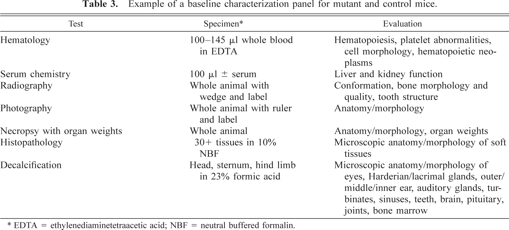
EDTA = ethylenediaminetetraacetic acid; NBF = neutral buffered formalin.
Tissues collected from mice for baseline histologic evaluation.
The cassette numbers correspond to those on glass slides to facilitate subsequent retrieval of specific tissues from archived specimens.
Clinical pathology
Baseline clinical pathology evaluations should include hematology, serum chemistry, and urinalysis. Blood specimens must be collected atraumatically, and the site of collection, preservative, anticoagulant, anesthesia, and handling of the animal can influence results. 21 Urine often can be collected as the animal urinates terminally or by open bladder puncture (during dissection) with a 25–27-ga needle and 1-ml syringe.
Radiography
Radiographs document gross skeletal abnormalities, e.g., fractures and unusual conformation, and can be used to evaluate bone quality (osteoporosis, osteopenia, or osteopetrosis). Lateral and ventrodorsal (VD) or dorsoventral (DV) views should be obtained with a permanent label and densitometric step wedge included in each radiograph. Animals should be positioned consistently and reproducibly, always facing the same direction, and with the label and wedge in same position, so that left/right are always obvious and radiographs are directly comparable. The permanent label is important for documentation. Inclusion of an aluminum step wedge in the radiograph will permit subsequent morphometric and densitometric evaluation. After euthanasia, positioning the animal for the DV view on a 5- × 7-in. note card, before onset of rigor, may facilitate the procedure. Kyphotic or hunched posture may be interpreted as a phenotype and may be related to osteoporosis or neuromuscular disease or vertebral anomalies 9 14 115 132 but is a common posture for sick mice and should be interpreted with caution. The posture interferes with obtaining a VD or DV radiograph that helps to evaluate related bone changes, and artifactual apparent scoliosis is common. Subjectively, the VD (dorsum down on the film) position with limbs outstretched seems to facilitate alignment of the vertebral column to obtain an interpretable VD film. With DV positioning, often there is apparent curvature of the vertebral column even in “normal” healthy mice. Nonradioopaque tape may facilitate positioning of limbs and head. Mammography and dental radiography machines have been used with success on mice. Most dental films are small and may not accommodate adult mice plus a step wedge and a label. Many dental machines are designed for very radiodense tissue, and it may be difficult to obtain appropriate exposures for mice on the available settings. Tabletop machines designed for human hands and feet are relatively inexpensive and simple to use with a fixed SID (source-to-image distance) that ensures uniformity in technique. Veterinary machines may suffice, and special films or screens may enhance resolution and image quality. Faxitron machines probably produce the highest quality radiographic images available and can provide several fixed magnifications, based on several fixed SIDs. Relatively long exposure times (up to 3 minutes) are not a great problem with cadaver specimens but may be an issue with anesthetized animals and can be reduced by use of special films and screens. In summary, many types of machines may be used to provide good information, but protocols or SOPs regarding positioning, film and screens, SIDs, and milliamp and kilovolt settings should be developed for optimal imaging of mice in each situation. Special techniques involving dissected limbs, vertebrae, or heads may be important for some studies, and uniform dissection techniques and positioning are critical to such evaluations. Other techniques that can provide quantitative information on bone quality and body composition include dual-energy x-ray absorptiometry (DEXA) 71 96 97 and microcomputed tomography or peripheral quantitative computed tomography. 13 71 136 These technologies may be more expensive initially but may yield the quantitative data that efficiently provide power or significance to a study.
Gross photography
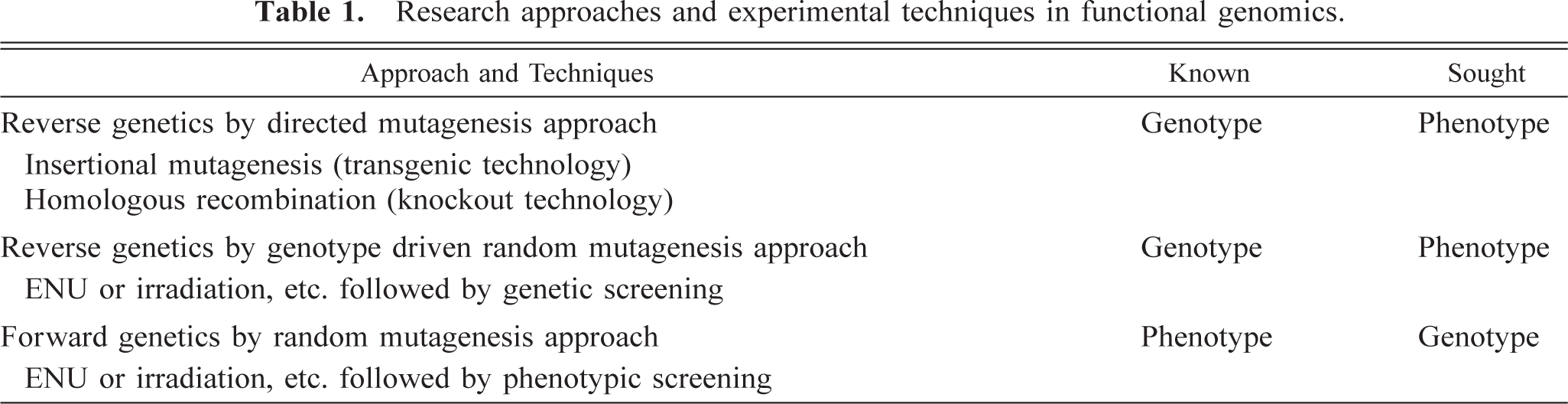
Photographs provide a permanent record of coat color, gross anatomy or conformation and any external lesions. Ideally, right lateral, left lateral, DV, and VD views would be obtained of every mouse evaluated, and additional photographs would be taken during dissection when gross abnormalities are discerned, but time and cost constraints may interfere with such efforts. Again, animals should be positioned consistently and reproducibly or in a specific position to demonstrate a lesion or feature of interest, with a label and ruler in the same position in each photograph (Figs. 1–4). The permanent label and the ruler are important for perspective and possible subsequent morphometric analysis of lesions or other features. A written or posted protocol or SOP regarding positioning, film, camera, lenses, working distance (distance from lens or camera to the subject), exposure, and F-stop settings may enhance uniformity, especially in situations where there are multiple users. Various types of light boxes, copy stands, and camera equipment can be used to provide good information. 88 127 Newer technologies involving video cameras or digital imaging may facilitate electronic capture and archiving. These technologies may be adequate and cost-effective for simple documentation purposes, although image quality seems not yet to be as good as with conventional films and 35-mm cameras.
Male transgenic mouse. With the skin removed, the abdominal distention can be seen to be due to a cystic mass, which is the hydronephrotic right kidney (R). The left kidney (L) is normal. This mouse has scant body fat.
Male transgenic mouse. With the skin removed, abundant body fat and prominent fat pads (arrows) are apparent.
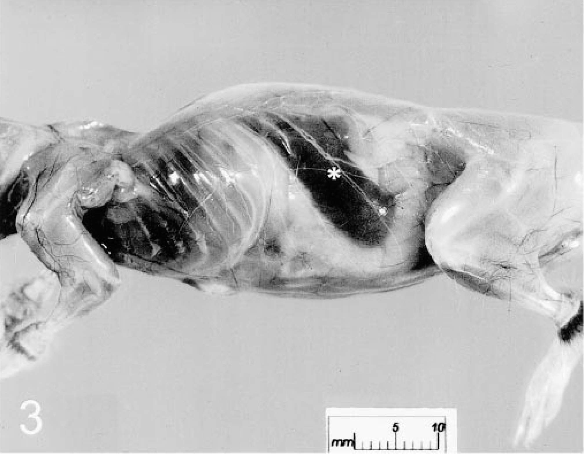
Female mouse, heterozygous for a targeted mutation. With the skin removed, the enlarged spleen (asterisk), scant body fat, and axillary lymphadenopathy are apparent. The body weight was 26.783 g, and the spleen weight was 0.393 g. The mouse was oriented in this direction to visualize the spleen.
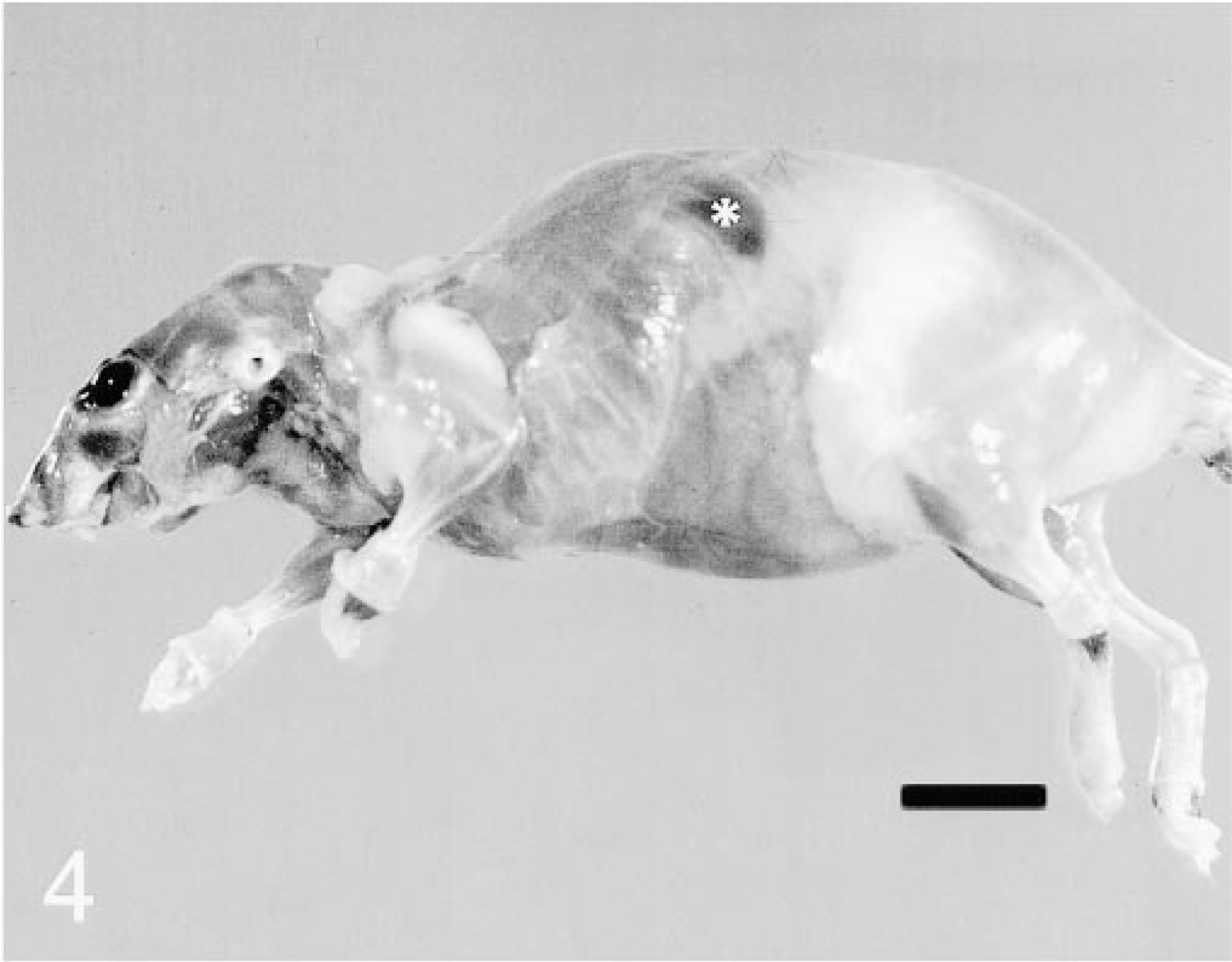
Female wildtype (+/+) “control” mouse. With the skin removed, ample body fat is apparent, and the spleen (asterisk) is difficult to discern. The body weight was 32 g, and the spleen weight was 0.07 g. The mouse was oriented in this direction to visualize the spleen.
Necropsy
A good necropsy technique for any animal should permit and facilitate evaluation and collection of all organs and lesions, provide qualitative and quantitative data, and be systematic, efficient, and simple so that it is reproducible in many animals and by many prosectors. Dissection, tissue collection, and reporting should be able to be performed relatively quickly, be taught relatively easily to individuals from a wide variety of backgrounds, and permit (require) visualization of all organs. Investigators that previously reported no lesions or a high incidence of missing kidneys or testes may suddenly find lesions and the appropriate number of organs when the necropsy technique is improved. Methods of dissection, fixation, and decalcification other than those outlined here can vary with preferences of the laboratory or investigator, and techniques to enhance evaluation of specific systems or conditions of interest can be incorporated into a baseline evaluation or developed into stage 2 or targeted evaluations.
Animals are weighed prior to dissection and should be weighed consistently before or after blood collection. Statistical evaluations of body weights could be impacted by removal of 1–1.5 ml of blood (approximately 1–1.5 g), from animals that generally weigh <25 g. During dissection, weights are obtained of heart, liver, spleen, right and left kidney, and testes or uterus and ovaries using the same weighing machine. Collected tissues are fixed in 10% neutral buffered formalin, except for the head, one hind limb, and sternum, which are fixed and decalcified simultaneously by 24-hour immersion in a 23% formic acid solution (e.g., TBD-2, Shandon Lipshaw, Pittsburgh, PA). This relatively gentle decalcifying solution usually permits trimming of bony tissues within 24 hours of collection, when formalin-fixed soft tissues are trimmed. Mice with high bone density (e.g., C3H mice) or osteopetrotic mutants may require a longer period of decalcification. Minimization of tissue exposure to aldehyde fixatives or acids that denature proteins (and epitopes) before paraffin processing reduces adverse fixation effects on antigens for subsequent immunohistochemical evaluations.
Subjective assessment of body condition should be included in the report, and a recently published scoring method 37 may be useful. Dissection is initiated by midventral skin incision and complete removal of the pelt to permit evaluation of subcutaneous fat, superficial lymph nodes, symmetry, and location and condition of most abdominal viscera (Figs. 1–4). The skin (entire pelt or selected specimens) should be laid flat on a paper towel or card stock 127 to ensure uniform flat fixation. Dissection is continued in a systematic manner, with complete collection of tissues, as listed in Table 4. Formalin infusion of lungs is achieved most easily in situ via injection into the cervical trachea before lungs are removed and airways potentially compromised. Removal of the heart, lungs, liver, and trachea (pluck) from the split mandible to the split pelvis is a simple means to ensure visualization and collection of all tissues. The oral cavity should be examined for dental and periodontal disease of the molars. Splitting the mandibular symphysis to remove the tongue facilitates examination of the oral cavity and removal of the pluck. During removal of the pluck, care should be taken to not compress the infused lungs, and they can be placed intact into a cassette at the time of dissection. Care should be taken to ensure removal of kidneys and adrenal glands, which may remain in the retroperitoneal space. The right kidney usually is marked (nicked with a blade) to facilitate identification. Even after removal of the pluck, the right kidney tends to remain cranial to the left kidney in a mouse that does not have situs inversus. There are many techniques for fixing and trimming the gastrointestinal tract, and special techniques (e.g., Swiss roll 127 ) can be used if the gastrointestinal tract is of particular interest to the investigator. Most prosectors can relatively quickly master a simple technique of gentle infusion of various segments of the intact gastrointestinal tract, followed by immersion fixation and trimming of representative cross sections for paraffin processing. Splitting the pelvic symphysis facilitates removal of the entire male or female reproductive tract intact. Reproductive tracts can be laid flat on paper for immersion fixation to facilitate identification of tissues and standardization of fixation and trimming.
Effort is made to maximize the number of tissues evaluated while minimizing number of glass slides and the processing costs. For example, four sections of a decalcified head on a single glass slide permits evaluation of brain and pituitary (for hydrocephalus, inflammation, neoplasia, degenerative changes), eyes (for retinal atrophy, degeneration or dysplasia, cataractous changes, inflammation, neoplasia), ears (Fig. 5; for otitis interna, media, externa, cholesteatomas), teeth (Fig. 6; for dental and periodontal disease, dysplasia, odontomas), bone and marrow, turbinates, olfactory and respiratory epithelium, salivary and Harderian glands, and other tissues (for inflammation, degeneration, neoplasia).
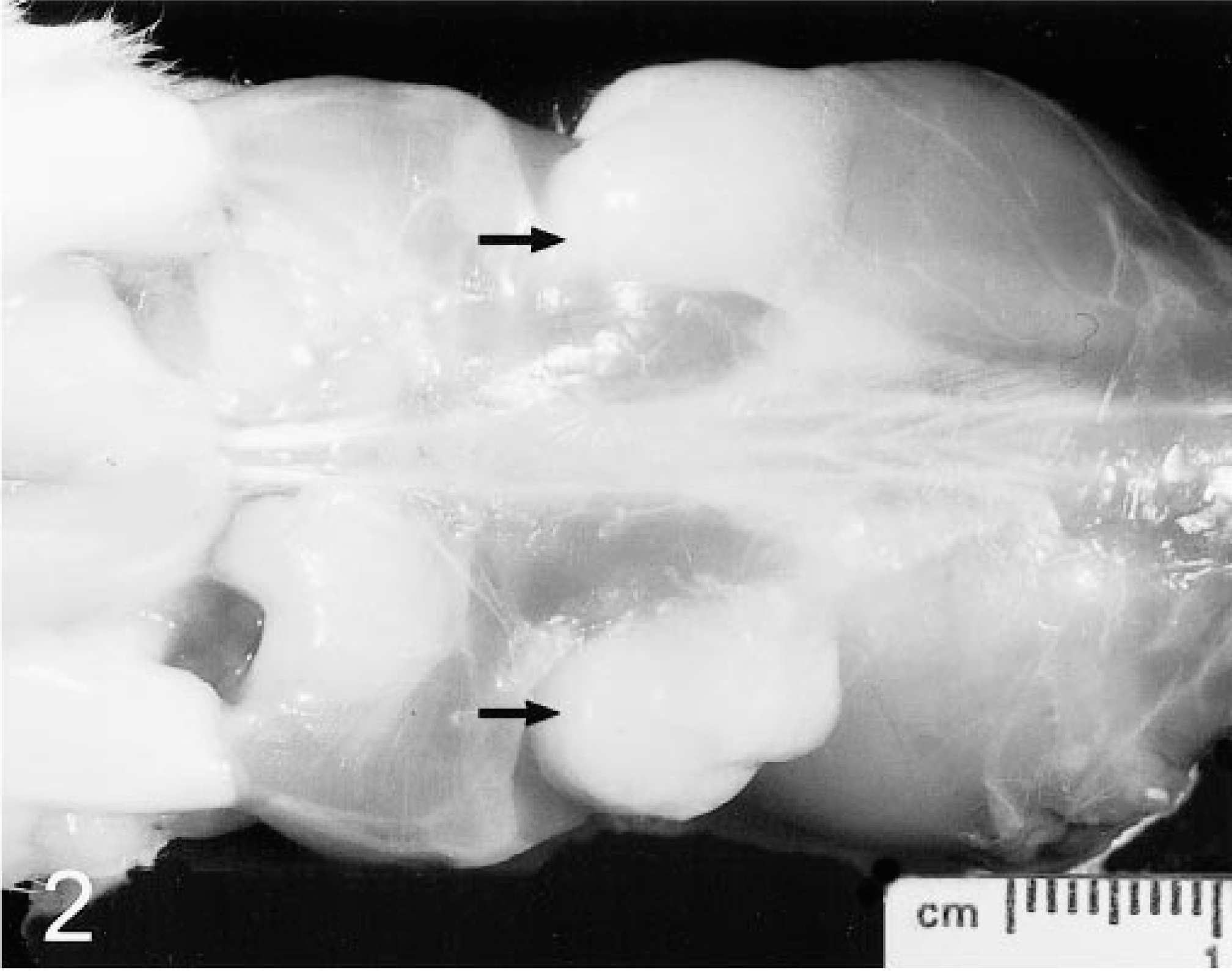
Oral cavity, decalcified head; 26-month-old mouse heterozygous for a targeted mutation. This cross section (coronal section) is rostral to the eyes and includes the nasal cavity and turbinates (T). There is marked erosion of the left molar tooth (ML). Antemortem absence of enamel (which is removed by the decalcification procedure) is indicated by the accumulation of debris on and adjacent to the remnants of the tooth. Inset: Same tooth stained with Goodpasture's tissue Gram's stain. The darkly staining plaquelike material (asterisks) is comprised largely of gram-positive cocci. MR = right molar tooth, which is relatively intact. Decalcification was by immersion in 23% formic acid for 24 hours. HE. Bar = 1.0 mm
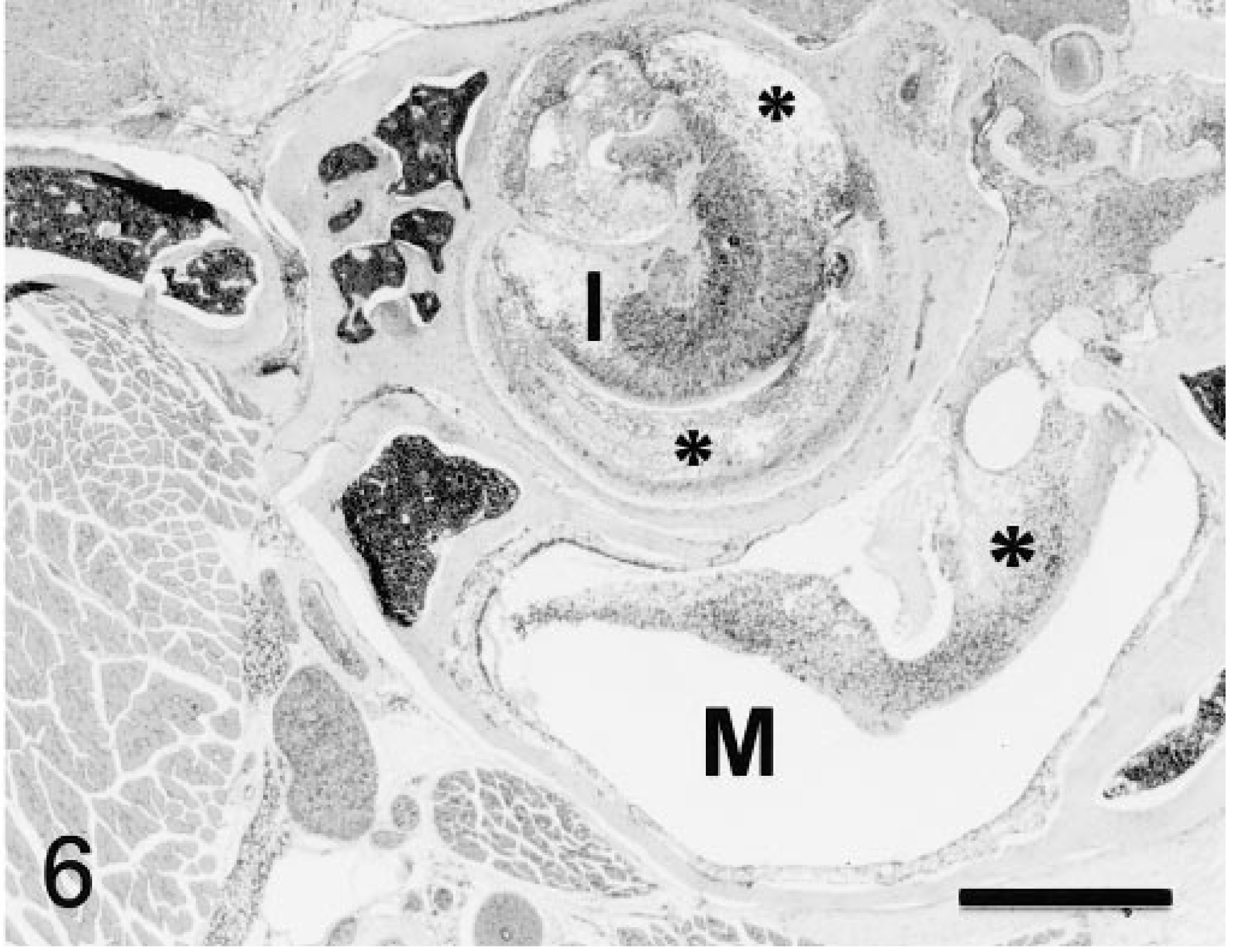
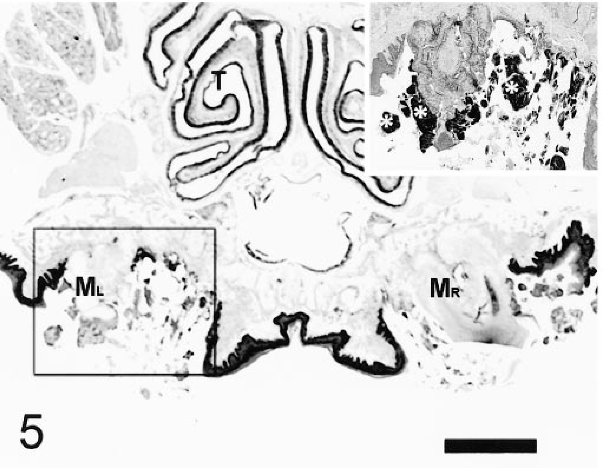
Internal ear and middle ear, decalcified head; 9-week-old mouse homozygous for a targeted mutation. There is suppurative otitis interna and media with suppurative exudate (asterisks) around the cochlea in the internal ear (I) and in the middle ear (M). Decalcification was by immersion in 23% formic acid for 24 hours. HE. Original magnification 6.6×. Bar = 0.5 mm.
Routine paraffin processing and staining with hematoxylin and eosin (HE) is employed for initial evaluations. Collected fixed tissues that are not processed immediately for histologic evaluation are archived in formalin in heat-sealed bags.
Evaluation of Early Embryonic Death and Perinatal Mortality
Generation and characterization of mutant mice frequently seems to be complicated by early embryonic death or perinatal mortality. Requests to evaluate placentae, feti, and neonates are common. Some suggestions for anatomic evaluations are provided in Table 5. Fetal and neonatal hematologic evaluations have been discussed elsewhere. 21 Information on normal and pathologic microscopic anatomy of placentae and embryos of mice is dispersed in the literature. Familiarity with mouse implantation, placentation, and development will facilitate evaluation. 55 68 69 In our laboratory, Bouin's solution is preferred over formalin or other fixatives for extraembryonic tissues, feti, and neonates. Bouin's solution effectively decalcifies pups up to 1 or 2 weeks of age without rendering tissues too soft and difficult to trim or section. Disadvantages of Bouin's solution include relatively poor penetration of tissues, hardening (brittleness) of tissues from overexposure, and its toxic and explosive properties. Injection of Bouin's soluion into the thorax and abdomen of feti or neonates can help to overcome its poor penetrating capabilities. Removal of specimens from Bouin's solution into 70% ethanol within 24–48 hours of immersion (depending on size of specimen), prompt trimming, and paraffin processing should prevent tissues from becoming too brittle. 144
Evaluation of early embryonic death and perinatal mortality in mice.
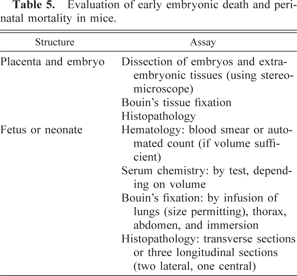
When examining feti and neonates, body weights, crown–rump lengths, and any gross external abnormalities should be recorded. Additionally, when examining neonates, skin or coat color(s), eye color, whether or not eyes are open, and whether or not there is milk in the stomach should be noted. Dead neonates may be cannibalized by the parents, resulting in apparently reduced numbers of offspring or in pup fragments that are insufficient for thorough evaluation.
Neonatal mortality can be complicated by a number of factors that are not related directly to the gene(s) of interest. Mothering issues should be considered early as a possible cause of pup demise. Observing dams and litters can reveal if the dam has no milk or is not feeding the pups (maternal neglect), if the pups move normally and attempt to nurse, or if they do not move normally and do not attempt to nurse, but effort should be made not to disturb the dam. Poor mothering may be influenced by strain, environment, and possibly by the pup itself. If litters are very large, dams may not produce sufficient milk, and weaker pups may not be able to nurse sufficiently. Removal of some pups or cross-fostering may promote survival of weaker pups. Small litters (one or two pups) may not stimulate the dam to produce sufficient milk. 121 Dams may choose not to nurse less viable pups, and cross-fostering onto a less discriminating dam may promote survival of these pups. Busy, loud environments and movement or mechanical vibration of the cage rack are not tolerated well in general, and some strains or dams seem to be especially sensitive to environmental factors. 151 Breeding mice may benefit from transfer to a quieter environment, from no disturbance of the cage for a few days after birth, and from nesting material, nest boxes, or breeding diets. Strains vary in their useful breeding lifespan, although many will breed until ≥9 months of age. Mice that are mated first when they are >3 months old may not breed. 45 Breeding a mutation into other mouse strains or into fecund, outbred stocks of mice also might promote survival of mutant pups and expose the effects of the mutation(s). 31
Stage 2 or Targeted Evaluations of Mutant and Control Mice
Stage 2 or targeted evaluations of mutant and control mice should be selected after evaluation of results of the baseline characterization and any other experimental information. Ideally, such evaluations are developed and performed during collaborations between pathologists and investigators with expertise in various disciplines. Examples of some system-specific characterizations are outlined in Table 6.
Examples of stage 2 characterization panels for mutant and control mice.
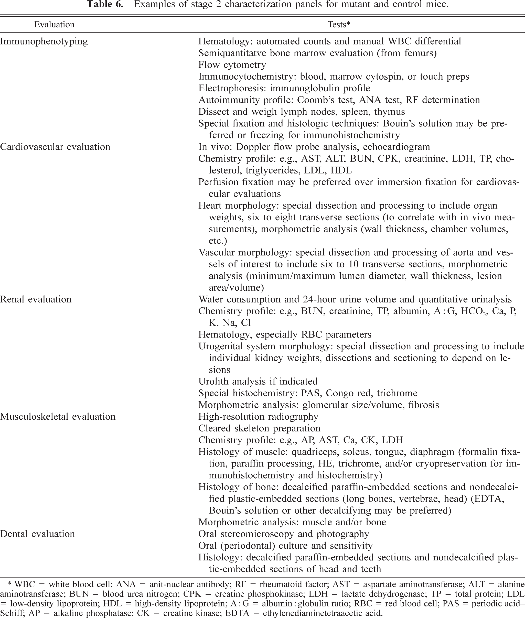
WBC = white blood cell; ANA = anit-nuclear antibody; RF = rheumatoid factor; AST = aspartate aminotransferase; ALT = alanine aminotransferase; BUN = blood urea nitrogen; CPK = creatine phosphokinase; LDH = lactate dehydrogenase; TP = total protein; LDL = low-density lipoprotein; HDL = high-density lipoprotein; A:G = albumin: globulin ratio; RBC = red blood cell; PAS = periodic acid-Schiff; AP = alkaline phosphatase; CK = creatine kinase; EDTA = ethylenediaminetetraacetic acid.
Impact of New and Emerging Technologies
New technologies, especially those that provide noninvasive, rapid, high-throughput assessments of mutant mice, are in development and use already. More widespread usage of various imaging, telemetry, and molecular techniques is inevitable and should be welcomed and taken advantage of by comparative pathologists. These technologies definitely aim to reduce dependence on costly, time-consuming, and animal-consuming techniques and to permit longitudinal studies on individual animals. Ultrasound and nuclear magnetic resonance imaging (MRI) techniques have been “downsized” for mice and have provided tremendous insight into cardiovascular function in vivo, even in pups. 37 46 113 152 MRI has been used to characterize mutant mouse models of muscular dystrophy, neuroanatomic abnormalities, and other pathologic changes in mutant mice. 34 40 73 125 Computed tomography techniques have been used primarily to evaluate bone quality and other skeletal characteristics, 13 71 and DEXA has shown utility in evaluating bone quality and body composition (lean versus fat). 71 96
Conclusions
Historically, research on mutant animals has been phenotype driven; an interesting phenotype was discerned, and experiments were undertaken to determine the genetic mechanisms. This approach is still valid and in use in experiments involving random mutagenesis by ENU or other means, where offspring of the mutated germ cells are evaluated for deviant phenotypes, the abnormalities are characterized, and the proximal genetic and molecular mechanisms are investigated. Transgenesis and targeted mutagenesis provide the opportunity to conduct genotype-driven research, where the genetic abnormalities are known, more or less, a specific phenotype is expected, and the more distal steps in the mechanism can be clarified. A resultant genetically engineered mouse with “no phenotype,” meaning no apparent phenotypic manifestation of the genetic abnormality, is a highly undesirable result. Comprehensive or appropriately targeted evaluations, longitudinal evaluations via modern imaging technologies, or evaluations in different mouse strains or stocks may help to expose a subtle phenotype. There has been a tendency for molecular biologists and geneticists, who play pivotal roles in the development of mutant mice, to underestimate the utility of pathology-based evaluation of mutant mouse models, and phenotypic analyses frequently and understandably are biased according to a research interest. Although targeted evaluations have great merit when based on critical analysis of sufficient data, comprehensive pathologic assessment of mutant mouse models is an important early step that can detect unanticipated changes related to the mutation(s), identify unexpected contributors to the phenotype related to mouse strain or environmental influences, and can help to target subsequent evaluations. Pathologists can take a proactive role in the field of functional genomics by developing and demonstrating the utility of comprehensive and targeted evaluation protocols. Our understanding of mouse strain peculiarities, the effects of intercurrent infections and other environmental influences, the genetic manipulations, and the molecular mechanisms of the human conditions that the mutant mice are intended to model will improve evaluation of these models, help to establish useful scientific collaborations, and contribute substantively to this new area of biomedical research.