
Abstract
A lesion was identified in the eye of a juvenile llama, and preliminary clinical findings included anterior uveitis and an exudative retinal detachment suggestive of infectious disease. However, histopathologic evaluation of the enucleated globe revealed an intraocular neoplasm composed of primitive neuroepithelium forming ribbons, cords, and rosettes, heteroplastic elements including spindle cells in a loose myxomatous matrix, and islands of well-differentiated hyaline cartilage. Immunohistochemically, neoplastic cells were positive for vimentin and neuron-specific enolase. Spindle cells were multifocally positive for desmin and muscle specific actin, indicating differentiation towards myofibers. These findings are consistent with a diagnosis of malignant teratoid medulloepithelioma, an extremely rare ocular neoplasm that affects children and young animals.
Keywords
The medulloepithelioma is a very rare ocular tumor that occurs primarily in children, but has also been described in several veterinary species, including horses, dogs, cockatiels, a goldfish, and a cat. 1 2 4–7, 11 13 16 17 19 Medulloepitheliomas can be classified as benign or malignant and as teratoid or nonteratoid. 2
A 1-year-old female llama was referred for ophthalmic examination because of a 1-month history of anterior uveitis with corneal edema of the left eye. The anterior uveitis had responded somewhat to topical corticosteroid therapy and topical atropine, but it returned with cessation of therapy. Complete ophthalmic examination revealed a lack of a menace response in the left eye, an intraocular pressure of 12 mm Hg using applanation tonometry, moderate aqueous flare, and complete retinal detachment. The subretinal area was filled with a light tan exudate or a mass (Fig. 1). The right eye was normal. Serology was negative for equine herpesvirus 1, positive for Toxoplasma gondii IgG, negative for Toxoplasma gondii IgM, and negative for Blastomyces dermatitidis and Histoplasma capsulatum. Results of a complete blood count and chemistry panel were normal, with the exception of a mild anemia. Thoracic radiographs revealed a mild interstitial pattern. Cytologic evaluation of a subretinal aspirate showed degenerate columnar epithelial cells, few lymphocytes and hemosiderin-laden macrophages, and two round basophilic objects of indeterminate origin on a background of proteinaceous fluid. Because of the positive toxoplasmosis titer, treatment with sulfadimethoxine was initiated. Twelve days later, the llama was returned for reevaluation because of worsening ocular signs. Keratoconus secondary to corneal edema had developed and the intraocular pressure had increased to 26 mm Hg. The other ocular signs remained the same. Because of the chronic ocular disease and inability to make a definitive diagnosis, an enucleation was performed.
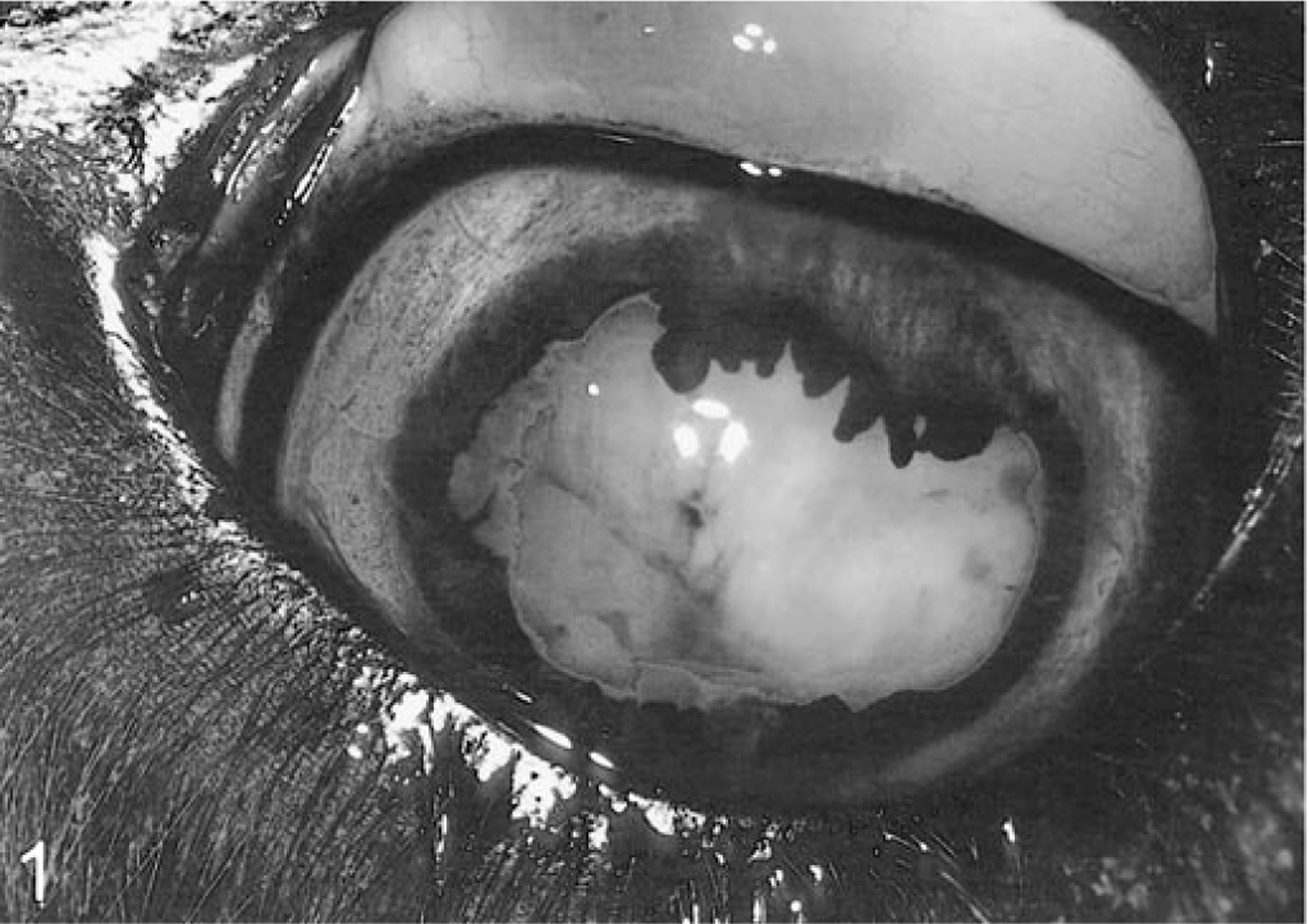
Eye; llama. Leukocoria was present in the eye as a result of the subretinal mass which is adjacent to and nearly abuts the posterior lens capsule.
The eye contained a large, semisolid mass of tissue in the shape of a partially flattened sphere, approximately 3 × 2 × 2.5 cm. The surface was brown, a result of hemorrhage, and the cut surface was mottled off-white, tan, and brown (Fig. 2). Precise location of the mass within the globe was not ascertained because only the mass, lens, iris, ciliary body, and cornea were submitted for histopathology.
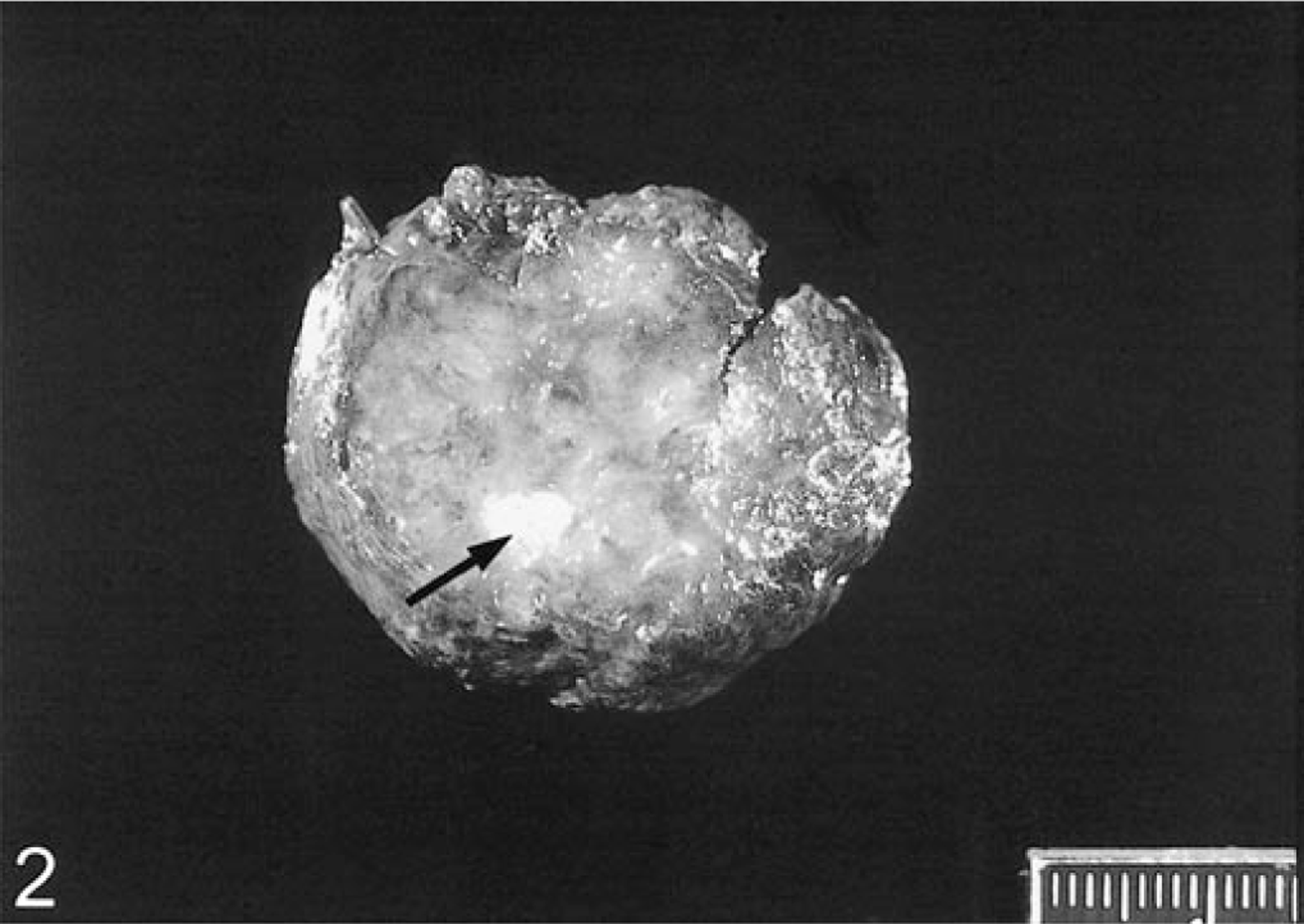
Malignant teratoid medulloepithelioma; llama. The cut surface of the semisolid mass removed from the eye was variably off-white, tan, and brown. An area of hyaline cartilage can be seen (arrow). Scale = millimeters.
The tissues were fixed in 10% neutral buffered formalin, embedded in paraffin, sectioned at 5 µm, and stained with hematoxylin and eosin (HE). Selected sections were probed using standard streptavidin–biotin immunoperoxidase methodology and the following primary antibodies: mouse anti-vimentin (BioGenex, San Ramon, CA), mouse anti-desmin (Dako, Carpinteria, CA), mouse anti-neuron-specific enolase (Dako), rabbit anti-S-100 protein (Dako), rabbit anti-glial fibrillary acidic protein (Dako), mouse anti-muscle-specific actin (Enzo, Farmingdale, NY), and a mixture of mouse anti-cytokeratin (Dako) and anti-AE1/AE3 (Boehringer Mannheim, Mannheim, Germany).
Histologically, the mass had no natural borders and varied from hypercellular to hypocellular in different areas. Neoplastic cells were grouped together in small and large sheets of cells and irregularly shaped, often interconnected streams, cords, and bundles. Many neoplastic cells resembled primitive neuroepithelium and formed ribbons and cords; Flexner–Wintersteiner rosettes were frequently present and were identified by a single layer of polarized, tall cuboidal/columnar cells encircling an open lumen (Fig. 3). Rosette-forming cells had basal, oval nuclei with one to three small nucleoli, finely stippled chromatin, and light eosinophilic apical cytoplasm. Fine filamentous eosinophilic and frequently tangled projections extended from the apices into the lumen of many of the rosettes. Less frequently, rosettes were smaller and lacked a central lumen (Homer–Wright rosettes). Mitotic activity in these and surrounding cells was robust; up to six mitoses/40X high-power field (HPF). Other larger rosettes did not fit the description of Flexner–Wintersteiner owing to their pseudostratified columnar arrangement (Fig. 4), and a few of these cells contained melanin in their cytoplasm. In other areas, the neoplastic tissue was comprised of either elongate “spindle” cells in a loose myxomatous pattern or densely cellular areas. These cells had a varied appearance, from round to oval to fusiform with nuclei of similar shape and light eosinophilic cytoplasm; cell borders were often indistinct. The mitotic rate ranged from zero to three mitoses/HPF. Additionally, there were numerous foci of small (0.1 mm, longest axis) to large (3.0 mm) irregularly shaped islands of hyaline cartilage scattered throughout the tumor; in a few foci cartilage was mineralized (Fig. 5). The tumor was very well vascularized, and numerous expansive regions of both recent and “old” hemorrhage were present. the latter identified by the presence of hemosiderin-laden macrophages. Interspersed between groups of neoplastic cells and often in association with areas of hemorrhage were numerous viable and degenerate neutrophils, fewer macrophages, and degenerate cells of uncertain origin. Examination of the iris and ciliary body revealed neoplastic cells that were either invading or extending from the nonpigmented ciliary epithelial cells (Fig. 6). The histologic findings were consistent with malignant teratoid medulloepithelioma.
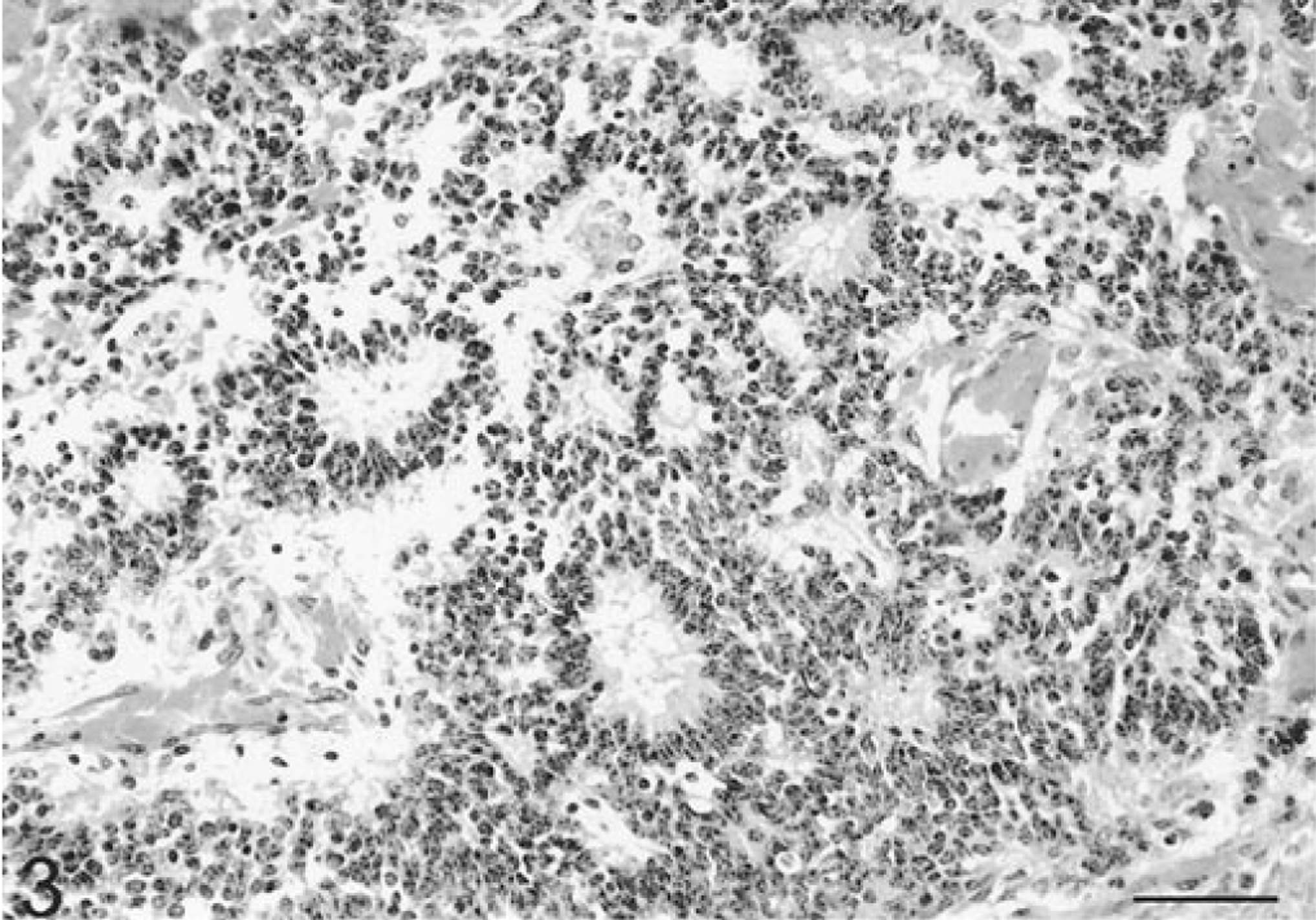
Eye, malignant teratoid medulloepithelioma; llama. Rosettes were present throughout the neoplasm intermixed with cords and sheets of cells. Many were Flexner–Wintersteiner rosettes, with a thin layer of polarized, tall cuboidal/columnar cells encircling an open lumen. Fine filamentous eosinophilic tangled projections extended from the apices into the lumen of many of the rosettes. HE. Bar = 50 µm.

Eye, malignant teratoid medulloepithelioma; llama. Other rosettes, especially larger ones, had a pseudostratified columnar arrangement and resembled embryonal medullary epithelium. HE. Bar = 25 µm.
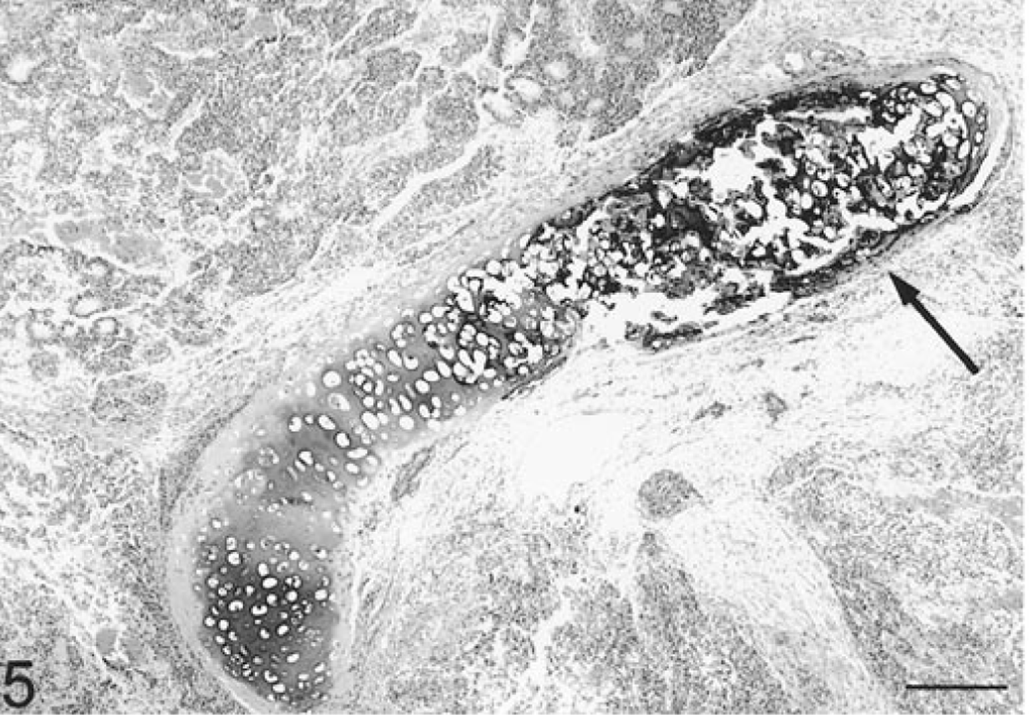
Eye, malignant teratoid medulloepithelioma; llama. Irregularly shaped islands of hyaline cartilage were scattered throughout the tumor, and some cartilage was mineralized (arrow). In other areas, neoplastic cells were grouped together in islands, larger sheets, and irregularly shaped, often interconnected streams, cords, and bundles. Numerous rosettes are at upper left. HE. Bar = 250 µm.
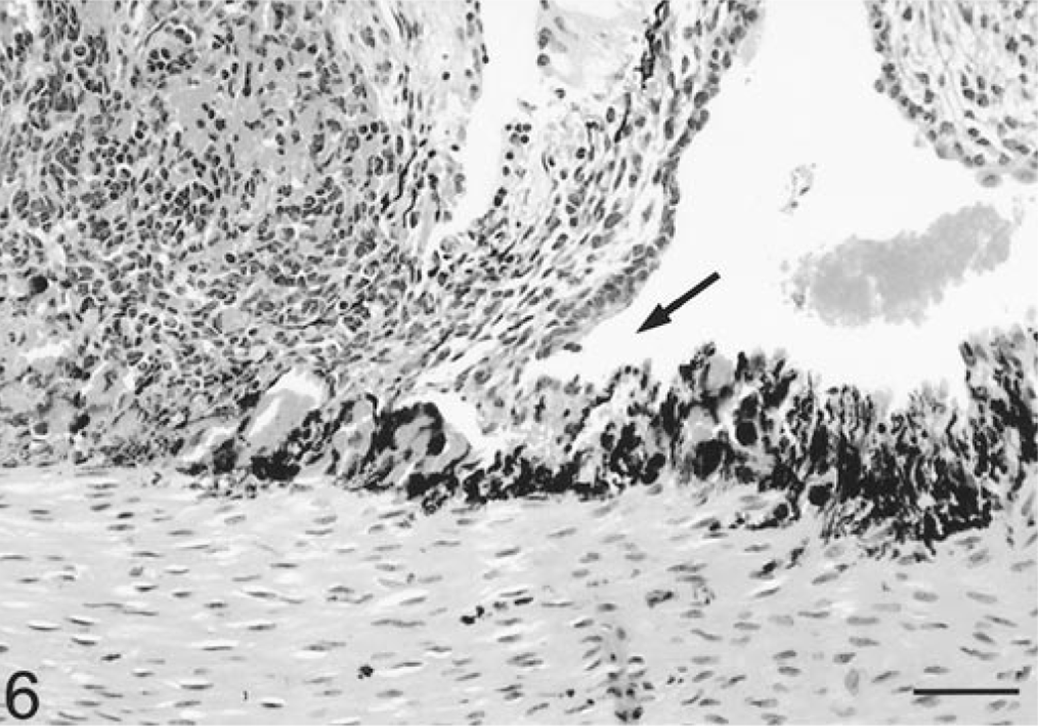
Eye, malignant teratoid medulloepithelioma; llama. Examination of histologic sections of the iris and ciliary body revealed neoplastic cells that were either invading or extending from the nonpigmented ciliary epithelium (arrow). HE. Bar = 50 µm.
Immunohistochemically, the neoplastic cells were diffusely positive for vimentin and neuron-specific enolase. In addition, there was multifocal positive staining of the spindle cell population for desmin and muscle specific actin. Spindle cells that stained for muscle-specific actin occasionally demonstrated cytoplasmic banding or cross-striations, supporting differentiation towards myocytes. The chondroid cells were positive for S-100 protein. All neoplastic cells were negative for cytokeratins and glial fibrillary acidic protein.
A literature search revealed only one immunohistochemical study on an ocular medulloepithelioma, a nonteratoid tumor variant in a human. 18 The llama and the human tumors were similar in both being positive for vimentin and neuron-specific enolase and negative for glial fibrillary acidic protein. The human tumor was negative for S-100 protein, but the chondrocytes in the llama stained positively. Cytokeratin staining was negative in the llama, however, some larger cells in the human tumor stained positively. The human neoplasm was not stained for desmin or muscle-specific actin.
Intraocular medulloepitheliomas are uncommon in humans and domestic animals. These tumors arise from the primitive medullary epithelium, or inner layer of the optic cup, before differentiation into adult tissues and are therefore classified as congenital tumors. 2 These tumors can be either benign or malignant. The cells retain the ability to differentiate; therefore, heteroplastic elements such as cartilage, skeletal muscle, and brain tissue may be seen. 2 There is some controversy as to whether the modifier “teratoid” should be used to describe medulloepitheliomas with heterologous elements. By convention, ocular medulloepitheliomas with heterologous elements are referred to as teratoid because the tumor contains tissues not normally found in the eye during embryonic or postnatal development. 8 9 15 22 However, medulloepitheliomas of the central nervous system are not referred to as teratoid. In this location, the mesenchymal stromal differentiation to cartilage, bone, and skeletal muscle is considered part of the spectrum exhibited by medulloepitheliomas. 3 10 12
Almost all medulloepitheliomas arise from the ciliary body, as probably occurred in this llama, but rarely they may arise from the optic nerve head or retina. 2 Medulloepitheliomas can invade the anterior and posterior segments but may be isolated to the anterior chamber, may be associated with the optic nerve or orbit, may be cystic, or may masquerade as persistent hyperplastic primary vitreous. 14
The most significant histopathologic finding in medulloepitheliomas is multilayered sheets and cords of poorly differentiated neuroepithelial cells that appear similar to embryonic retina and ciliary epithelium. 2 20 21 These sheets and cords of medullary epithelium show polarization of the cells very similar to that of the normal embryonal retina and have a basement membrane on one surface and a series of terminal bars on the other. Homer–Wright and Flexner–Wintersteiner rosettes, as seen in retinoblastomas in humans, may be present, but the most frequently observed rosettes in medulloepitheliomas are larger and surrounded by more than a single layer of cells (Fig. 4). 2
Findings on initial examination of this llama and the apparently low incidence of intraocular tumors in this species were suggestive of infectious disease as the most likely cause of the anterior uveitis. However, the poor response to therapy and the results of the cytologic examination of the subretinal aspirate did not support infectious disease as a likely etiology and histologic examination of the enucleated globe confirmed the presence of neoplasia. This malignant teratoid medulloepithelioma is the first intraocular tumor of any type to be described in a llama.
Footnotes
Acknowledgements
We thank the pathologists at the Armed Forces Institute of Pathology, Washington, D.C., for their review of case material and Dr. D. Laviola for her help in case management.