
Abstract
Tissues from 10 adult California sea lions (Zalophus californianus, seven females and three males) that had metastatic carcinoma in sublumbar area lymph nodes were examined histologically. A distinctive epithelial proliferative lesion interpreted as intraepithelial neoplasia was found in genital tracts of all ten animals; in vagina (5/7), cervix (7/7), uterus (3/7), penis (3/3) and prepuce (3/3). Intraepithelial neoplasia closely resembled metastatic carcinomas and was directly contiguous with invasive carcinoma in one animal. Rare eosinophilic intranuclear inclusion bodies were found in penile and preputial intraepithelial neoplasia (one animal), cervical intraepithelial neoplasia (one animal), invasive cervical carcinoma (one animal) and metastatic carcinoma (two animals). Electron microscopic examination of tissues from two sea lions (one with intraepithelial neoplasia and one with metastatic carcinoma) demonstrated viral particles consistent with a herpesvirus. An immunohistochemical stain for the latent membrane protein of Epstein-Barr virus was positive in intraepithelial neoplasia in one sea lion. Herpesvirus DNA sequences were detected by consensus primer polymerase chain reaction (PCR) in metastatic carcinomas from all four sea lions from which unfixed tumor samples were available. Results of sequencing were consistent with a novel gammaherpesvirus in the genus Rhadinovirus. DNA extracted from the four metastatic carcinomas also was tested for papillomavirus by Southern blot and PCR with consensus papillomavirus primers; all samples were negative by both methods. These findings support the genital origin of the sea lion carcinoma and implicate a novel gammaherpesvirus as a possible cause.
Keywords
There have been several reports of metastatic carcinoma of undetermined origin in California sea lions (Zalophus californianus). 1 7 11 In a recent study, 66 of 370 (18%) sexually mature California sea lions that were necropsied at a California marine mammal rehabilitation center from 1979 to 1994 had metastatic carcinomas of undetermined origin. The lymph nodes of the sublumbar area contained carcinoma in all affected animals. Most of the carcinomas had squamous differentiation, and glandular differentiation was present in some animals. Transitional epithelium of the urinary tract was suspected to be the site of origin based on the histologic appearance of the neoplasms. 7
In the present study, tissues from seven female and three male California sea lions that had metastatic carcinoma present in sublumbar area lymph nodes were examined histologically in an attempt to identify the primary sites of the carcinomas. Histopathology, electron microscopy, immunohistochemistry, and molecular techniques were used to investigate the possibility that viruses might be involved in the pathogenesis of the lesions.
Materials and Methods
Necropsy, Histology and Immunohistochemistry
Ten adult California sea lions (seven females and three males) that were found stranded on the central California coast and subsequently died or were euthanized at the Marine Mammal Center in Sausalito were necropsied. In addition to samples of all major organs, the genital and urinary tract organs were collected, immersed in 10% neutral buffered formalin, and dissected. Formalin-fixed tissue specimens were embedded in paraffin, sectioned at 5 µm, and stained with hematoxylin and eosin (HE) for light microscopic examination. Selected sections were stained with periodic acid–Schiff (PAS) and mucicarmine.
On sections that contained inclusion bodies, immunohistochemistry was performed with the avidin–biotin–peroxidase complex method using a commercial mouse monoclonal antibody against the latent membrane protein of Epstein-Barr virus (EBV) code M 0897, Dako, Carpinteria, CA) at a 1:80 dilution. Sections were counterstained with Mayer's hematoxylin.
Transmission electron microscopy
For ultrastructural studies, tissues from sea lion Nos. 3 and 5 that contained inclusion bodies were deparaffinized, hydrated, postfixed in 1% osmium tetroxide, dehydrated, cleared and embedded in epoxy resin. One-micrometer sections were cut and stained with toluidine blue for preliminary light microscopic examination. Thin sections (80–90 nm) were cut, stained with uranyl acetate and lead citrate, and examined with a Zeiss EM10 transmission electron microscope.
Herpesvirus consensus primer polymerase chain reaction
DNA was extracted from available sections of frozen, unfixed metastatic carcinomas (sea lion Nos. 1, 2, 5, 7), and consensus primer polymerase chain reaction (PCR) was performed in reactions designed to amplify conserved regions of herpesviral DNA polymerase and terminase genes. 9 22 The reaction products were sequenced directly and compared with published herpesvirus sequences contained in GenBank using BLASTP software on the National Center for Biotechnology Information (NCBI) website (www.ncbi.nlm.nih.gov). Phylogenetic trees showing the sequence similarity relationships between herpesviral species were carried out using CLUSTAL followed by a tree algorithm (MegAlign program of DNAStar package, Madison, WI).
Papillomavirus Southern blot and PCR
Genomic Southern blots were performed on HaeIII digested DNA extracted from metastatic carcinomas of sea lion Nos.1, 2, 5, 7 with 32P-labeled probes. Probes consisted of constructs containing complete human papillomavirus (American Type Culture Collection), canine papillomavirus, bovine papillomavirus type 1, deer papillomavirus and cottontail rabbit papillomavirus genomes. Consensus PCR primers (with IUPAC mixed base codes) were designed for conserved regions of the papillomavirus E1 gene: 5′-TATGTDTCAAADTABYTCCAK-3′ and 5′-GGBCCTCCAAAYASWGGVAAD-3′. These primers amplify a 175-bp target starting at nucleotide 2,145 (as aligned to the bovine papillomavirus type 1 genome). PCR was performed on DNA extracted from metastatic carcinomas as previously reported 21 except that PCR cycling conditions were as follows: 9 minutes at 94 C; 40 cycles at 94 C for 30 seconds, 42 C for 30 seconds, 72 C for 30 seconds; then 5 minutes at 72 C.
Results
Histopathologic findings
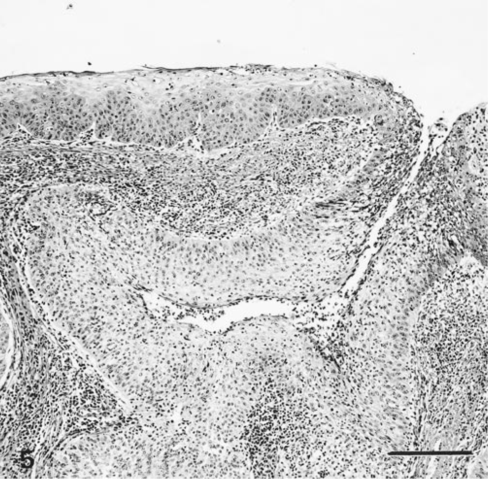
A distinctive microscopic lesion was found in the penis and prepuce of all three males and in vagina (5/7), cervix (7/7), and uterus (3/7) of the females. The normal mucosal epithelium of the tissues was greatly thickened by many layers of epithelial cells that were larger and had larger nuclei than their normal counterparts (Fig. 1). The basal layer of cells appeared to palisade along the basement membrane. The cells had distinct borders, ranged from polygonal to round or elongate, and had moderate amounts of eosinophilic cytoplasm. Nuclei were oval and had irregular profiles. Chromatin was generally fine, and single prominent nucleoli were present in most cells. Mitotic figures were occasionally encountered. Areas in which the cells had undergone squamous differentiation and keratinization were often present. The degree of nuclear pleomorphism and the mitotic rate varied among the samples. Plasma cells and lymphocytes were often present in the connective tissue adjacent to the epithelium and, in some animals, were interspersed among the epithelial cells.
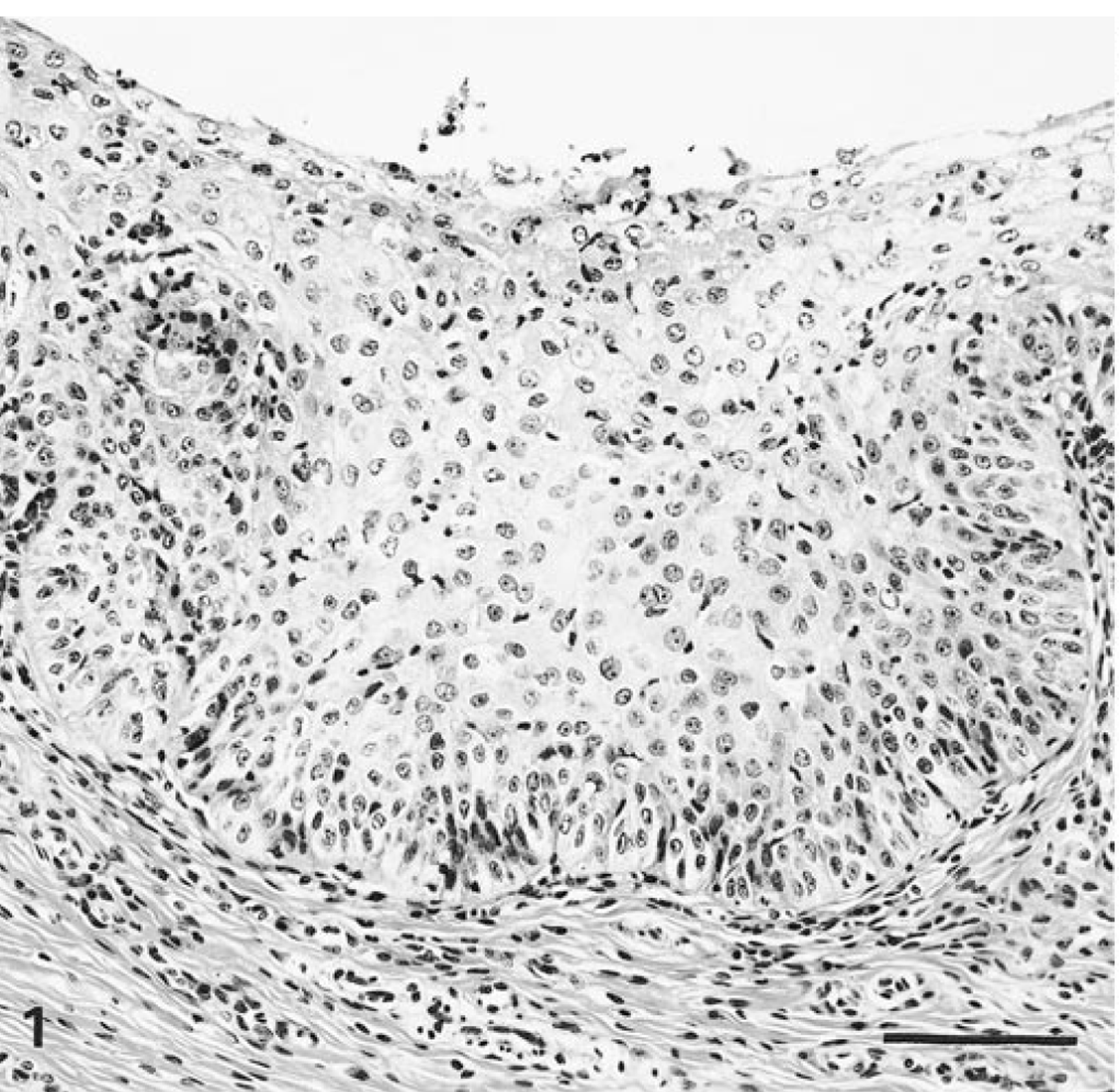
Cervix; sea lion No. 2. Mucosal epithelium is markedly thickened by increased numbers of enlarged, incompletely differentiated epithelial cells. Normal cervical epithelium in this location is two cell layers thick. An inflammatory infiltrate is present. The epithelial lesion was interpreted as intraepithelial neoplasia. HE. Bar = 100 µm.
The genital epithelial lesions were similar in all sites, with one exception. In all seven females, there were scattered areas of glandular differentiation characterized by lumen formation within cervical and uterine epithelial lesions (Fig. 2); none of the males had glandular differentiation.
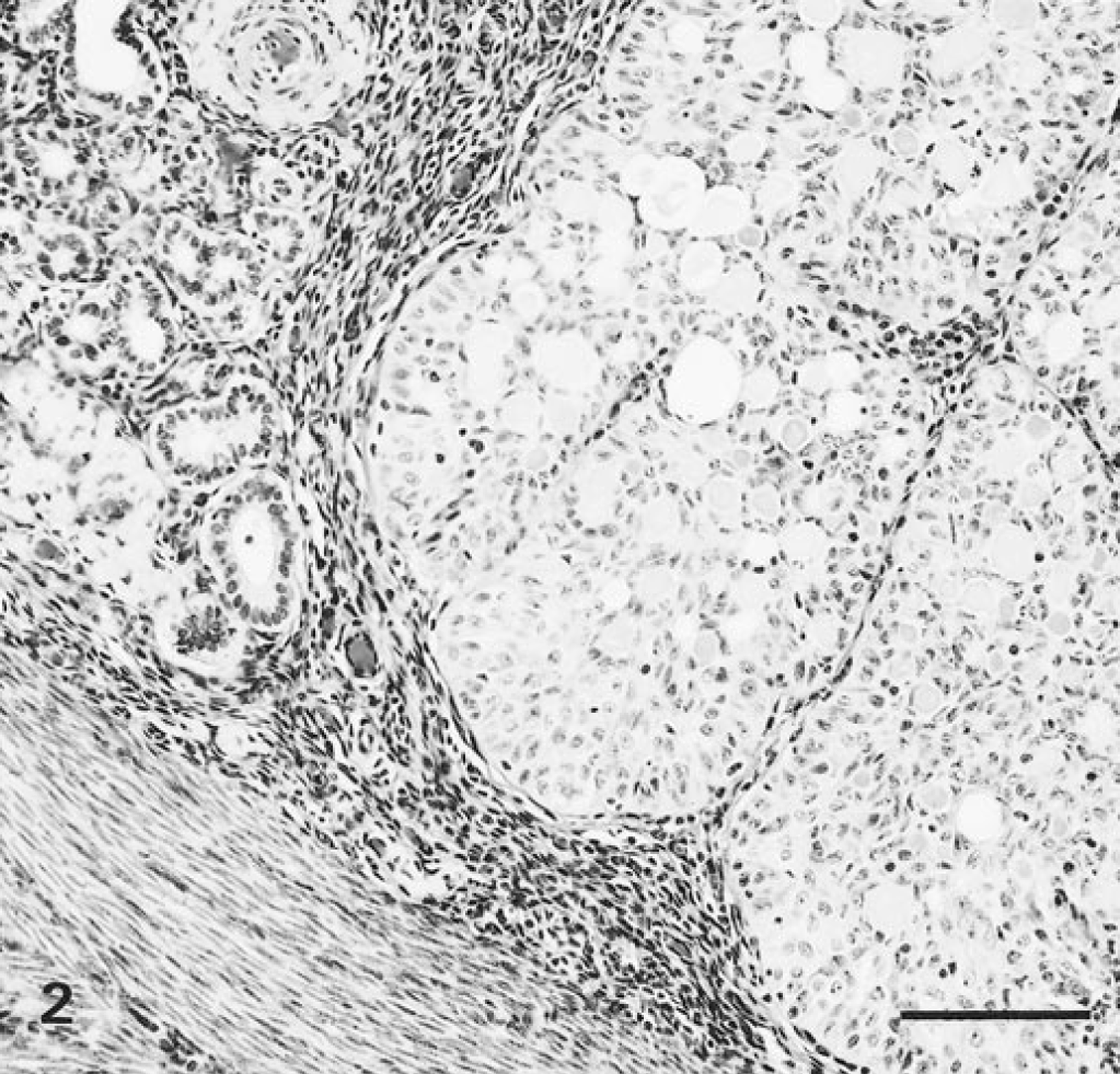
Uterus; sea lion No. 4. Normal uterine glands are at left. At right, glands have been replaced by epithelial lesion with glandlike differentiation (intraepithelial neoplasia). HE. Bar = 100 µm.
The histologic appearance of these genital epithelial lesions closely resembled that of the carcinomas. In some cases, the lesions were virtually identical. Sea lion Nos. 2, 3, and 5 had carcinomas composed of cords and lobules of epithelial cells, as described for the genital epithelial lesions. Squamous differentiation and keratinization of the cells were present multifocally (Fig. 3). Sea lion Nos. 1, 4, and 10 had carcinomas composed of similar epithelial cells that often formed glandlike structures, creating a cribriform pattern (Fig. 4). The lumens frequently contained mucin (PAS positive and carminophilic when stained with mucicarmine). Sea lion Nos. 1–4, 8, and 9 had carcinomas with varying proportions of both glandlike structures and areas of squamous differentiation. Thus, sea lions 1–4 had some carcinomas with either squamous or glandlike differentiation as well as other carcinomas with both types of differentiation. The carcinomas of the three males (sea lion Nos. 5, 6 −7) had only squamous differentiation. All seven females had carcinomas with areas of glandlike differentiation. Invasion of adjacent tissue, scirrhous reaction, vascular invasion, necrosis, and a high degree of nuclear pleomorphism were common features of the carcinomas but were absent from the genital epithelial lesions.
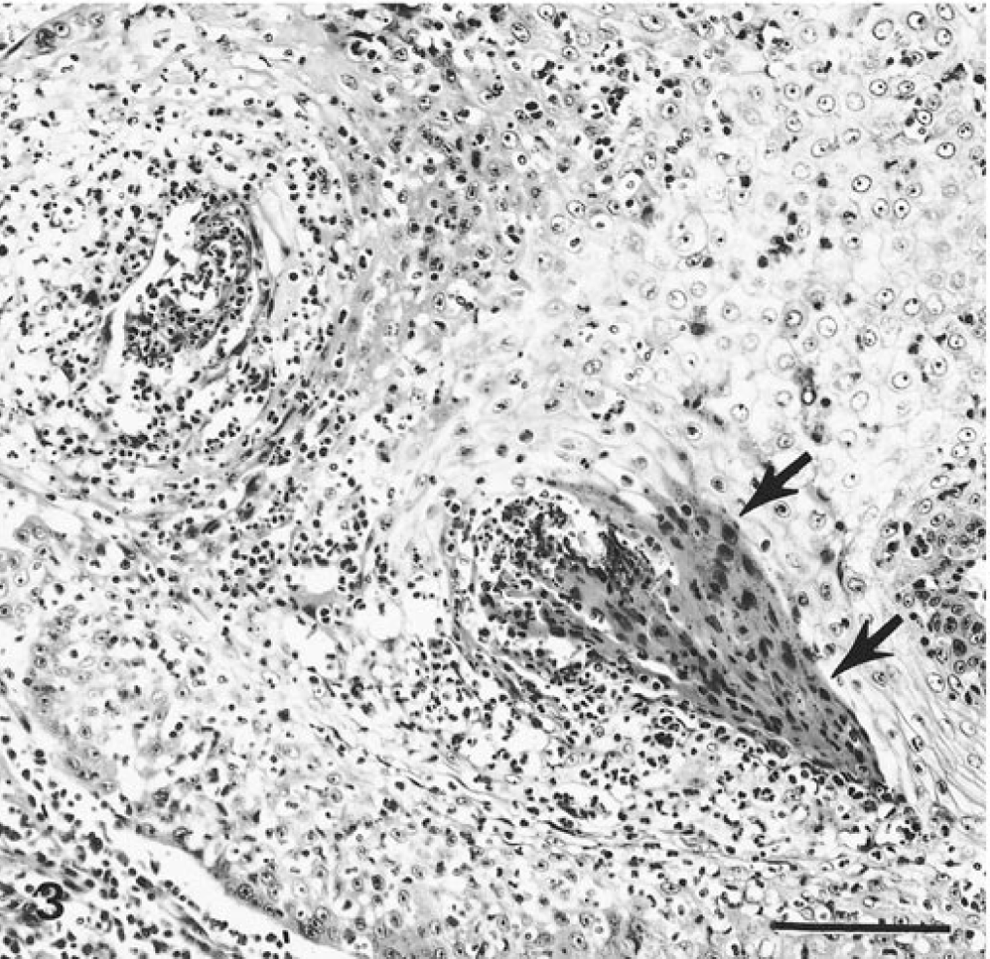
Mesovarium; sea lion No. 3. Metastatic carcinoma contains keratinized cells (arrows). HE. Bar = 100 µm.
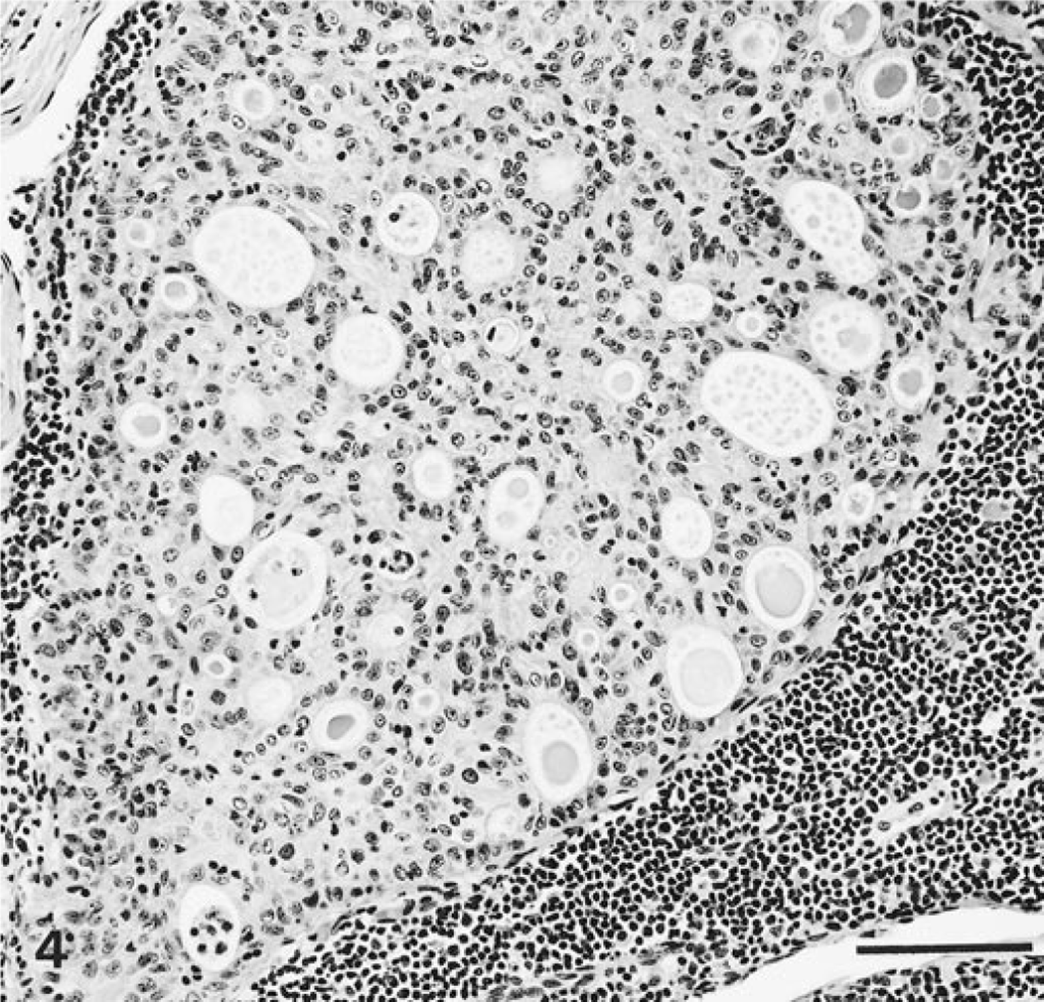
Sublumbar area lymph node; sea lion No. 8. Metastatic carcinoma with cribriform pattern. HE. Bar = 100 µm.
In one of the females (sea lion No. 2), the genital epithelial lesion in the cervix was directly contiguous with invasive carcinoma (Fig. 5). Thus, there appeared to be transition from the genital epithelial lesion to invasive carcinoma. In two other cases (sea lion Nos. 1 and 9), the genital epithelial lesions were in very close proximity to similar invasive carcinoma, but no direct connection was found.
Cervix; sea lion No. 2. Epithelial lesion (intraepithelial neoplasia) at upper left is directly contiguous with, and transitions into, invasive carcinoma. HE. Bar = 200 µm.
Eosinophilic intranuclear inclusion bodies were observed rarely in tissues of five sea lions. They were found in preputial and penile epithelial lesions (Fig. 6) of one sea lion, in cervical epithelial lesion of another sea lion, in invasive cervical carcinoma of a third sea lion, and in metastatic carcinoma (Fig. 7) of two additional sea lions. Similar intranuclear inclusion bodies also were found in hyperplastic stratified squamous epithelium of the oropharynx in the sea lion that had inclusions in invasive cervical carcinoma (Table 1). The inclusion bodies were homogeneous, round to oval, and 4–10 µm in diameter and either were surrounded by a clear space or completely filled the nucleus.
Histopathologic findings and results of herpesvirus consensus primer PCR testing of unfixed metastatic tumors for California sea lions with metastatic carcinoma (CA) involving sublumbar area lymph nodes (ln).
IEN = intraepithelial neoplasia.
NF = none found.
NS = no sample available.
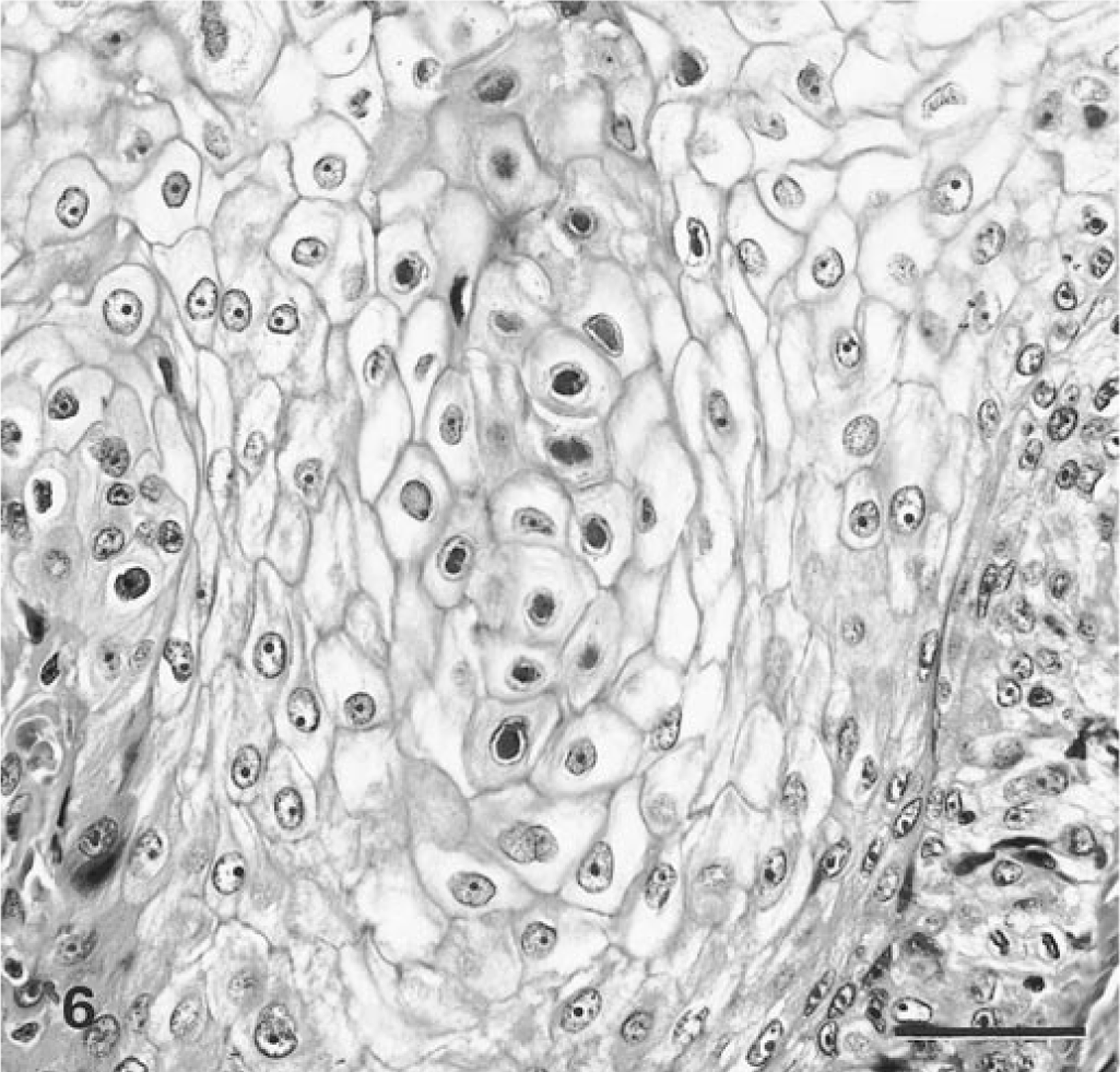
Prepuce; sea lion No. 5. Epithelial lesion (intraepithelial neoplasia) contains intranuclear inclusion bodies at center. HE. Bar = 50 µm.
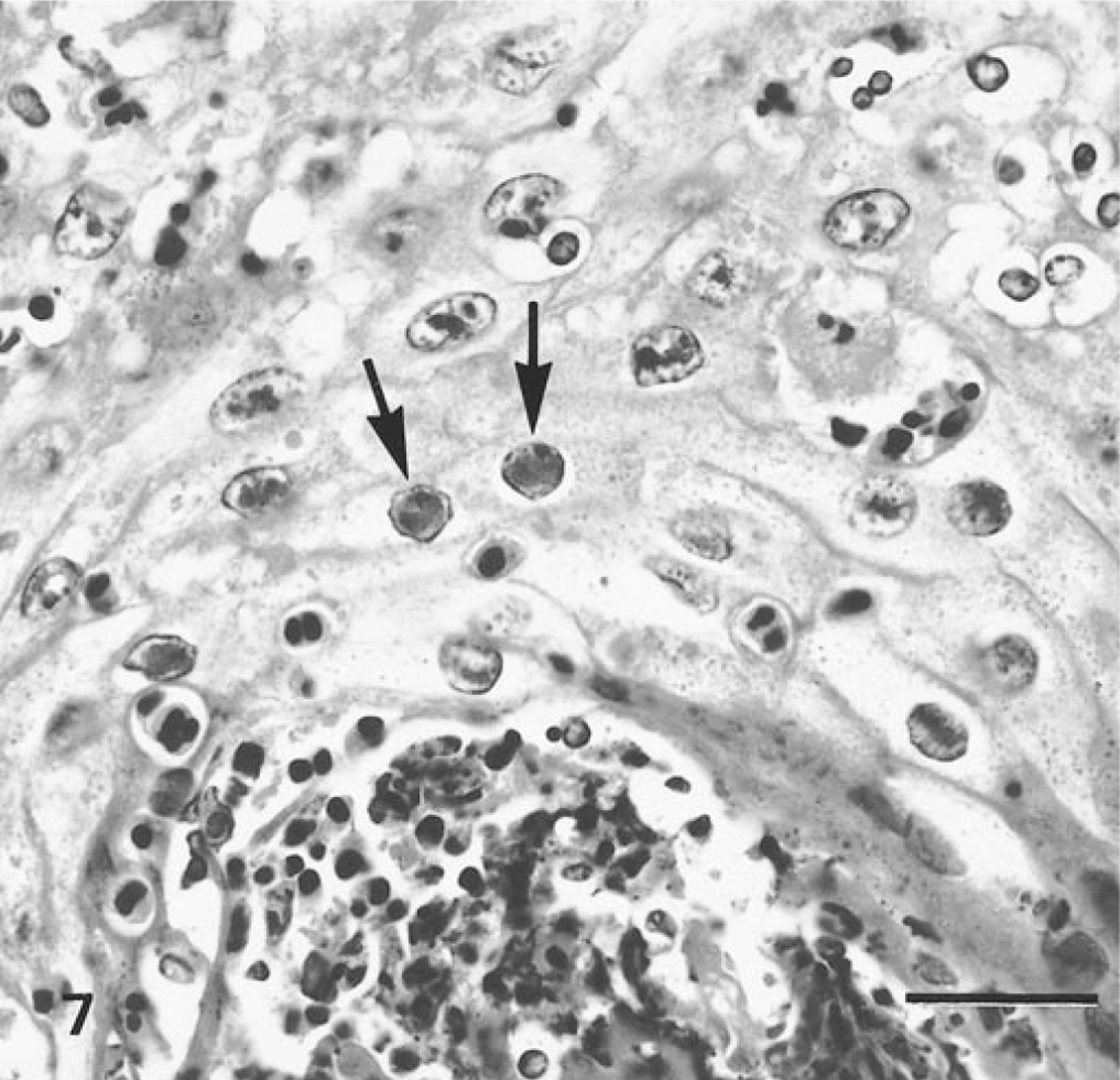
Mesovarium; sea lion No. 3. Higher magnification of the field shown in Fig. 3. Metastatic carcinoma contains intranuclear inclusion bodies (arrows) in focus of squamous differentiation. A cluster of more darkly stained keratinized cells is present at lower right. HE. Bar = 25 µm.
Ultrastructural findings
Transmission electron microscopic examination of formalin-fixed tissues that contained the genital epithelial lesion with inclusion bodies (sea lion No. 5) revealed 92–115-nm-diameter nonenveloped intranuclear viral nucleocapsids that had round to hexagonal profiles (Fig. 8); 150–200-nm-diameter enveloped extracellular viruses also were observed. Similar viral particles were found in a metastatic carcinoma that contained inclusion bodies (sea lion No. 3).
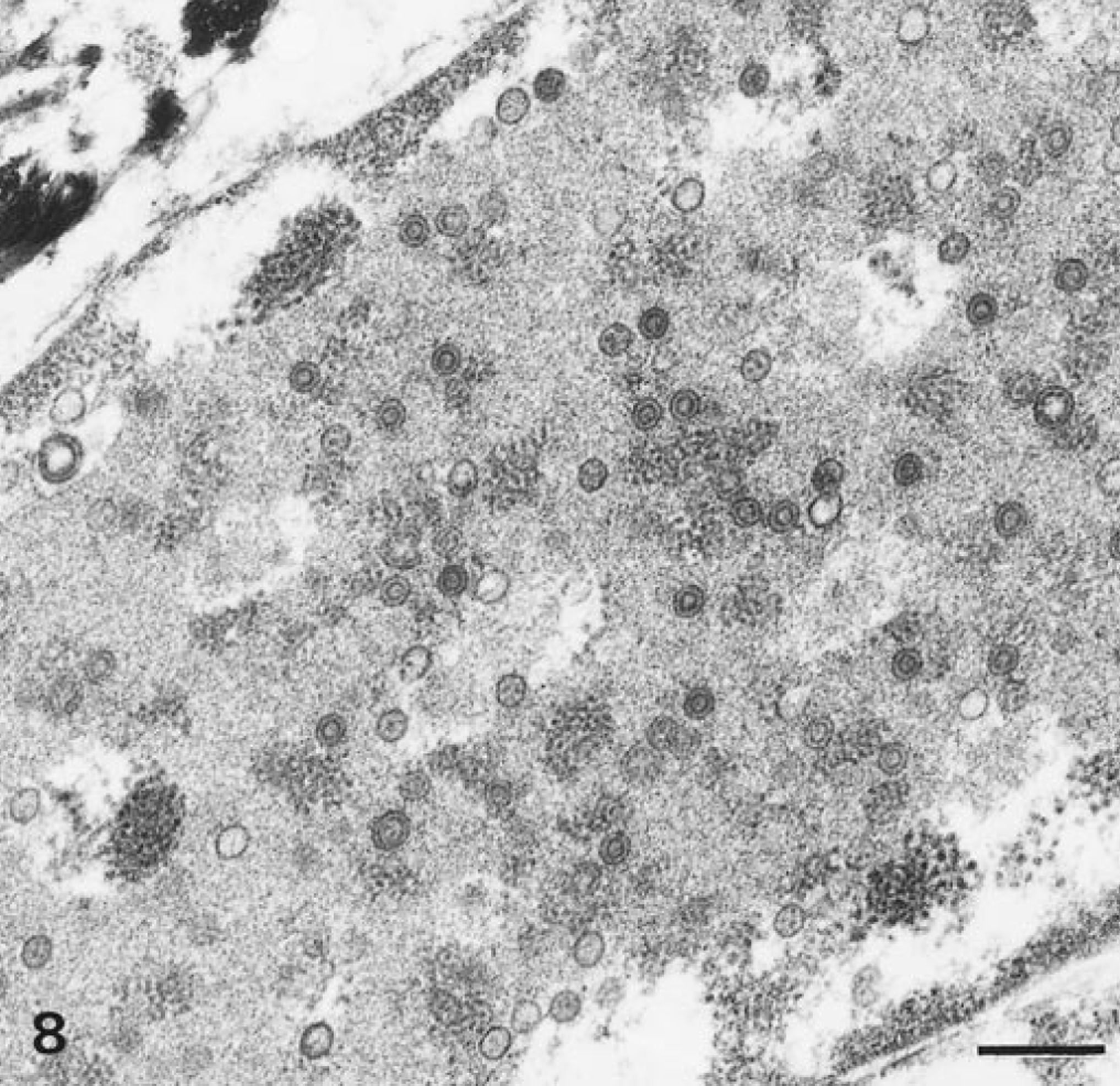
Transmission electron micrograph. Prepuce, epithelial lesion (intraepithelial neoplasia); sea lion No. 5. Nucleus of cell contains numerous 92–115 nm diameter viral nucleocapsids consistent with those of herpesviruses. Bar = 500 nm.
Immunohistochemical findings
In one sea lion (No. 5), an immunohistochemical stain for the latent membrane protein of EBV stained preputial epithelial lesion and intranuclear inclusion bodies (Fig. 9) but not normal adjacent preputial epithelium. The inclusion bodies in the hyperplastic oropharyngeal lesion (sea lion No. 2) also stained positively. Lesions with inclusion bodies from sea lion Nos. 2, 3, 6, and 9 did not stain positively.
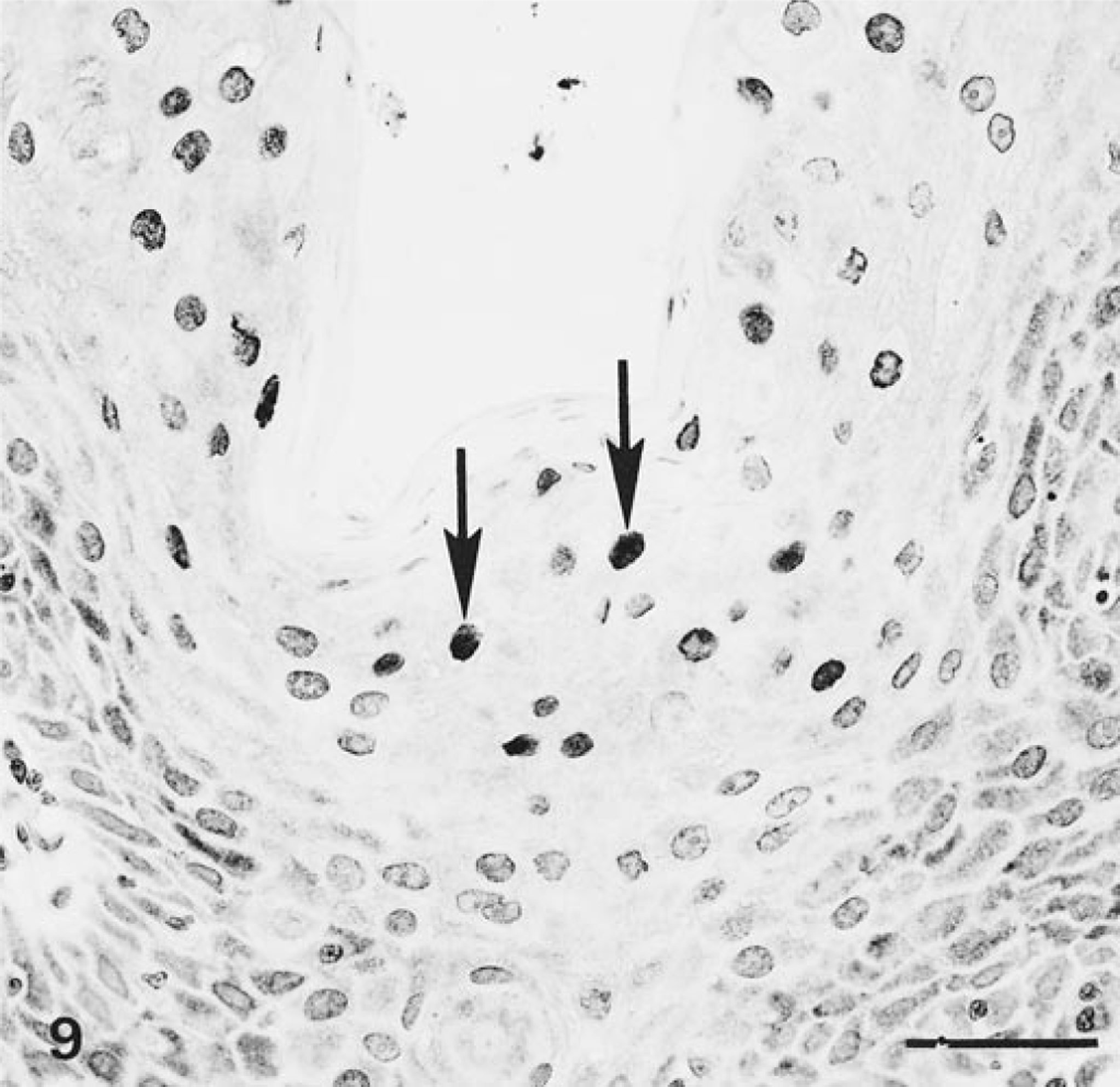
Penis; sea lion No. 5. Epithelial lesion (intraepithelial neoplasia) has positive immunohistochemical staining of intranuclear inclusion bodies (arrows) for latent membrane protein of Epstein-Barr virus or a closely related protein. Avidin–biotin–peroxidase complex method for latent membrane protein of Epstein-Barr virus counterstained with Mayer's hematoxylin. Bar = 50 µm.
PCR findings
Unfixed frozen samples of metastatic carcinomas were available from four of these sea lions (Nos. 1, 2, 5, and 7). Herpesvirus consensus primer PCR followed by DNA sequencing 9 22 was used to demonstrate the presence of herpesvirus DNA and to generate information about portions of the putative DNA polymerase and terminase genes. The four sea lion carcinoma samples yielded identical sequences (no DNA polymorphisms) (Figs. 10, 11). Similarity analyses using BLASTP software on the NCBI website and the GenBank database suggested that these DNA sequences were different from those of any herpesvirus published to date and were derived from a previously unknown member of the gammaherpesvirus genus Rhadinovirus. This relationship for the sea lion herpesvirus was again demonstrated using phylogeny mapping software and the set of GenBank herpesvirus sequences for which data on both genes are available (Fig. 12). The sea lion carcinoma herpesvirus sequences were unlike those from phocid herpesvirus 1, an alphaherpesvirus associated with pneumonia in harbor seals. 8 Isolation of a putative gammaherpesvirus from a California sea lion has been reported, 8 but because the DNA polymerase and terminase sequences from that isolate are not available, a comparison with the sea lion herpesvirus reported here is not possible.
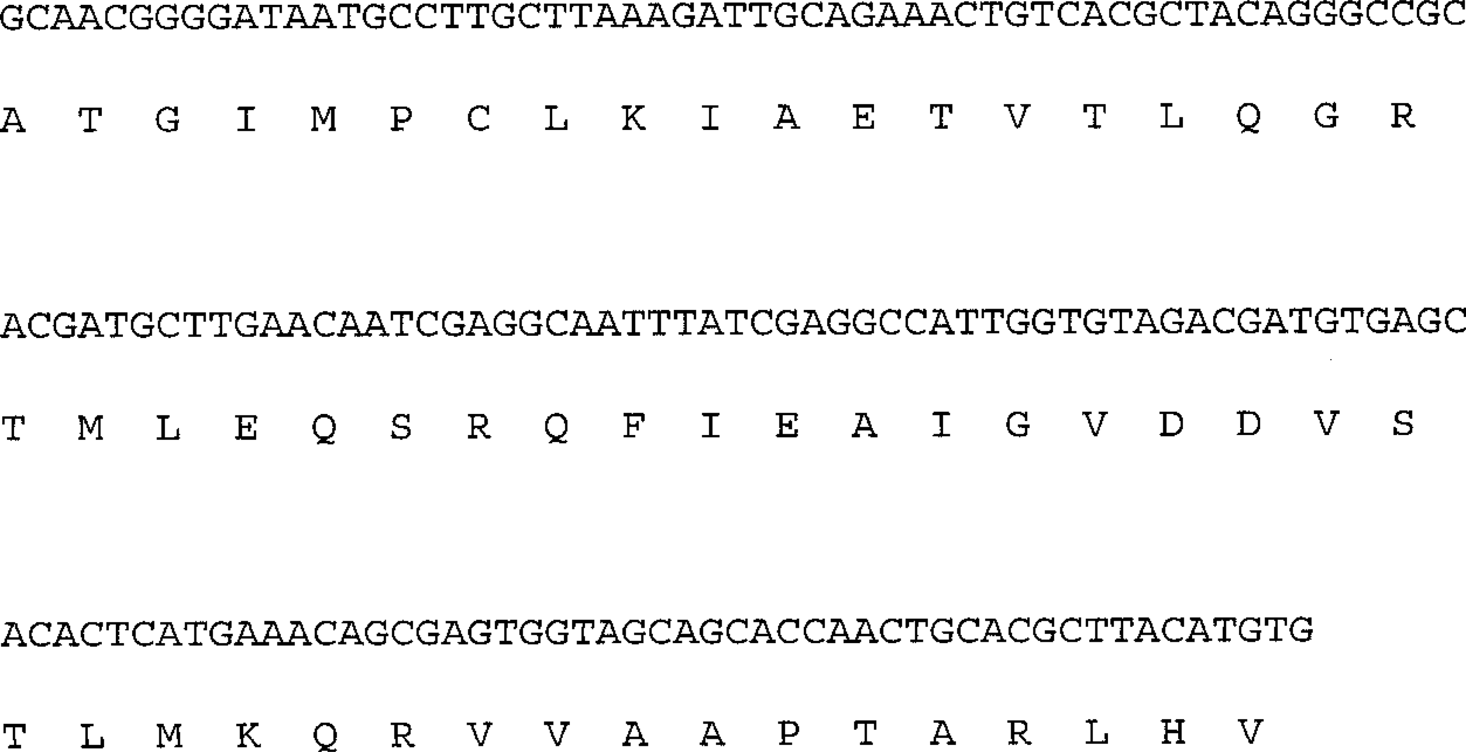
DNA sequences of sea lion herpesvirus that were amplified by consensus primer PCR. Protein products are from conceptual translation of internal region of the DNA polymerase gene (sequence deposited in GenBank, accession no. AF193617).
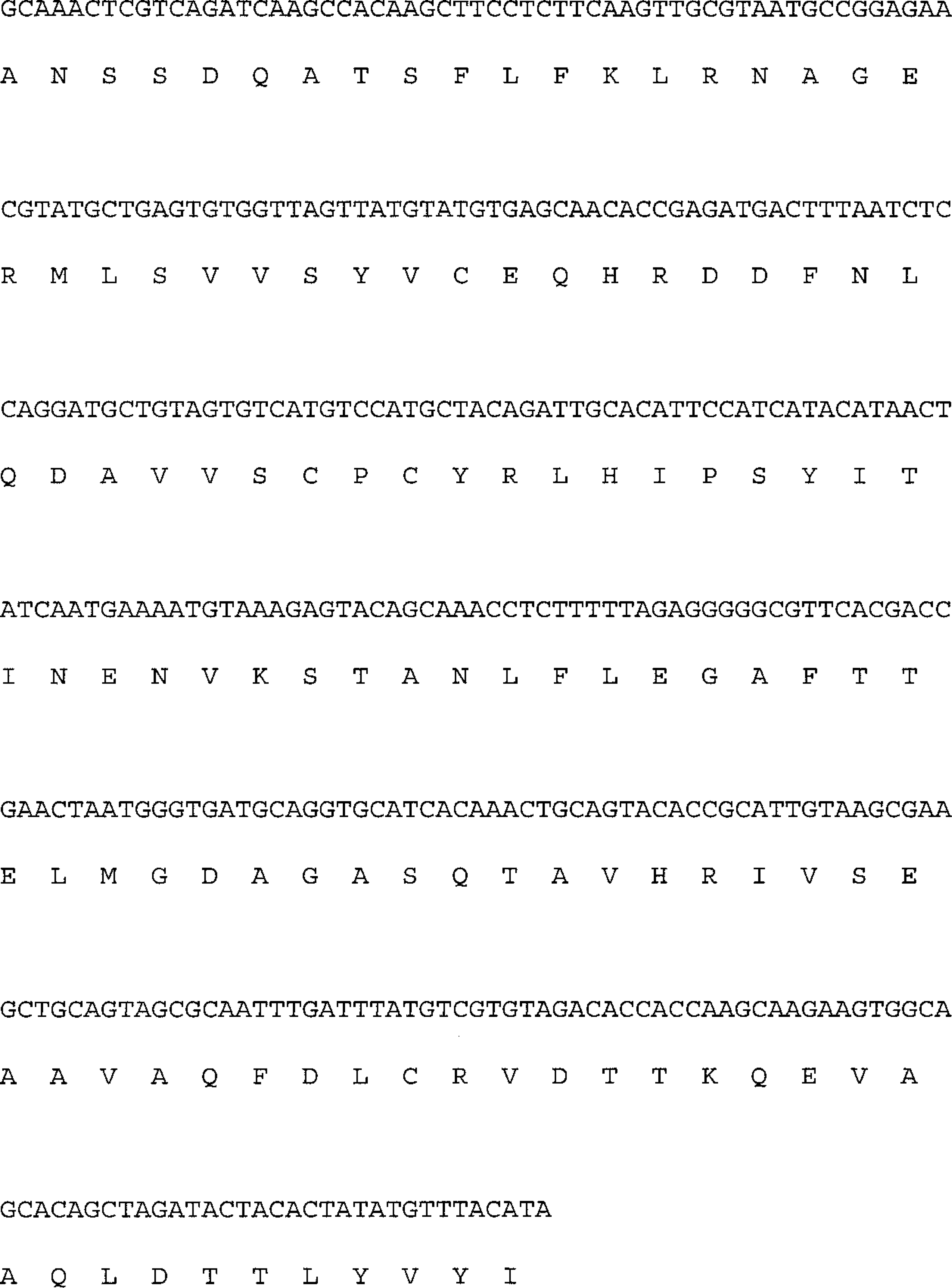
DNA sequences of sea lion herpesvirus that were amplified by consensus primer PCR. Protein products are from conceptual translation of internal region of the terminase gene (sequence deposited in GenBank, accession no. AF193618).
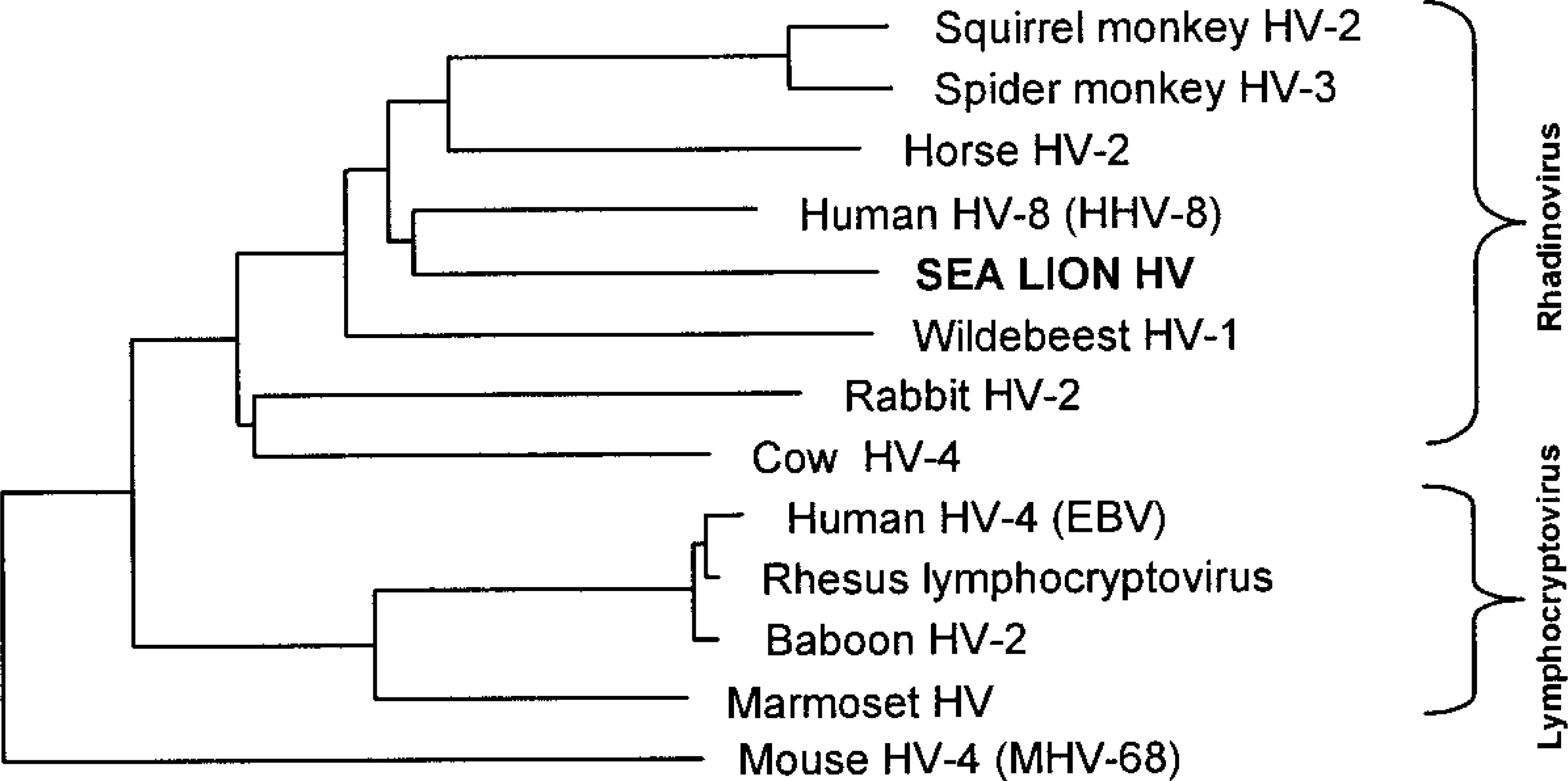
Phylogenetic tree placing the sea lion herpesvirus in the gammaherpesvirus genus Rhadinovirus based on comparisons using corresponding portions of the terminase protein. The relationships are derived from aligning the set of herpesvirus protein sequences in GenBank for which both the terminase and DNA polymerase genes have been determined. The tree based on DNA polymerases is similar (not shown). HV = herpesvirus; HHV = human herpesvirus; EBV = Epstein-Barr virus; MHV = mouse herpesvirus.
DNA extracted from the four metastatic carcinomas was tested for genetic evidence of papillomavirus by genomic Southern blot using five different papillomaviruses as probes and by PCR with consensus papilloma E1 gene primers. All samples were negative by both methods.
Discussion
The similarity of the cases reported here to the previously reported cases of metastatic carcinoma of undetermined origin in California sea lions makes it likely that they represent the same disease. 1 7 11 The histopathologic findings support the conclusion that genital epithelium is the site of origin of the metastatic carcinomas. The presence of the distinctive genital epithelial lesion in all 10 sea lions that had metastatic carcinoma, the strong histologic similarity between the genital epithelial lesions and the carcinomas, and the observation of a zone of transition from genital epithelial lesion to carcinoma support the interpretation that the genital epithelial lesions represent intraepithelial neoplasia (IEN; a noninvasive neoplastic proliferation of epithelium that is present in its site of origin and is a precursor to invasive, metastasizing carcinoma). 19 The presence of glandlike differentiation in the genital epithelial lesions and carcinomas of all of the females and its absence in the corresponding lesions of the males mimics normal genital epithelial morphology of sea lions in which glands are present in the cervix and uterus but absent in the prepuce and penis. IEN was not observed in any nongenital tissues, including transitional epithelium of the urinary tract. IEN similar to that found in these sea lions has been observed in genital epithelia of California sea lions that did not have carcinomas (T. P. Lipscomb, personal observation).
The presence of characteristic eosinophilic intranuclear inclusion bodies and the electron microscopic finding of typical herpesviral particles 3 in IEN and carcinoma indicate herpetic infection of these lesions. In a previous study, herpesvirus-like particles were detected by electron microscopy in a lesion described as a hyperplastic plaque on the penis of a California sea lion. 7 Positive immunohistochemical staining for latent membrane protein of EBV in IEN suggests the presence of an EBV-related herpesvirus. The failure of immunohistochemistry to detect latent membrane protein or a related protein in the other affected sea lions may be a result of prolonged fixation of the tissues or lack of expression of the protein. Results of PCR and sequencing indicate the presence of a novel gammaherpesvirus in metastatic carcinomas from all four sea lions for which unfixed tumor samples were available. The combination of these findings indicates an association between the herpesvirus and the carcinomas.
Cervical carcinoma in human beings is similar to this sea lion disease in that it arises as IEN of the cervix and has similar histologic features. Infection of cervical epithelium by certain human papillomaviruses has been implicated as an important factor in the development of cervical cancer. 13 To investigate the possibility of papillomavirus involvement, genomic Southern blots and papillomavirus PCR were performed on DNA extracted from the four metastatic carcinomas. The results were negative.
Herpesviral infections are associated with several animal cancers including renal adenocarcinoma of frogs 6 and lymphoma of chickens 5 and monkeys. 10 In humans, EBV has been implicated in the pathogenesis of several tumors, including nasopharyngeal carcinoma 23 and the African form of Burkitt's lymphoma. 18 Recently, human herpesvirus 8 (HHV-8) was implicated as the possible cause of Kaposi's sarcoma. 2 Like this sea lion virus, EBV and HHV-8 are gammaherpesviruses. The sea lion virus is most closely related to HHV-8 (Fig. 12).
The sea lion carcinoma is particularly similar to EBV-associated nasopharyngeal carcinoma (NPC). Although rare in most parts of the world, there is a high incidence of NPC in certain areas of southern China 24 and in Eskimos in Alaska and Greenland. 14 In situ hybridization studies have demonstrated EBV RNA in almost 100% of NPC suggesting a significant causal role, 16 although the localization to the geographic areas of high incidence in spite of the ubiquity of EBV infection indicates that other factors are involved. Both the sea lion carcinoma and NPC arise in mucosal epithelium as IEN, 17 often metastasize as squamous cell carcinoma, and contain gammaherpesvirus nucleic acids. 16
The presence of virus-infected lesions in genital epithelium suggests the possibility of venereal transmission, as occurs in several herpesviral diseases of human beings 4 and animals. 12 The polygynous behavior of California sea lions 20 might facilitate spread of the infection.
Regarding the association of viruses with human cancers, it has been stated that “prevalence of infection is always much higher than the incidence of the associated tumour.” 15 For example, EBV is a ubiquitous infectious agent endemic in all human populations, yet the incidence of EBV-associated cancers is quite low. 4 With metastatic carcinoma present in 18% of stranded, sexually mature animals that were necropsied, 7 this cancer is one of the most important diseases of California sea lions. If the virus is the primary cause, infection results in cancer in a remarkably high proportion of these animals. This high prevalence could be useful in future studies of herpesviral carcinogenesis. Investigations of this disease also may provide insights into human cervical carcinoma, another virus-associated genital cancer. Additional research is needed to elucidate the role of the herpesvirus and investigate potential cofactors in the sea lion genital carcinoma.
Footnotes
Acknowledgements
This study was partially funded by the National Marine Fisheries Service and the Marine Mammal Center and by a grant from the American Registry of Pathology. We thank J. Jenkins for electron microscopy, R.-A. V. Ferris for photomicroscopy, W. Inskeep for editorial comments, and B. Jenson and C. Baker for providing, respectively, canine and rabbit papillomavirus genomes. T. P. Lipscomb is a lieutenant colonel and D. P. Scott is a major in the US Army. The opinions or assertions contained herein are the private views of the authors and are not to be construed as official or as reflecting the views of the Department of the Army or the Department of Defense.