
Abstract
Six genetically related Shorthorn calves were affected with the tibial hemimelia syndrome. The lesions included bilaterally malformed or absent tibia and abdominal hernia in all animals, a long shaggy haircoat, retained testicles in males, and meningocele in three animals. The malformations were similar to those described previously in Galloway calves. Pedigree analysis demonstrated a mechanism by which a recessive allele in a homozygous state could be responsible for the disorder. The condition in these calves was considered the result of a recurrence of a genetic mutation affecting a putative hemimelia locus.
Keywords
A syndrome of multiple congenital anomalies, including tibial hemimelia, abdominal hernia, and cranial defects, has been described initially in a beef calf of unspecified breed 7 and later in Galloway cattle. 5 Both tibias were shortened, malformed, or absent, there was cranioschisis involving the frontal bone leading in most cases to formation of one or two meningoceles, and a large defect in the ventral abdominal midline was associated with abdominal hernia. Additional lesions also included reproductive system anomalies (nonfusion of Müllerian ducts in females, bilateral cryptorchidism in males), internal hydrocephalus, and sometimes nonfusion of the pelvic symphysis. Pedigree analysis and breeding trials in the Galloway cattle indicated that this combination of anomalies was inherited as a simple autosomal recessive trait. 3 It was estimated at the time that 1–2% of purebred Galloway calves born between 1965 and 1975 were affected, originating from 47% of the herds. 6 Here, we describe a similar syndrome observed in Shorthorn cattle.
Six purebred Shorthorn calves were examined shortly after birth. All six were born between February and August 1999. The pathologic findings are summarized in Table 1. Calf 1 was female, from a herd of purebred Shorthorn cattle in Quebec, Canada. It was examined by a field veterinarian, who observed two meningoceles on either side of the forehead, a large abdominal hernia, and hind legs that appeared flaccid and shortened. The hair coat was long and curly. The animal was alert but unable to stand. It was euthanatized, and no necropsy was performed.
Congenital anomalies detected in six Shorthorn calves.
Calf 2 was a male born at the same farm and was submitted to a Quebec provincial animal pathology laboratory. The hair coat was long and curly. Two meningoceles were present on either side of the head, about 6 cm in diameter, protruding through fusion defects between the occipital and parietal bones. There was a large abdominal hernia through a defect in the ventral abdominal midline. The hind limbs appeared shorter than normal, and there was absence of the tibial diaphyses; bony masses were present distal to the femur and proximal to the tarsus, interpreted as malformed tibial epiphyses.
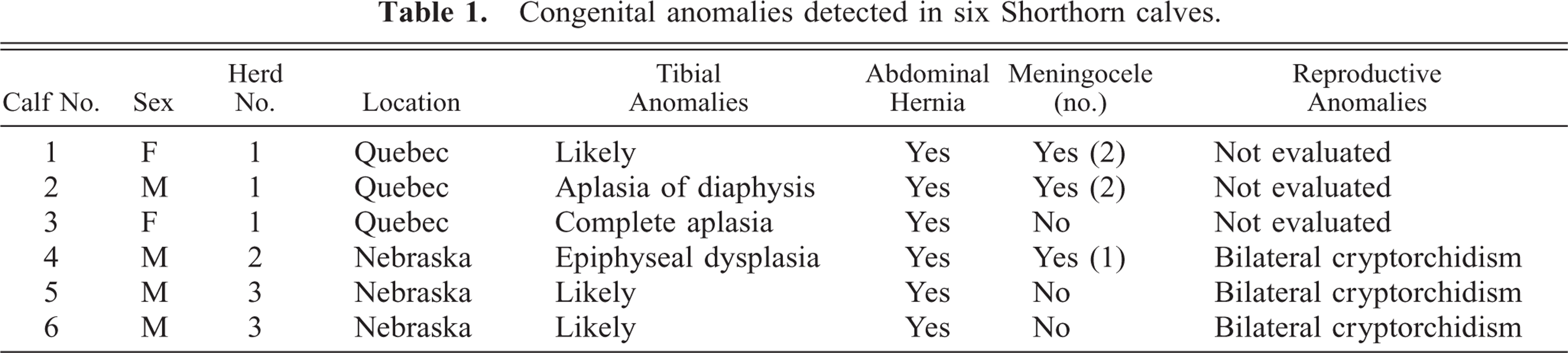
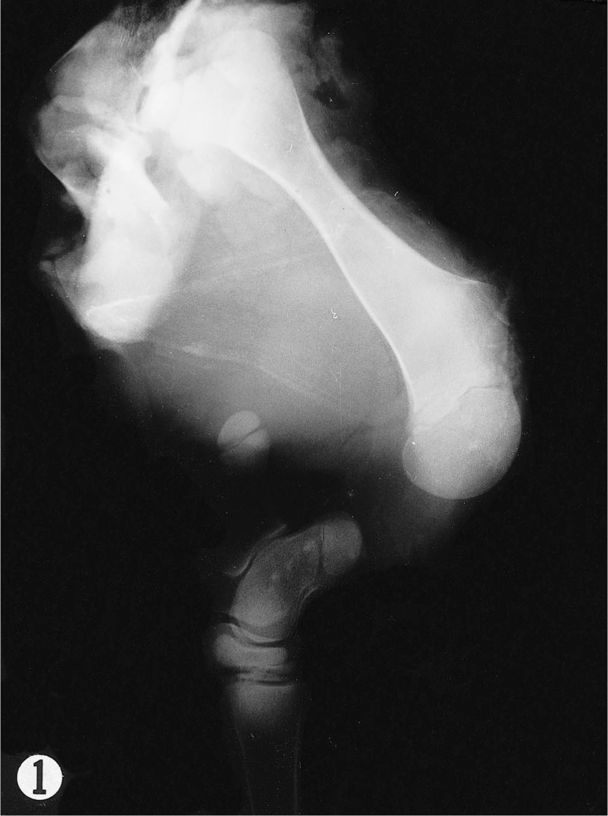
Calf 3 was a female from the same farm and was sent to the pathology service of the Faculté de Médecine Vétérinaire of the Université de Montréal. The animal was alert with a suckle reflex and attempted but was unable to stand. It had a long and curly hair coat, and abdominal viscera were herniated through a 20-cm-long defect in the caudal abdominal wall on the ventral midline. The hind limbs were shortened, and both had complete absence of the tibia, confirmed by radiographs (Fig. 1). The distal femoral epiphyses bore only a single large rounded condyle (Fig. 2). The patellae were absent. The ilia were shortened and malformed, and the acetabular cavities were shallow. No abnormality was noted in the skull bones. The brain showed hydrocephalus, with moderate dilation of all ventricular chambers. On the roof of both lateral ventricles, two to four rounded sessile nodules up to 1.5 cm in diameter extended from the neuropil into the ventricular lumina; histologically, these nodules consisted of gray matter of normal appearance, without an ependymal covering.
Radiograph. Hind limb (lateral view); calf No. 3. Complete aplasia of the tibia.
Femur; calf No. 3. The distal epiphysis is large and rounded, without normal development of the condyles and intercondylar fossa.
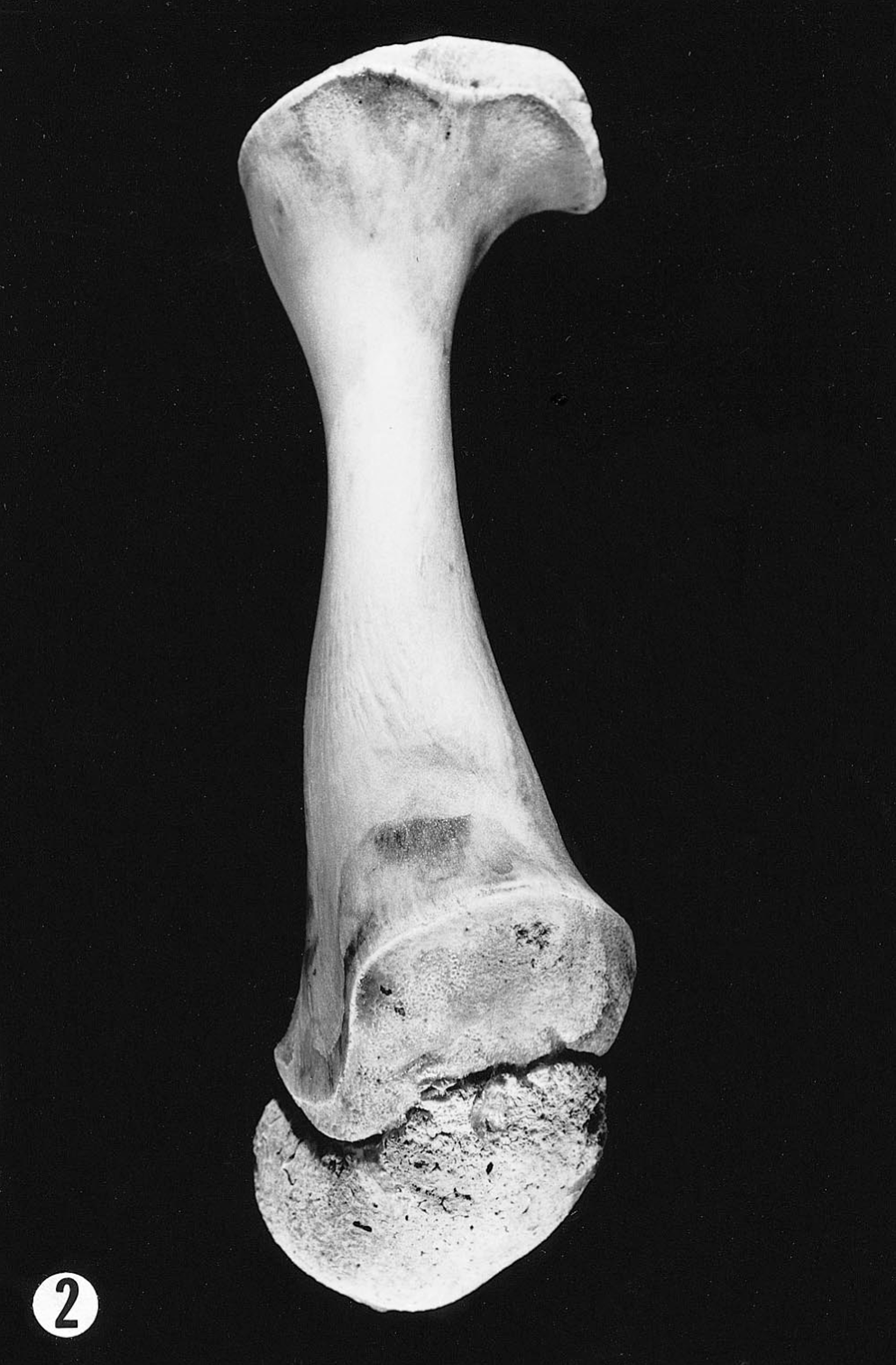
Calf 4 was a male from a Nebraska herd consisting of two Shorthorn and 20 crossbred cows. Its dam was a Shorthorn and had been artificially inseminated with semen from a Shorthorn bull. The calf was presented directly to the Nebraska Bovine Congenital Disease Investigation Center. It had a meningocele, 2 cm in diameter, in the dorsal occipital region (Fig. 3). An abdominal hernia extended from the pubis to near the umbilicus. The hair coat was long and shaggy. Both testes were retained near the caudal poles of the kidneys. The pelvic symphysis was not fused and the pelvis was misshapen, with shallow acetabula and irregular bony surfaces. The table and ramus of the ischium were absent, and no obturator foramina were formed. The pubis was also absent. The wings of the ilium were small and underdeveloped. The distal femurs were malformed, their ends consisting of an irregular mass of chondrosseous tissue, which radiographically appeared as an osseous shadow replacing the femoral condyles, patella, and proximal tibial epiphysis (Fig. 4). The tibia was rotated 60–80 degrees medially. The distal tibia was flared.
Skull; calf No. 4. There is cranioschisis and formation of a meningocele.
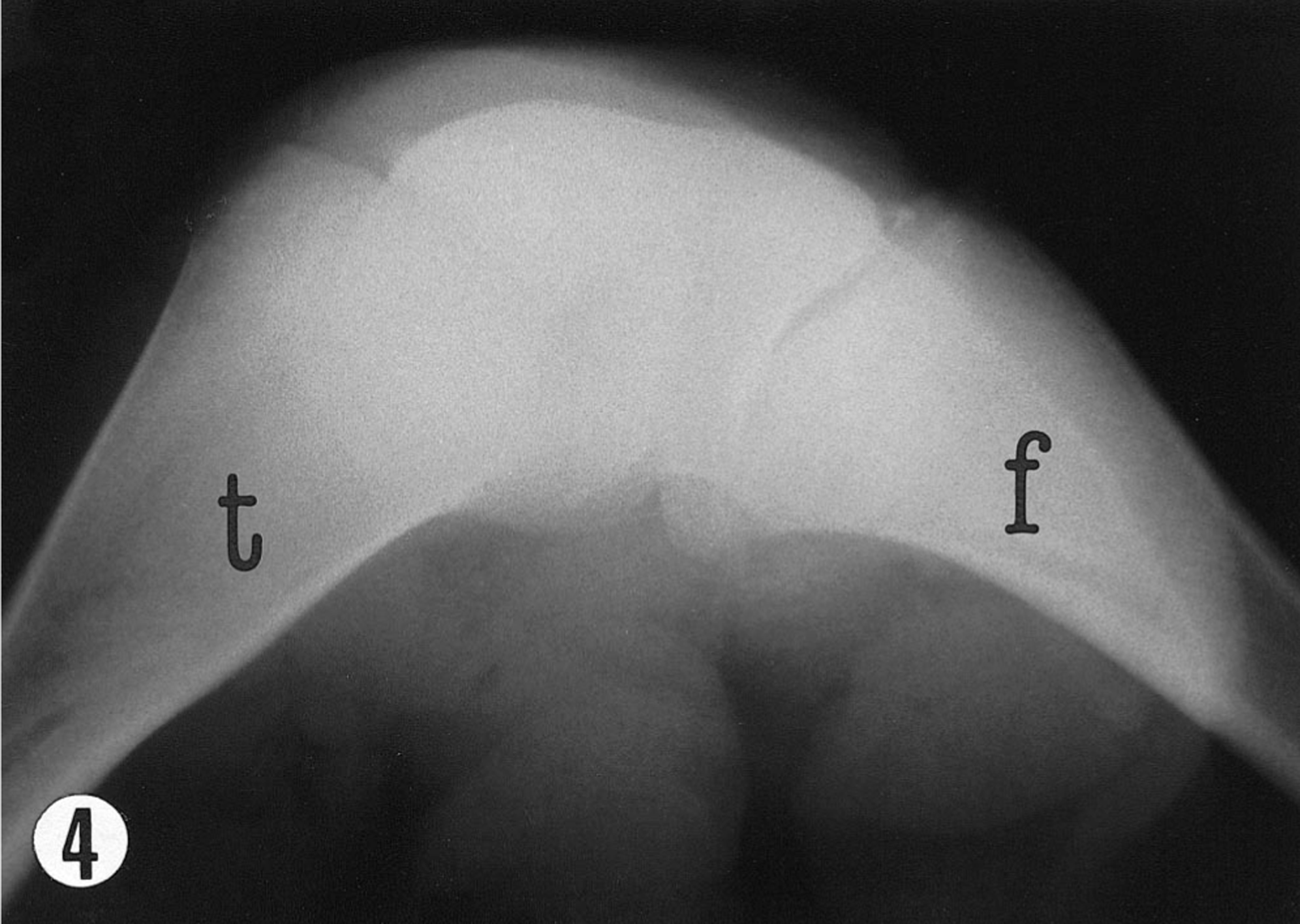
Radiograph. Stifle joint (lateral view); calf No. 4. The femoral condyles, patella, and proximal tibial epiphysis are fused in a large osseous mass (t = tibia; f = femur).
Calves 5 and 6 came from another small herd of purebred Shorthorns in Nebraska. Necropsies were not performed, but their condition was reported to the Nebraska Bovine Congenital Disease Investigation Center. Based on photographs and descriptions provided by the owners, both were diagnosed with the same syndrome. Both calves were males and were recumbent at birth. They had small hind quarters, shortened rear legs, ventral abdominal hernias, long shaggy hair, and retained testicles (Fig. 5).
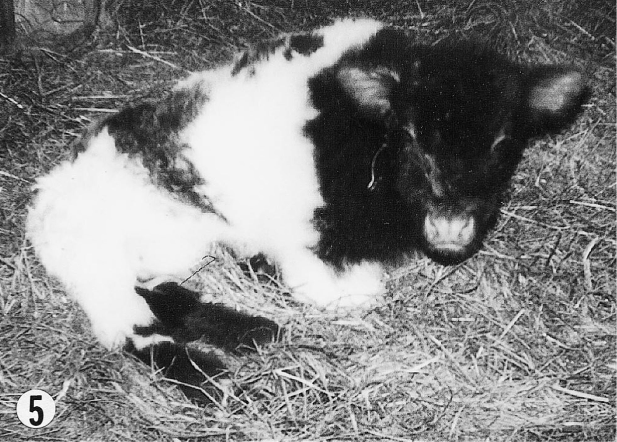
Calf No. 6. Note short malformed hind limbs and long curly hair.
Pedigrees were collected for all affected animals. Analysis demonstrated a common maternal and paternal ancestor within seven generations in all six calves (Fig. 6).
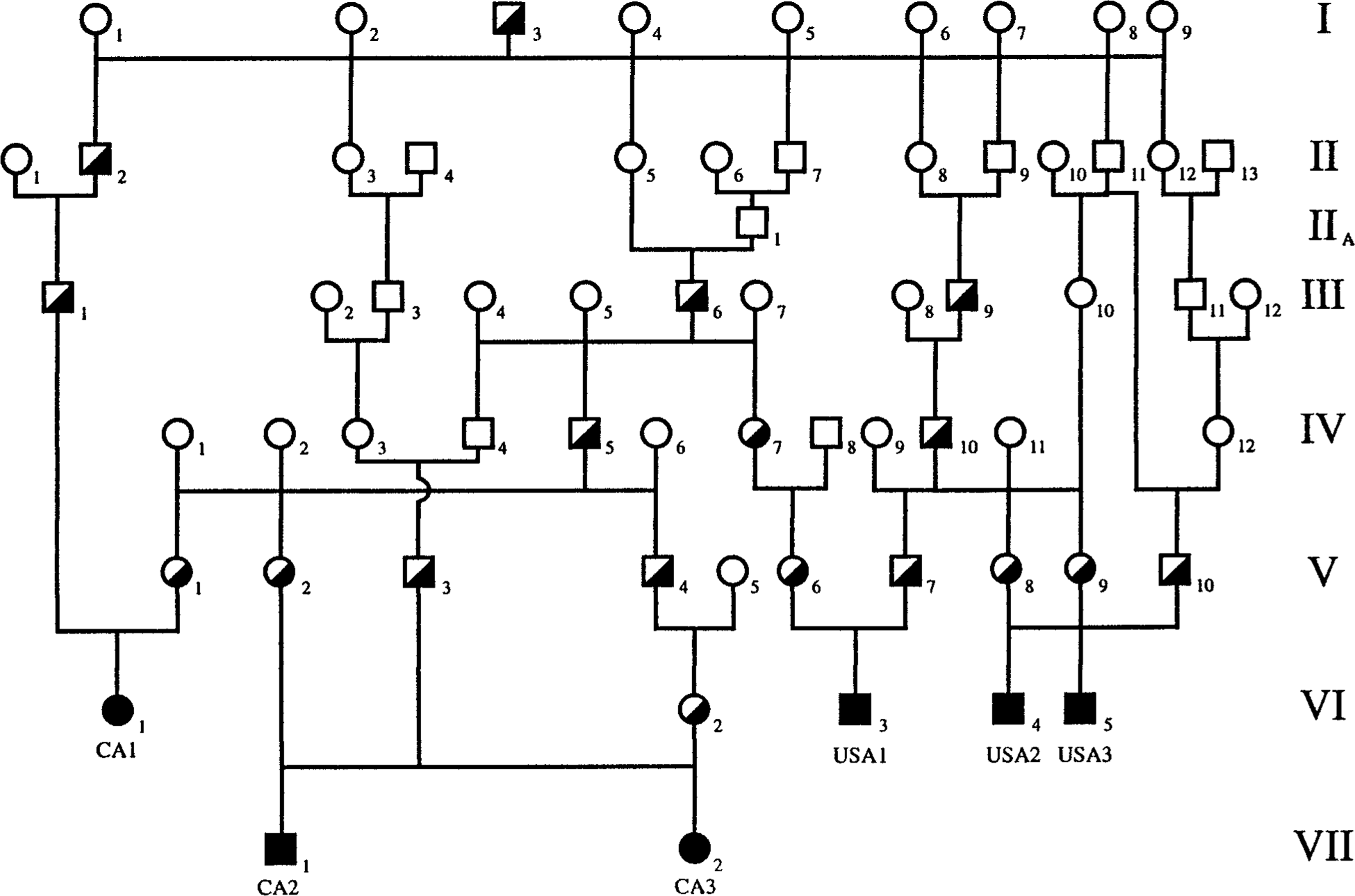
Familial relationships of the six calves; CA1, CA2, CA3, USA1, USA2, and USA3 refer to calf Nos. 1–6, respectively (squares = males, circles = females; darkened figures = affected animals; half-darkened figures = obligate heterozygotes).
Although there were variations in the skeletal anomalies among individuals, the combination of tibial hemimelia, abdominal hernia, and meningocele is strikingly similar to the condition previously described in Galloway cattle. All calves had common ancestors; the familial relationships provide a mechanism by which a recessive allele could have been transmitted from a heterozygous sire (sire 3 of generation I, Fig. 6) and led to a homozygous state for all six calves. These relationships and the phenotypic similarity to a proven genetic disorder in Galloway cattle strongly suggest this to be a simple autosomal recessive trait. A definitive confirmation would require breeding trials between suspected carriers or identification of a defective gene. Anomalous tibial development without an apparent genetic basis has been reported in cattle, but the lesions were not bilateral and were not associated with abdominal hernia or cranial lesions. 1 2 No infectious or toxic agent is known to cause such a combination of defects, and the distance between the affected herds argues against a common environmental influence.
Past experience with this disorder in Galloway cattle and with other recessive traits suggests that numerous carriers may be present in the North American Shorthorn cattle population. Other affected calves might have appeared previously but were unreported because they were considered isolated cases, without an apparent hereditary basis. The appearance of multiple cases within a short time period made possible the investigation and discovery of familial relationships that revealed the probable hereditary basis behind these defects. The sudden occurrence of the syndrome in multiple animals within a period of a few months on three different farms could have been due to linebreeding and high inbreeding coefficients in those herds.
The similarity of the affected Shorthorn phenotype to the Galloway phenotype suggests that the same gene locus may be mutated. The common ancestral Shorthorn sire is not known to be related to the historic carriers in the Galloway breed. Because mutation rates are estimated in the range of 104–106 per generation, one might expect mutations to recur at regular intervals and similar mutations to occur independently in separate breeding populations. However, recurrent mutations are not often documented in the veterinary literature. Syndactyly is a recent example of a mutation of the same gene that emerged in two independently bred cattle populations. Crossbreeding trials demonstrated the mutation to be allelic, but the phenotypes were distinct, suggesting unique mutations. 4 Similarly, the appearance of the tibial hemimelia syndrome in purebred Shorthorns 20 years after its identification in Galloways is most likely due to recurrence of a similar mutation. It is not clear how a single mutation could lead to concurrent anomalous development of skull, pelvic, and hind limb bones, abdominal wall, brain, reproductive system, and hair. Perhaps the mutation affects a homeobox gene disrupting fetal development in a generalized fashion. The minor differences in phenotype with the Galloways (variability in tibial anomalies, presence of pelvic anomalies and long haircoat, lower frequency of meningocele) suggest that the mutation is unique or that different modifying genes are present in the Shorthorns.
Reasonable medical certainty allows this syndrome to be considered a simple autosomal recessive trait until substantial evidence to the contrary is presented. The recessive inheritance must be considered when counseling owners of affected calves, and control programs should be encouraged by national Shorthorn cattle breed registries.
Footnotes
Acknowledgements
We are indebted to Dr. Denys Turgeon for pathologic examination of one animal and Dr. Ted Burnside for help with pedigree analysis.