
Abstract
We report here on a case of a Holstein-Friesian male calf with the congenital total absence of thoracic limbs (amelia). cytogenetic study showed a high rate of chromosome instability, represented by chromosome or chromatid breaks and gaps in 46% of the analyzed metaphase spreads. Moreover, 12% of the spreads appeared to be polypolid. The number of micronuclei also was significantly higher when compared to control animals. This paper discusses the association between chromosome instability and limb malformation.
Amelia—the total absence of limbs—is a rare congenital malformation diagnosed in domestic animal newborns. In cattle, only cases of hemimelia (the absence of a portion of a limb) and deformities of hind limbs have been reported recently. 11, 22
The etiology of limb malformation includes hereditary factors, environmental factors, or a combination of both. In humans, some limb anomalies are inherited, and genes responsible for the anomalies have been identified. 12, 20 There also are reports indicating that chromosomal aberrations are associated with congenital limb malformations. 13 In the present study we describe a case of a Holstein-Friesian newborn male calf with a total absence of thoracic limbs associated with chromosomal instability.
A newborn male Holstein-Friesian calf lacking thoracic limbs was subjected to clinical examination (Fig. 1). Thoracic limbs were not present, and scapular spines were poorly palpable. During external examination other anomalies were not detected. The thorax and ribs had normal constitution. The head, trunk, and pelvic limbs were normally developed. The animal could take food normally, but at day 10 the calf was euthanatized.
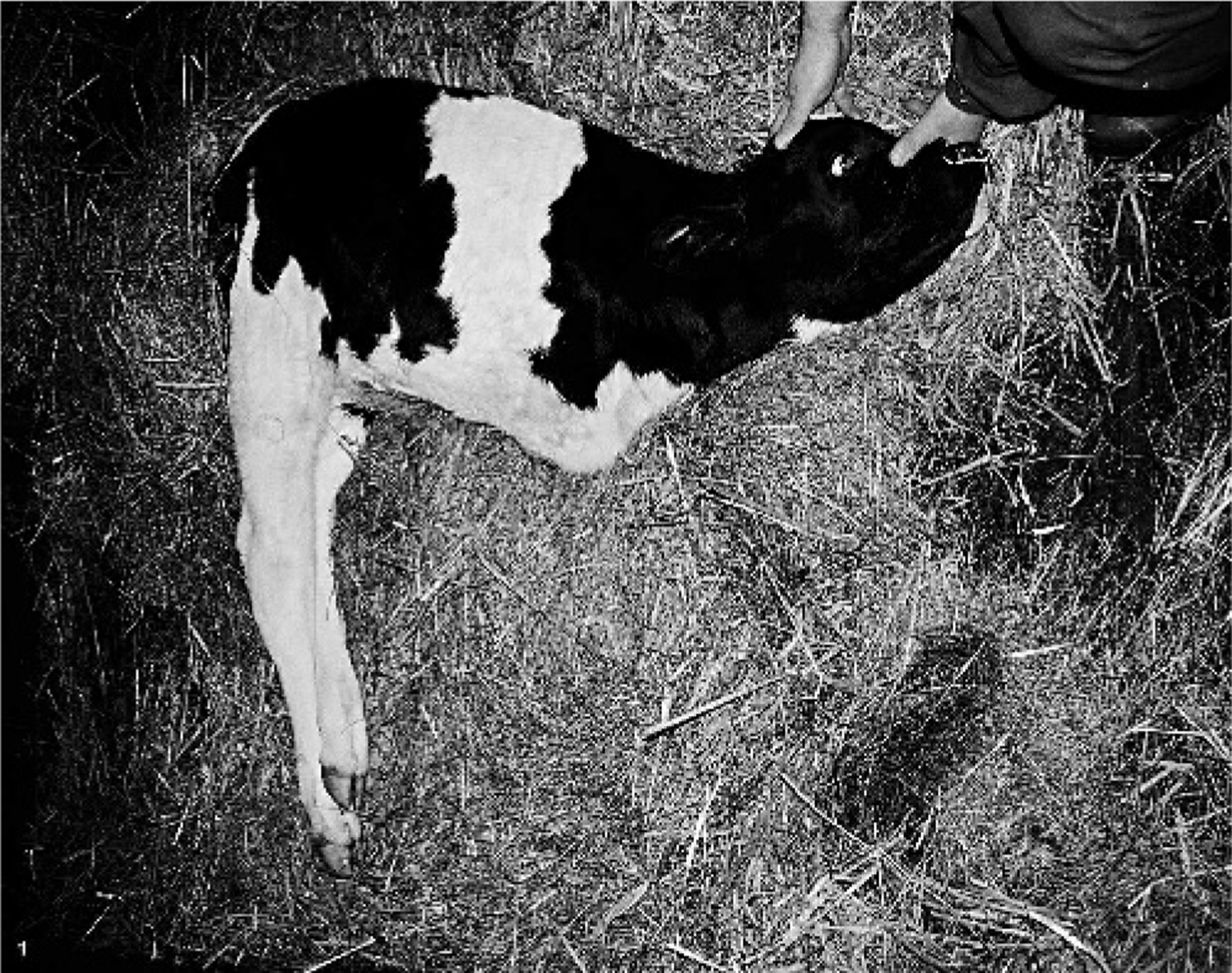
Male calf without thoracic limbs.
Chromosome spreads were obtained from a short-term (72-hour) lymphocyte culture, and the preparations were examined with the use of Giemsa staining and C- and G-banding techniques. International bovine chromosome nomenclature was followed. 10 Frequency of the micronuclei was estimated among 1,000 interphase nuclei. As a control, frequencies of chromosome/chromatid breaks and polyploid spreads, as well as the presence of micronuclei were also analyzed for 3 healthy bulls. Microscopic evaluation was carried out under a Nikon Eclipse 600 microscope (Melville, NY), equipped with a cooled CCD digital camera and Lucia software (Laboratory Imaging, Ltd., Prague, Czech Republic).
Postmortem examination revealed the presence of chondral scapulas and a lack of distal skeleton. Soft tissues of the thoracic limbs and the latissimus muscle of the back were not present. X-ray examination confirmed only a shadow of chondral scapulas on the level of spinous processes of thoracic vertebrae (Fig. 2).
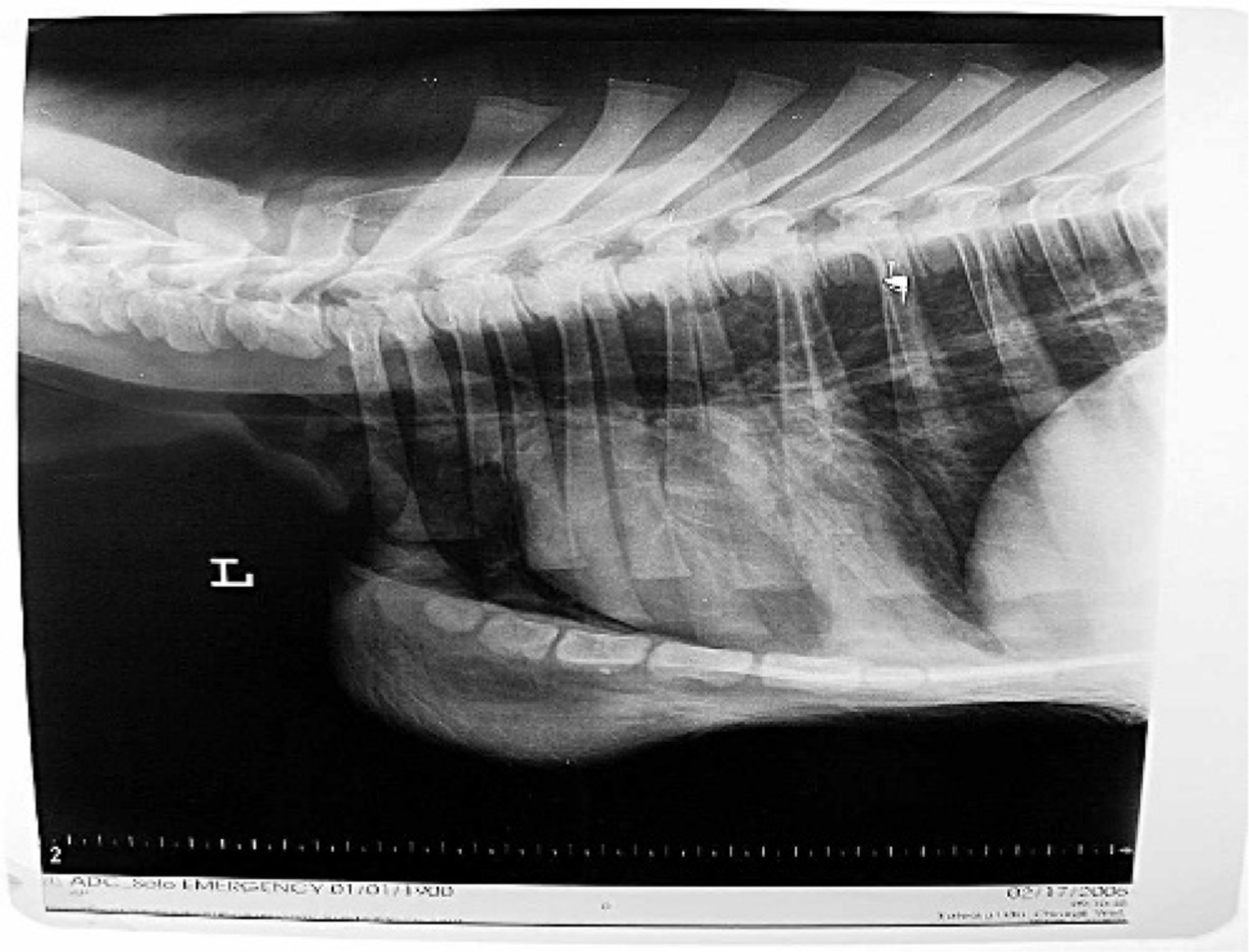
Radiograph of the lower part of the neck and upper part of the chest of the amelic male calf.
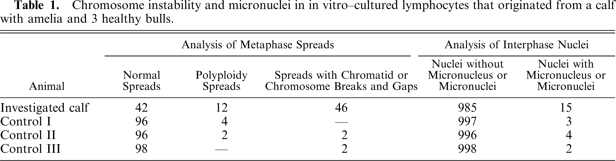
Cytogenetic analysis of 100 Giemsa-stained metaphases revealed that 58% were abnormal (Table 1): 12% were polypolid (Fig. 3a), and 46% showed chromosome and/or chromatid breaks and gaps (Fig. 3b, c). No aneuploid spreads were found. A single break or gap occurred in 8 and 17 spreads, respectively. Moreover, 21 spreads with 2 chromosomes with a break or gap were observed. The most common chromosome abnormality was a chromatid break or gap of a small autosome; this phenomenon was observed in 70% of the abnormal spreads. Unfortunately, it was not possible to identify this small chromosome with the use of the G-banding technique. Length measurement of the chromosome with a broken chromatid indicated that it could be chromosome No. 23, 24, or 25. Chromatid breaks in the X chromosome also occurred quite often (26% of abnormal spreads). The breakage was always observed at the same site, presumably Xq24-25. A chromatid gap was also observed at the same site on the X chromosome.
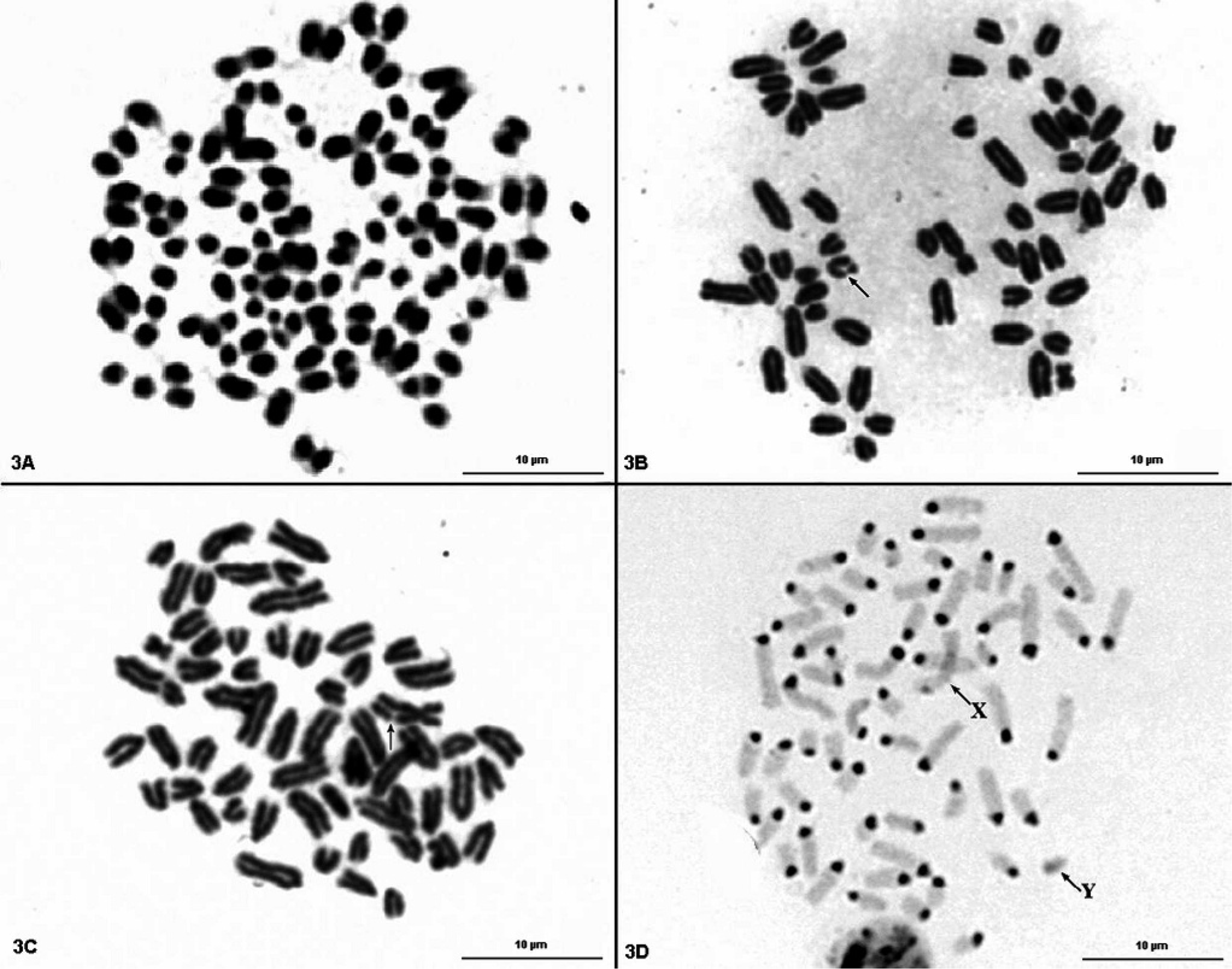
Cytogenetic preparations obtained from in vitro–cultured lymphocytes of the amelic calf:
Chromosome instability and micronuclei in in vitro–cultured lymphocytes that originated from a calf with amelia and 3 healthy bulls.
In addition, the C-banding technique was also applied, since it is known that the Roberts syndrome (RS)—a human genetic disorder characterized by craniofacial anomalies and limb defects—is related to an abnormal appearance of centromeric heterochromatin, e.g., puffing and splitting. 21 In the studied case the distribution and appearance of the constitutive heterochromatin was normal (Fig. 3d).
Analysis of micronuclei frequency in lymphocyte cultures derived from the calf also was performed (Table 1, Fig. 4). It was shown that the number of nuclei with at least one micronucleus was higher (15/1,000) compared with lymphocyte cultures carried out under the same conditions for 3 healthy bulls (on average, 3/1,000).
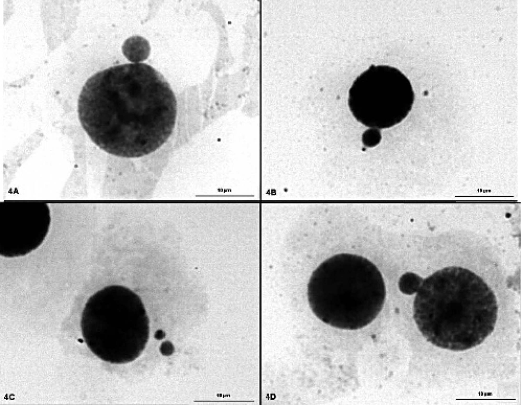
Cytogenetic preparation obtained from in vitro–cultured lymphocytes of the amelic calf: a single micronucleus connected with the nucleus (
It is difficult to determine the cause of the observed limb malformation. There are several reports of amelia in humans associated with chromosomal aberrations. A case of autosomal monosomy in a human fetus with total amelia, 17 mosaic trisomy 8 in a patient with tetraamelia/total amelia, 6 the deletion of interstial 7q in an infant with ectroamelia, 13 a deleted ring chromosome 4 in a baby with amelia, 8 or karyotype 46, XY with bilateral upper amelia 16 were described. However, many of these cases had multiple congenital anomalies and thus the chromosome mutations mentioned above might not be responsible for limb anomalies only.
Increased genomic instability also was connected to limb malformations. Spontaneous chromosomal breakage was reported in a child with limb defects. 2 Chromosome instability also was diagnosed in a calf affected by congenital malformation (lack of the distal left anterior leg and right anterior leg ended with a hook-shaped, naillike structure); namely, high rates of structural chromosome aberrations and increased yields of sister chromatid exchanges were reported. 4 Another case of a malformed calf affected with polymelia and a high incidence of chromosome breaks was described recently. 15 On the other hand, chromosome instability can be analyzed in relation to the distribution of fragile sites, which are divided into two categories: rare and common. 1, 19 These sites are expressed under specific conditions during lymphocyte culture. The common ones are induced by mutagens, such as aphidicolin, whereas the rare ones are folate sensitive. An association between the rare fragile and abnormal phenotype was reported in a girl with brachydactyly and a short stature. 18 Mutagen-induced chromosome instability also was analyzed in cattle, and the most expressive fragile sites in cows were observed in chromosomes 1 and X. 3 The fragile site in the X chromosome is present in the same chromosome band (Xq24) as the chromatid break in the amelic calf.
Since the mechanism of limb development is common in higher vertebrates, mutations of the homeotic and other genes should also be considered as potential causes of limb malformations. 5, 12 It was reported recently that a mutation of the WNT3 gene causes tetra-amelia. 14 In the present case a gene mutation seems to be unlikely, because pedigree data did not indicate an inherited background of this abnormality.
Chromosome instability similar to that reported in this paper was found in sheep exposed to dioxins during pasturage. 9 It is also very well documented in humans that a high incidence of limb deformity in newborns was an effect of thalidomide. 7 Genotoxicity also can be evaluated with the use of the micronucleus test. In the present study no information about the exposure of the pregnant cow to teratogens was available. However, the increased micronuclei frequency observed in the studied animal indicates that an environmental cause of the observed malformation cannot be excluded.
Footnotes
Acknowledgements
IS is a holder of the Young Scientists Fellowship (Foundation for Polish Science, contract 88/2004).