
Abstract
An extramedullary plasmacytoma was found in a 10-year-old sheep. The tumor involved the mediastinum, where a 25 × 15 × 10-cm encapsulated mass was found. The lungs had multiple metastases ranging from 0.5 to 2 cm in diameter, and the portal vein contained a 10-cm-long mass. The cytologic and histopathologic analyses were consistent with a moderately differentiated plasmacytoma. The immunophenotype of the tumor cells was lambda light chain IgG+, CD79a-, and CD3-. Occasional granulomas were observed at the periphery of the mediastinal and pulmonary tumors. Microbiologic culture yielded growth of Corynebacterium from these granulomas. This is the first report of plasmacytoma in sheep. The tumor most likely arose from mediastinal lymph nodes and metastatized to the lungs and portal vein.
Plasma cell tumor (plasmacytoma or multiple myeloma) is an immunoglobulin-producing tumor reported more often in the dog but rare in the cat, horse, pig, and cow. These tumors arise primarily from bone marrow, with or without involvement of other organs. 6 Extramedullary (solitary) plasmacytoma (EP) is less common and has been reported primarily in dogs and cats. 1 6 In the dog, EP is usually found in the skin and the oral cavity, 1 whereas in humans the upper respiratory tract is more often involved. To our knowledge, neither plasmacytoma nor multiple myeloma has been reported in sheep. Here, we describe the gross, histopathologic, and immunohistochemical features of an EP located in the mediastinum, lungs, and portal vein of a sheep.
A 10-year-old female Merino sheep was presented with progressive weakness, severe dyspnea, and seromucous nasal discharge. Because of the severe respiratory signs, euthanasia was elected by the owner. Necropsy revealed Oestrus ovis parasitism in paranasal sinuses. A 25 × 15 × 10-cm encapsulated mass present in the mediastinum compressed both lungs (Figs. 1, 2). The mass was composed of a yellowish white firm tissue with multiple hemorrhages and necrotic foci of different sizes (Fig. 2). Both lungs had numerous firm white nodules from 0.5 to 2 cm in diameter. The majority of these nodules appeared similar to the mass within the mediastinum; however, some of them contained a yellowish white fluid. A 10-cm-long lobulated mass partially occluded the portal vein within the liver. This mass was similar to the mediastinal mass. Bone marrow and other organs had no significant changes.
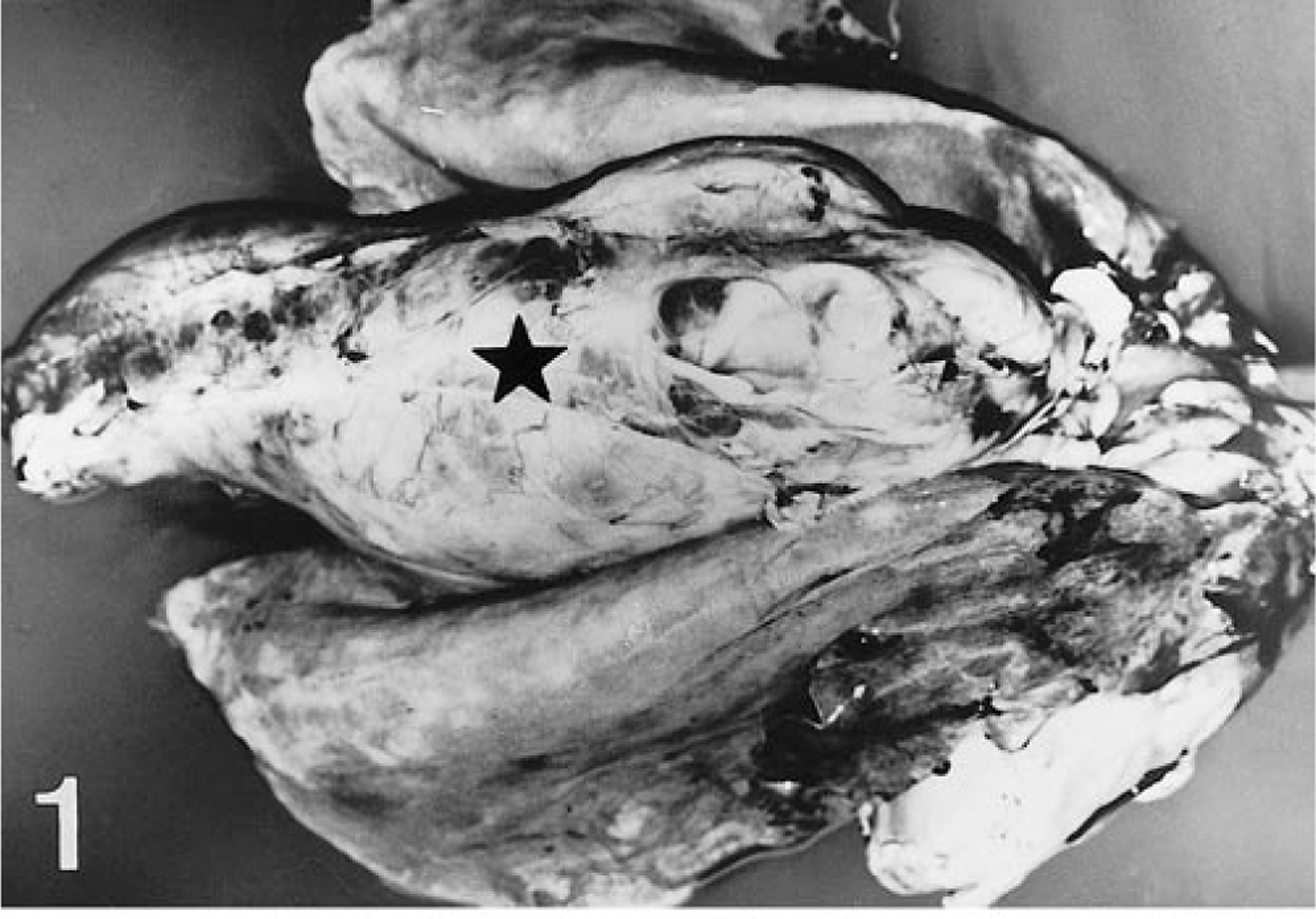
Mediastinal mass; sheep. Gross appearance of the encapsulated mass (star), which was larger than the lungs.
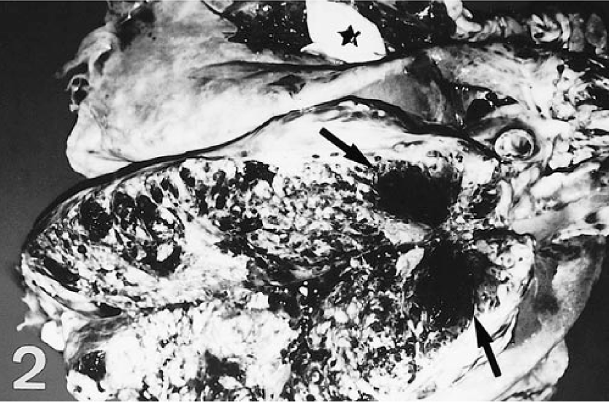
Mediastinal mass; sheep. Section revealing a whitish tissue with numerous hemorrhages (arrows). Note purulent fluid from a pulmonary nodule (star).
Smears of the masses taken during necropsy were stained by the Giemsa method. Tissue samples from the mediastinal mass, lungs, heart, bone marrow from femur and humerus, spleen, hepatic and mesenteric lymph nodes, liver, pancreas, gastrointestinal tract, kidney, urinary bladder, and brain were fixed in 10% formol saline for 24 hours and embedded in paraffin. Four-micrometer-thick tissue sections were stained with hematoxylin and eosin (HE) and by Gram's method. The avidin–biotin–peroxidase complex (ABC) method was used for immunohistochemical staining. Endogenous peroxidase activity was inactivated by incubation with 3% hydrogen peroxide in methanol for 30 minutes at room temperature (RT; 20–25 C). Tissue sections for detection of CD3 and lambda light chain IgG (λ-IgG) were incubated with 0.1% pronase (Sigma Chemical, St. Louis, MO) for 10 minutes at RT. Tissue sections for the CD79a monoclonal antibody (mAb) were processed with 0.1 M citric acid, pH 6.0, in a microwave oven at 100 C for 5 minutes. After three 10-minute rinses in phosphate-buffered saline (PBS; pH 7.4, 0.01 M), normal goat serum (Vector Laboratories, Burlingame, CA) diluted 1:10 was applied for 30 minutes at RT. Anti-human CD3 polyclonal antibody (pAb) diluted 1:200 in PBS (Dako, Glostrup, Denmark), anti-human λ-IgG pAb diluted 1:1,500 in PBS (Dako), and anti-human CD79a mAb diluted 1:50 in PBS (Dako) were applied to different tissue sections for 18 hours at 4 C. After three 10-minute rinses in PBS, a biotinylated goat anti-rabbit IgG (Vector Laboratories) diluted 1:200 was applied for 30 minutes at RT as a secondary reagent for primary pAbs, and a biotinylated goat anti-mouse IgG (Dako) diluted 1:25 was applied for 30 minutes at RT as a secondary reagent for the CD79a mAb. After three 10-minute rinses in PBS, an avidin–biotin complex (Vector Laboratories) diluted 1:50 was applied for 1 hour at RT as a third reagent. Sections were incubated for 1 minute with 3-3′-diaminobenzidine tetrahydrochloride (Sigma Chemical) diluted 0.035% in Tris-buffered saline (pH 7.6) containing 0.01% hydrogen peroxide as chromogen. The specific primary antibodies were replaced by PBS and by rabbit or mouse nonimmune sera in negative control tissue sections for pAbs and mAbs, respectively. Ovine and human lymph nodes were used as positive controls to evaluate the cross-reactivity of the anti-human antibodies.
Bacteriologic culture from tissue samples of the masses located in mediastinum and lung yielded growth of Corynebacterium spp.
The cytologic examination revealed an elevated monomorphous cell population with marked plasma cell differentiation admixed with lymphocytes. Tumor cells were variable in size, ranging from 15 to 70 μm, with well-defined cytoplasmic borders and eccentric rounded and hyperchromatic nuclei. Binucleated and multinucleated giant tumor cells were also frequent. The cytoplasm was basophilic and of variable amount and frequently showed a pale juxtanuclear area (negative Golgi staining) (Fig. 3).
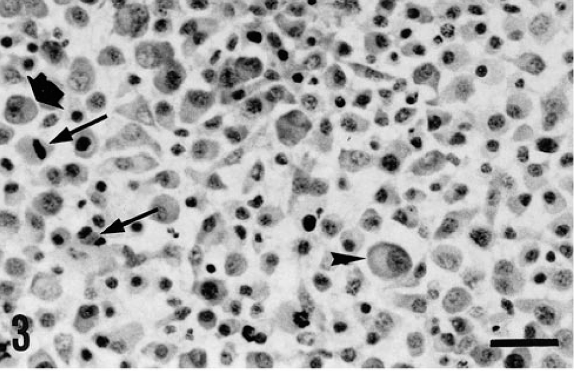
Mediastinal mass; sheep. Smear showing a predominant cell population with plasma cell differentiation, often showing negative staining of the Golgi (arrowhead). Mitoses (thin arrows) and binucleate tumor cells (thick arrow) were also frequent. Giemsa. Bar = 40 μm.
Histopathology revealed a mesenchymal tumor composed of a dense population of round to oval cells arranged in diffuse sheets with a fine fibrovascular stroma (Fig. 4). Intratumoral hemorrhages with foci of necrosis and mineralization were frequently observed. Tumor cells had one or more hyperchromatic, often eccentric nuclei, with one or more prominent nucleoli. Well-differentiated tumor cells had abundant eosinophilic and finely granulated cytoplasm (Fig. 4). Some tumor cells, isolated or in small groups, had acidophilic translucent cytoplasm with a hyaline appearance and eccentric irregular nuclei with condensed chromatin (Fig. 5). In some areas of the mediastinal mass, tumor cells were highly pleomorphic, with large, hyperchromatic nuclei, several prominent nucleoli, and small amounts of cytoplasm (Fig. 4). Mitoses were frequently observed (6/high-power field).
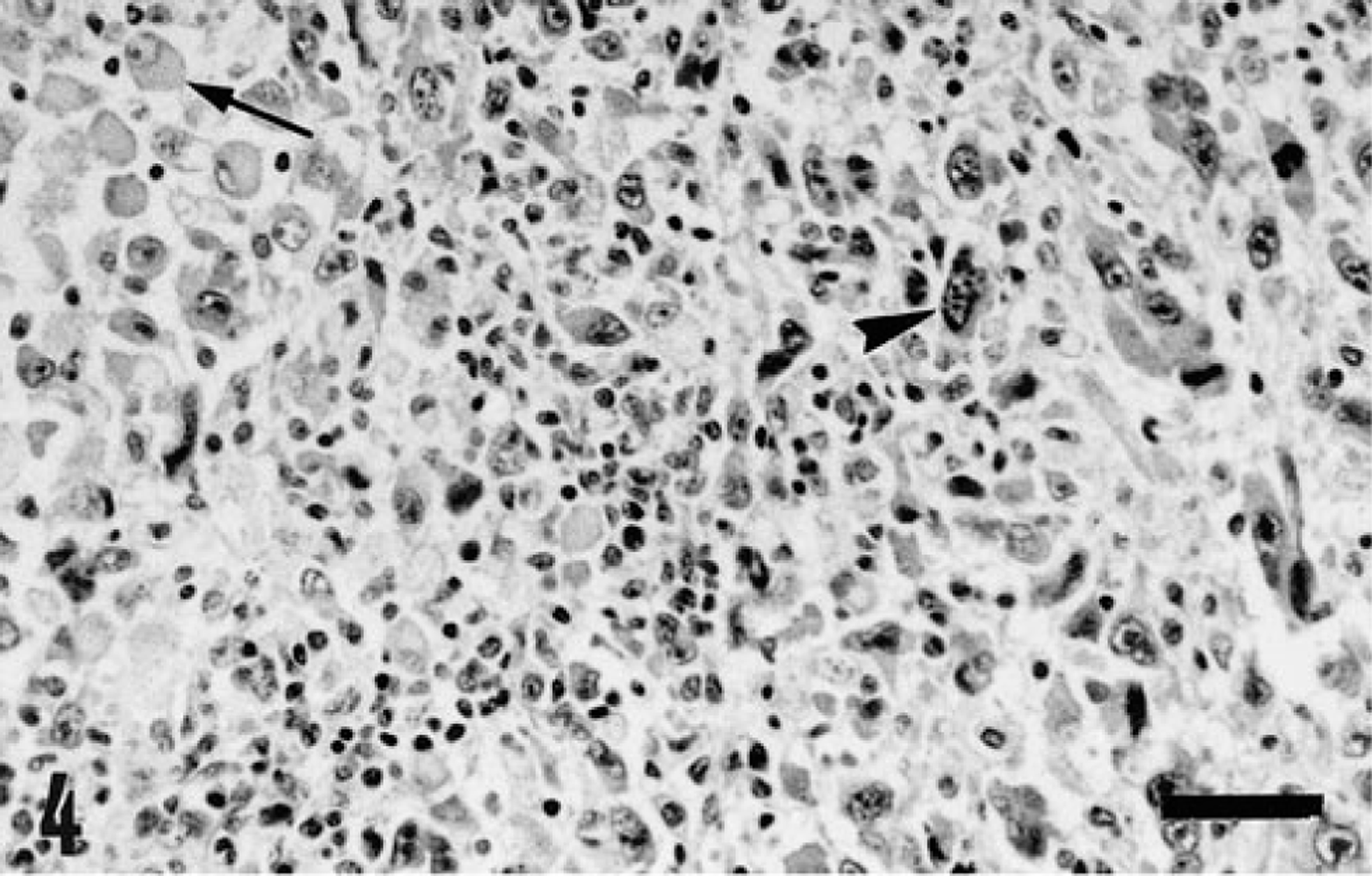
Mediastinal mass; sheep. Well-differentiated tumor cells resemble plasma cells (arrow). Highly anaplastic tumor cells have large pleomorphic nuclei and small amounts of cytoplasm (arrowhead). HE. Bar = 70 μm.
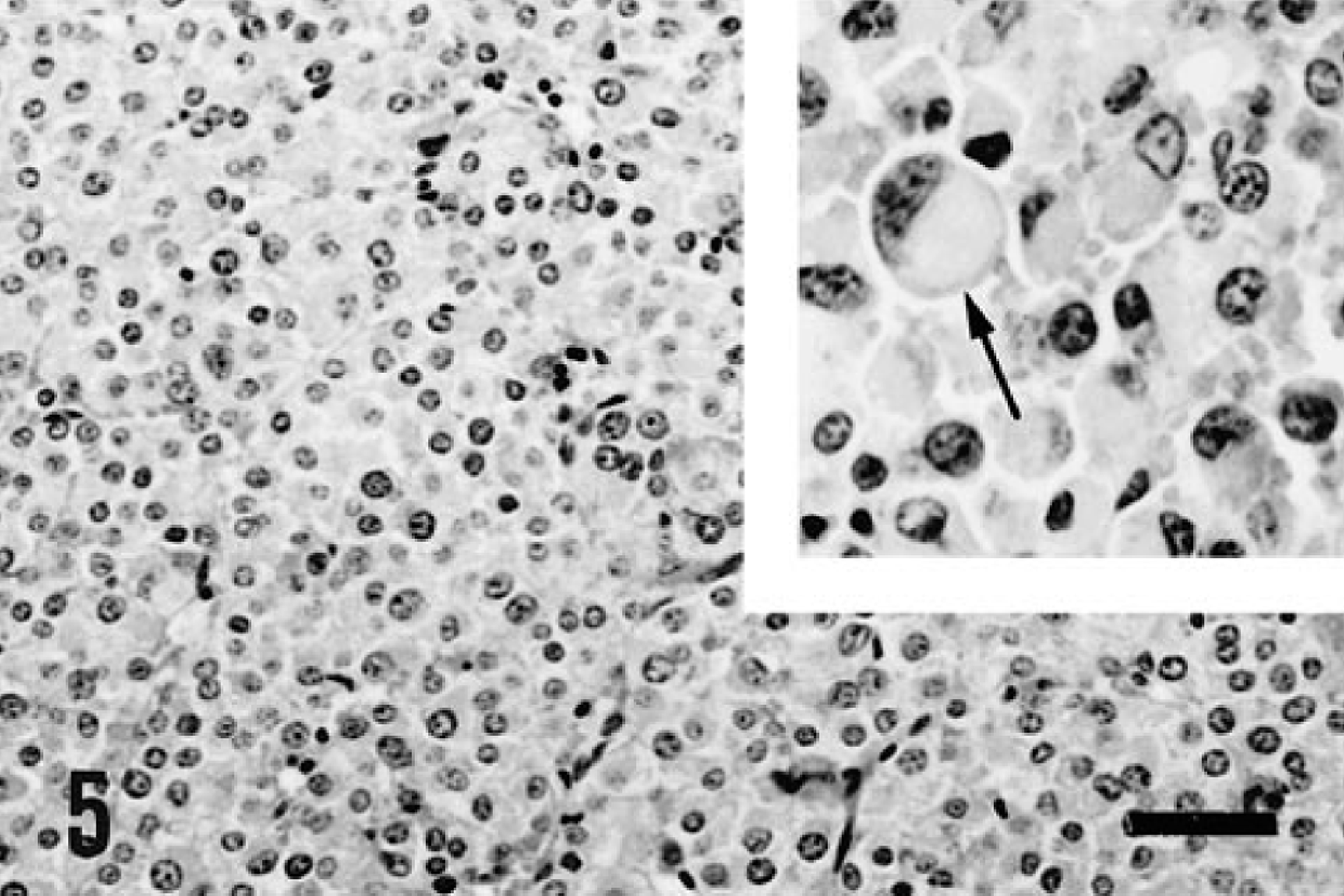
Pulmonary metastasis; sheep. Poorly differentiated round tumor cells are arranged in diffuse sheets and nests. Bar = 80 μm. Inset: Tumor cells have translucent cytoplasm (arrow). Bar = 35 μm.
The mediastinal mass was encapsulated by a thick band of fibrous connective tissue, and numerous calcified granulomas contained multinucleated giant cells, macrophages, lymphocytes, and plasma cells. Tumor metastases within the lungs were not encapsulated, and occasional granulomas were observed at the periphery. The pulmonary granulomas were encapsulated by a thick fibrous capsule and contained abundant necrotic material and cellular debris, with foci of mineralization surrounded by granulation tissue admixed with neutrophils, macrophages, multinucleated giant cells, lymphocytes, and plasma cells. Gram-positive bacilli compatible with Corynebacterium spp. were observed within these granulomas.
Bone marrow from the femur and humerus was composed of extensive fatty tissue with isolated hematopoietic foci. Tumor cells were not found in sections of bone marrow and other organs examined, except in the lungs and portal vein.
Immunohistochemical evaluation revealed numerous tumor cells (40–80%) from the mediastinal, pulmonary, and portal vein masses with a diffuse cytoplasmic positive staining for anti-human λ-IgG (Fig. 6). Numerous plasma cells within the medullary cords of sheep control lymph nodes were also stained with the anti-human λ-IgG pAb. Positive staining was also present in some giant tumor cells and in plasma cells contained within the granulomas at the periphery of the mediastinal tumor. Anti-human CD79a mAb reacted with isolated apparently normal plasma cells and lymphocytes, but tumor cells were consistently negative. The majority of lymphoid cells within the lymphoid follicles and variable numbers of lymphocytes and plasma cells within the medullary cords of sheep control lymph nodes were also stained with CD79a mAb. Anti-human CD3 pAb stained numerous lymphocytes within granulomas, paracortical areas of lymph node control sections, and isolated lymphocytes infiltrating the tumor but did not react with tumor cells.
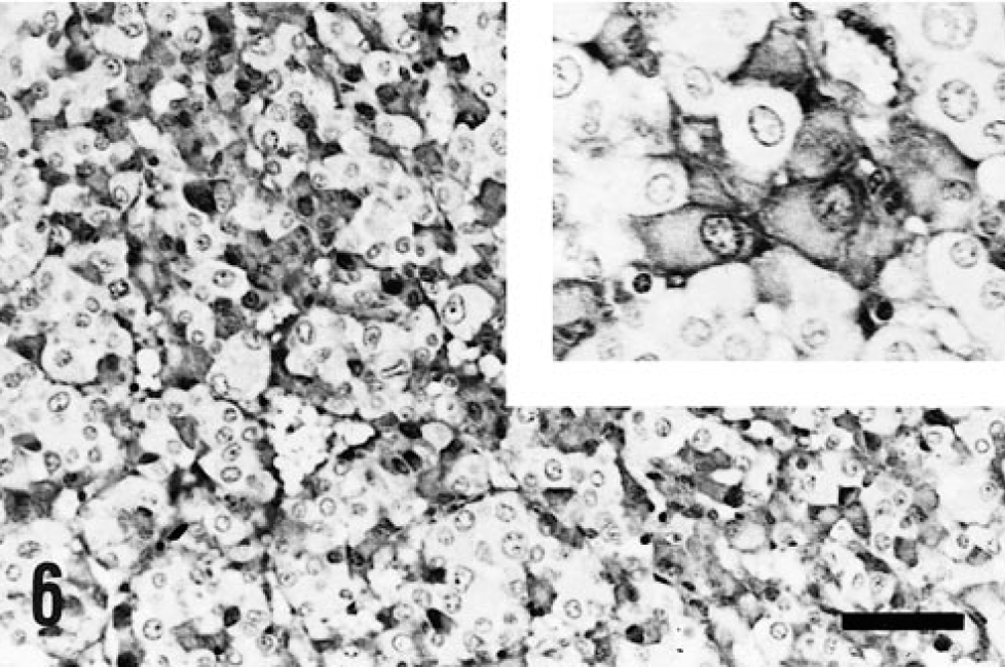
Pulmonary metastasis; sheep. Cytoplasmic immunoreactivity for the anti-human γ-IgG pAb is evident in numerous tumor cells. Bar = 80 μm. Inset: Immunoreactive tumor cells with pale paranuclear staining. ABC. Bar = 35 μm.
The large size of the mediastinal mass compared with that of the pulmonary nodules and the portal vein mass suggests that the mediastinum was the site of origin of the tumor. The mediastinal tumor probably arose from a mediastinal or tracheobronchial lymph node, as has been previously reported in human EP. 4 However, lymph node tissue was not identified in the mediastinal mass, probably because of its large size. The involvement of the lung and tracheobronchial lymph nodes has been often reported in human EP, 4 whereas in the dog only one EP has been reported in the lung. 1 Portal vein involvement has also been reported in ovine and caprine lymphomas 10 but not in EP.
The expression of λ-IgG by tumor cells of the ovine EP was consistent with the results previously obtained in EP of the dog and cat. 1 CD79a is a pan-B-cell marker in humans 5 and other species, including sheep, in which a B-cell lymphoma was detected with this antibody. 7 However, in the present case neoplastic cells did not express this antigen. This finding is not unexpected because plasmacytomas in the dog are often negative 2 or produce variable immunoreactions with the CD79a mAb. 8 In the pig, lymphomas with plasmacytic differentiation were also negative, 9 and human plasmacytomas show variable immunostaining with this antibody. 5
In the dog, cutaneous and oral plasmacytomas often arise from areas with chronic inflammation. 8 In the present case, necrotizing and mineralized granulomas were observed at the periphery of the mediastinal tumor and in the lungs and were associated with Corynebacterium infection. This finding suggests that the plasma cell tumor may have arisen from chronic inflammation. However, this association may be incidental; Corynebacterium infection is common in sheep, but plasma cell tumors are very rare.
The large size of the mediastinal mass was responsible for the severe lung compression and the respiratory signs observed. Solitary plasmacytomas are usually benign, 3 8 but some authors have suggested that benign plasmacytomas may eventually progress to malignancy. 3 This progression may have occurred in the present case; most of the areas of the tumor were well differentiated, but foci of marked anaplasia were found and multiple metastases occurred in the lung and portal vein.
EP are very rare tumors in farm animals, and this is the first report of EP in a sheep. The tumor arose from the mediastinum and metastatized to the lungs and portal vein. The cytologic, histopathologic, and immunohistochemical features were similar to those of in EP of other species, such as the dog and cat.