
Abstract
A 13-year-old female mongrel dog had a pleomorphic adenoma of the lacrimal gland in the right upper orbit. The tumor measured 3.8 X 3.0 X 3.3 cm, appeared white, round, and firm, and pressed the right globe and surrounding tissues. Histopathologically, the tumor had a thin connective tissue capsule and was composed of tubules with two cell types, some resembling luminal epithelial cells making up the tubular structures and the other of myoepithelial cells. Epithelial tubules were disposed in an adenomatous fashion and separated from each other by proliferating pleomorphic myoepithelial cells. Immunohistochemically, large numbers of the luminal epithelial cells revealed an immunopositive reaction against keratin/cytokeratin (AE1/AE3), and some epithelial cells reacted against cytokeratin 14. Spindle-shaped myoepithelial cells revealed an immunopositive reaction against cytokeratin 14, α-smooth muscle actin, and vimentin. A small number of myoepithelial cells reacted against desmin. S-100 protein immunopositivity was frequently found in luminal epithelial cells and rarely in the pleomorphic myoepithelial cells. Glial fibrillary acidic protein positivity was commonly found in myoepithelial cells, myxoid matrices, and intracystic materials, but not in luminal epithelial cells.
In the dog, neoplasms of the lacrimal gland are rare. 4 , 15 Most primary epithelial tumors are lacrimal gland adenocarcinomas. These neoplasms are locally invasive, recur after attempted resection, but are not known to metastasize following what is usually a brief postoperative follow-up period. 7 , 11 , 15 In this report, the characteristics of a pleomorphic adenoma derived from the lacrimal gland in a dog were described histopathologically and immunohistochemically.
The animal was a 13-year-old female mongrel dog that had a neoplastic mass in the right upper orbit (Fig. 1). The tumor was surgically removed. Tissue samples for histologic and immunohistochemical investigations were taken from the mass and fixed in 10% neutral buffered formalin. Histologic sections were cut 4 µm thick from paraffin-embedded blocks and stained with hematoxylin and eosin (HE). Sections of the neoplasm were processed with alcian blue (pH 2.5) and periodic acid–Schiff (PAS) reaction with and without pretreatment of testicular hyaluronidase and silver impregnation stain. Serial sections from the neoplasm were immunohistochemically examined by the avidin–biotin–peroxidase complex (ABC) procedure (Vectastain Elite ABC Kit; Vector Laboratories, Burlingame, CA). Specific antisera used consisted of anti-human keratin/cytokeratin (AE1/AE3) (Nichirei, Tokyo, Japan), anti-human cytokeratin 14 (Biomeda Corp, Foster City, CA), anti-human α-smooth muscle actin (SMA) (Sigma, St. Louis, MO), anti-human vimentin (Progen, Heidelberg, Germany), anti-human desmin (Progen) monoclonal antibodies, and anti-bovine glial fibrillary acidic protein (GFAP) and anti-bovine S-100 protein (Dako, Carpinteria, CA) polyclonal antibodies. Deparaffinized sections were blocked for endogenous peroxidase in 3% H2O2 for 10 minutes. All sections were incubated with primary antibody at 4°C for 16 hours, with biothylated secondary antibody for 30 minutes at room temperature, and with avidin-peroxidase conjugate for 30 minutes. Finally, sections were developed in 0.05% 3,3′-diaminobenzidine solution and counterstained with hematoxylin.
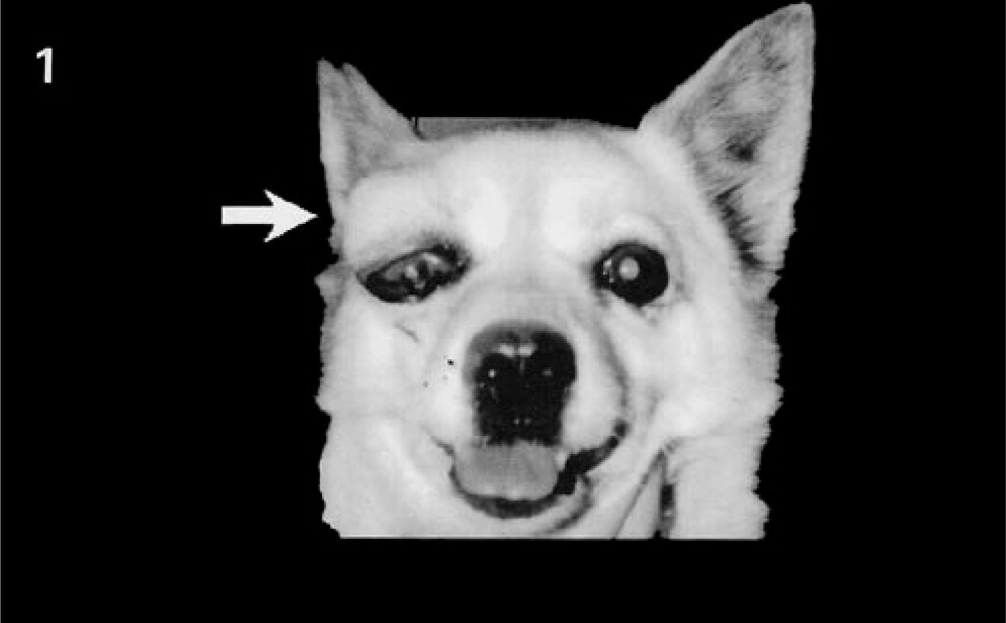
Lacrimal gland tumor; dog. The neoplastic mass measured 3.8 × 3.0 × 3.3 cm and shows dorsolateral location to the right globe (arrow).
On gross examination at surgical excision, location of the neoplastic mass was dorsolateral to the right globe (Fig. 1). The neoplastic mass measured 3.8 × 3.0 × 3.3 cm and appeared white, round, and firm. The right globe and surrounding tissues were compressed by the mass. Histopathologically, the neoplasm had a thin connective tissue capsule and did not infiltrate into the right globe and surrounding tissues. The neoplasm was composed of tubules with two cell types. These cells consisted of cuboidal luminal epithelial cells and peripheral myoepithelial cells (Fig. 2). Luminal epithelial cells showed tubular structures resembling normal ducts and were lined with a double layer of cells: an inner row consisting of cuboidal or columnar cells and a less well-defined outer layer of commonly vacuolated spindle-shaped myoepithelial cells. The tubular structures ranged from small to large and were irregular in outline. The epithelium occasionally underwent squamous differentiation with keratinization. Epithelial tubules were disposed in an adenomatous fashion and separated from each other by proliferating myoepithelial cells (Fig. 2). Often within the tubular lumina there was a homogeneous or sometimes granular, eosinophilic materials that frequently reacted positively with PAS and not with alcian blue with or without the hyaluronidase treatment. Multiple cystic structures lined by myoepithelial cells contained eosinophilic fine granular or fibrillar material (Fig. 3) that reacted positively with alcian blue and not with PAS. In different portions of the tumor, proliferation of the myoepithelial cells with alcian blue–positive extracellular matrices predominated over the luminal epithelial cells. Alcian blue positivity of intracystic materials and extracellular matrices disappeared after the hyaluronidase treatment.
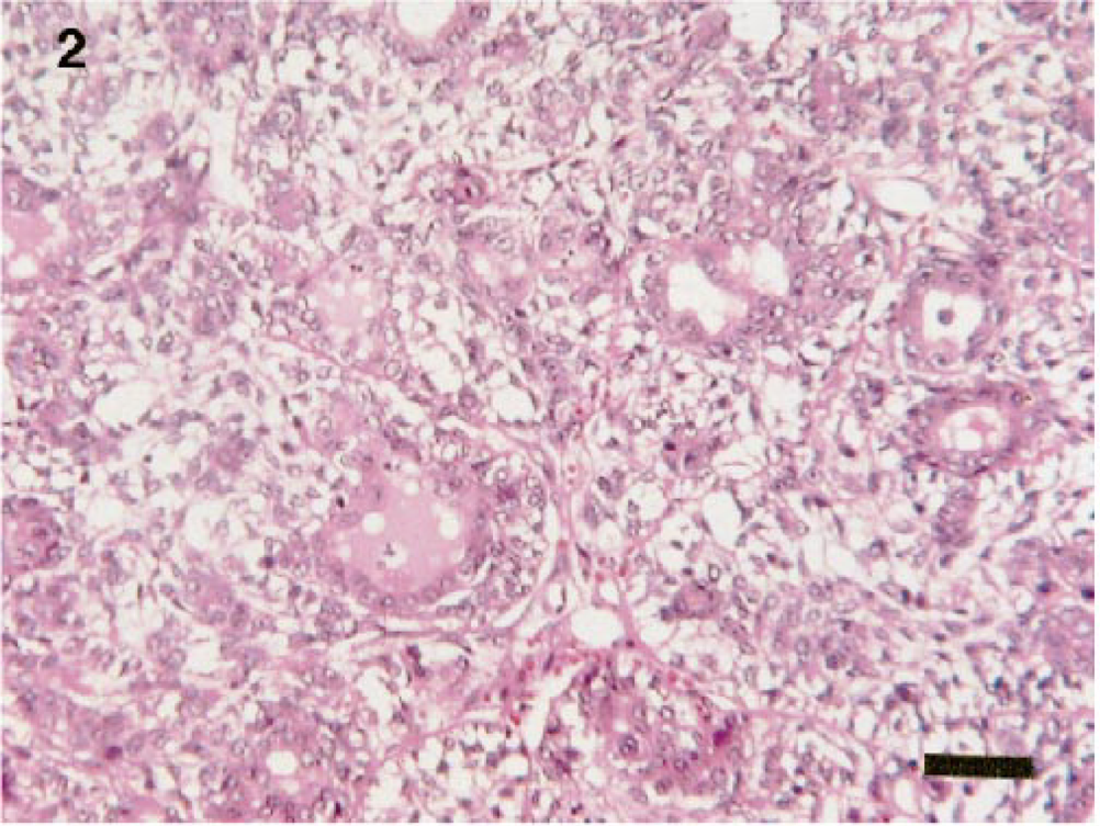
Lacrimal gland tumor; dog. The tumor is composed of cells of two types, some resembling luminal epithelial cells and the other resembling myoepithelial cells. Epithelial tubules are disposed in an adenomatous fashion and separated from each other by proliferating myoepithelial cells. HE. Bar = 50 µm.
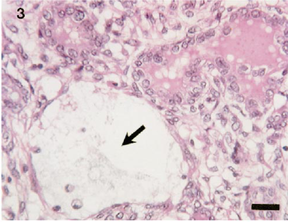
Lacrimal gland tumor; dog. Multiple cystic structures lined by myoepithelial cells contained eosinophilic fine granular or fibrillar materials (arrow). HE. Bar = 25 µm.
The immunohistochemical properties of neoplastic cells are summarized in Table 1. Most of the luminal epithelial cells had a strong immunopositive reaction against anti-human keratin/cytokeratin (AE1/AE3) (Fig. 4) and anti-human cytokeratin 14 monoclonal antibodies. Spindle cells resembling myoepithelial cells had a strong immunopositive reaction against anti-human cytokeratin 14, anti-human α-SMA (Fig. 5), and anti-human vimentin monoclonal antibodies. Some myoepithelial cells had an immunopositive reaction against anti-human desmin antibody. Positivity against anti-bovine GFAP polyclonal antibody appeared in the myoepithelial cells and surrounding extracellular matrices and in the fine granular or fibrillar materials in the multiple cystic structures (Fig. 6). GFAP immunopositivities remained intact in the sections with the hyaluronidase treatment. The luminal epithelial cells were frequently positive against anti-bovine S-100 protein polyclonal antibody (Fig. 7). In some areas, some pleomorphic myoepithelial cells also showed a positive reaction against S-100 protein antibody.
Results of immunohistochemical examination of neoplastic and normal lacrimal gland. ∗
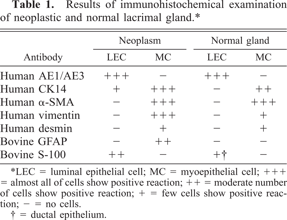
∗LEC = luminal epithelial cell; MC = myoepithelial cell; +++ = almost all of cells show positive reaction; ++ = moderate number of cells show positive reaction; + = few cells show positive reaction; - = no cells.
† = ductal epithelium.
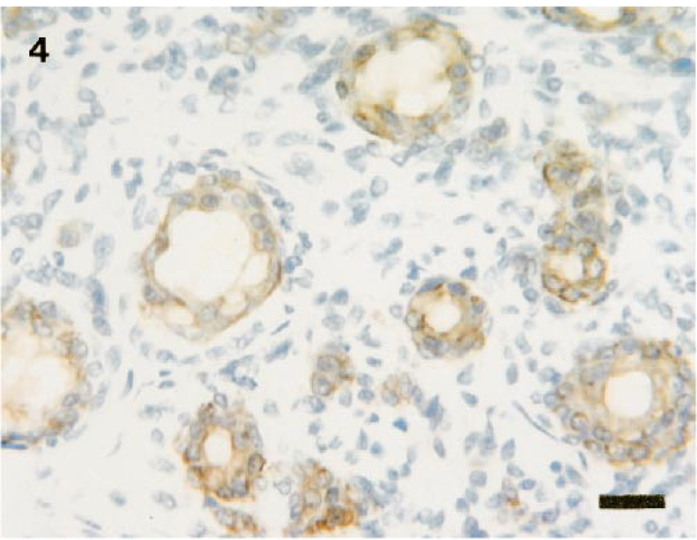
Lacrimal gland tumor; dog. Luminal epithelial cells had a strong immunopositive reaction against anti-human keratin/cytokeratin (AE1/AE3). ABC complex method, Mayer's hematoxylin counterstain. Bar = 25 µm.
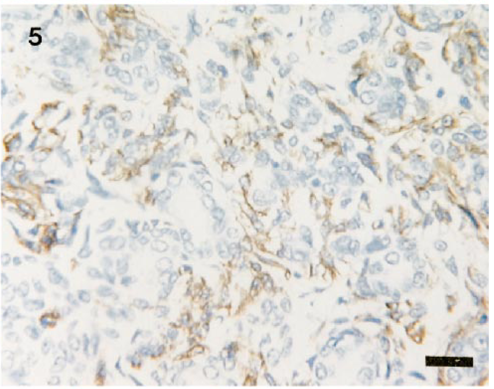
Spindle cells resembling myoepithelial cells show a strong immunopositive reaction against anti-human α-SMA. ABC complex method, Mayer's hematoxylin counterstain. Bar = 25 µm.
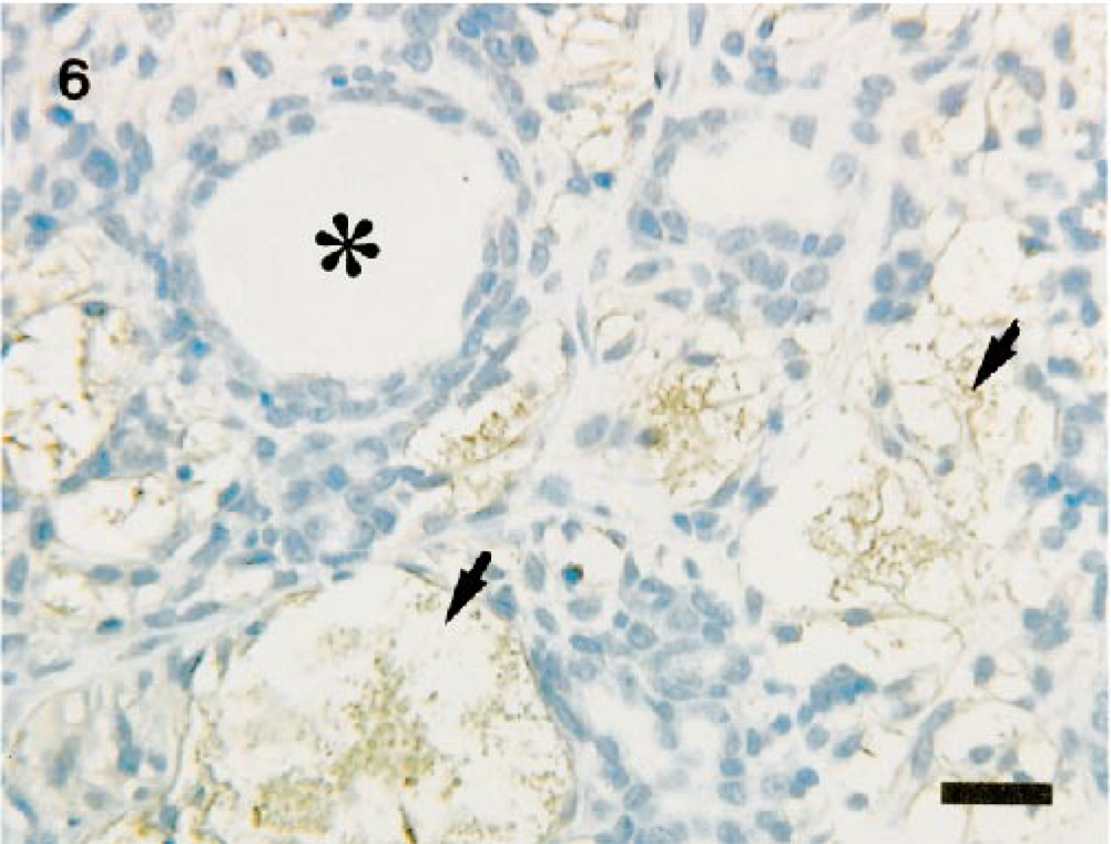
Lacrimal gland tumor; dog. Positivity against anti-bovine GFAP polyclonal antibody appears in the myoepithelial cells and surrounding extracellular matrices (arrows) and not in the luminal epithelial cells (∗). ABC complex method, Mayer's hematoxylin counterstain. Bar = 25 µm.
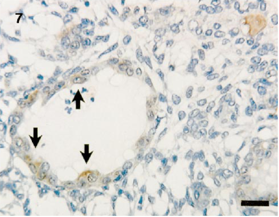
Lacrimal gland tumor; dog. The luminal epithelial cells were frequently positive against anti-bovine S-100 protein polyclonal antibody (arrows). ABC complex method, Mayer's hematoxylin counterstain. Bar = 25 µm.
Few lacrimal gland tumors have been reported in humans and animals. 3 , 5 , 11 , 13–15 Lacrimal adenomas were most frequently found in human patients. Histologically these neoplasms revealed a similarity in structure to mixed pleomorphic adenomas of the salivary glands 13 , 14 with some showing an adenocystic pattern. 3 , 5 Malignant tumors of the lacrimal gland are rare. Malignant lacrimal gland tumors include adenocystic carcinomas, squamous cell carcinomas, mixed malignant tumors, adenocarcinomas, and lymphomas. 5 , 10 , 11 To our knowledge, in the dog, only three cases of adenocarcinoma were described previously, 7 and other types of tumor, e.g., adenomas or mixed tumors, have not been reported. In the present study, histologic and immunohistochemical examinations revealed that this neoplasm was a mixed tumor similar in structure to the salivary pleomorphic adenomas of the dog. 4 , 15 A salivary pleomorphic adenoma is usually composed of a mixture of different proportions of luminal epithelium, myoepithelium, hyaline/myxoid/chondroid material, and bone. 4 The epithelium appears as ducts lined with cuboidal, columnar, or occasionally squamous cells. 4 The luminal epithelial cells in our tumor appeared positive when immunostained with keratin/cytokeratin (AE1/AE3) and cytokeratin 14, and the lumina contained PAS-positive materials. These findings suggest that the neoplastic epithelial cells were derived not from the acinous epithelium but from the reserve cells of intercalated and striated ducts of the lacrimal gland. 2 , 13 Spindle cells resembling myoepithelial cells reacted against cytokeratin 14, α-SMA, and vimentin. 6 Therefore, the tumor was diagnosed as a pleomorphic adenoma of the lacrimal gland. In some areas, epithelial tubules were disposed in an adenomatous pattern and separated from each other by proliferating myoepithelial cells. Such a pattern is often called adenomyoepitheliomatous. Although chondroid material or bone frequently associated with salivary pleomorphic adenoma were not detected in the present case, proliferating myoepithelial cells in major parts of the tumor were surrounded by myxoid matrices stained with alcian blue and digested with hyaluronidase. Therefore, the myxoid matrices may contain hyaluronic acid and chondroitin sulfate. 2
In humans, GFAP and S-100 protein are commonly studied in the salivary gland. Immunohistochemically, ductal epithelial cells in the normal salivary gland are positive against both antibodies, but myoepithelial cells are negative. In pleomorphic adenoma of the salivary gland, GFAP and S-100 protein are occasionally identified in myoepithelial and luminal epithelial cells. 1 , 8–10, 12 The immunoreactivity of GFAP is closely related to myxomatous and early chondromatous differentiation in pleomorphic adenoma and is not associated with another type of salivary gland tumor. 8 , 9 In normal lacrimal gland tissues, S-100 protein is identified in myoepithelial cells and ductal epithelial cells, 6 but GFAP was not identified in any part of the gland. 13 GFAP and S-100 protein are identified in pleomorphic myoepithelial cells in the myxoid and/or chondroid areas in the pleomorphic adenoma of the lacrimal gland. 13 , 14 The expression of GFAP is present as a sign of stromal transformation into myxoid and chondroid matrices in this tumor. 1 , 10 , 13 Thus, it is considered that pleomorphic adenomas of the lacrimal gland presumably develop from mesenchymal metaplasia of the myoepithelial cells, as is the case with pleomorphic adenomas of the salivary gland. 1 , 6 , 8–10, 12 In the present study, histologic, histochemical, and immunohistochemical characteristics agreed with those of pleomorphic adenomas of the lacrimal and salivary glands in human patients. 1 , 6 , 9 , 10 , 12–14 Immunohistochemical expression of GFAP localized in the myoepithelial cells, myxoid matrices, and intracystic materials could provide new evidence for a differential diagnosis of neoplastic lesions arising in the lacrimal gland of the dog.