
Abstract
A 1-mo-old Ivesi male lamb was presented with 2 large red masses on the skin of the left ear. The tumors were removed using gentle dissection and submitted for histologic evaluation. The tumors consisted of numerous thin-walled capillaries lined by endothelial cells and nests of stromal cells. Immunohistochemically, the endothelial cells were positive for CD45, and the stromal cells were positive for neuron-specific enolase. GFAP-positive cells were occasionally present within the tumor. Endothelial and stromal cells were negative for S100, CD34, CD31, and factor VIII–related antigen. The tumor had strong gross, microscopic, and immunohistochemical similarities with human extraneural hemangioblastoma.
Hemangioblastoma (HB) is a rare vascular neoplasm that occurs in the central nervous system (CNS) of humans, 4 and is rarely reported in veterinary medicine. 1 HB is a benign, highly vascular tumor whose histologic origin is not yet fully defined. It occurs most frequently in the cerebellum, and sometimes in the meninges, spinal cord, and corpus callosum. 27 HB cases that develop outside the CNS are exceptional in humans 13 and may be found in the liver, 22 lungs, 16 bones, 20 and forearm (around the ulnar nerve). 27 Human HBs comprise 1–2% of intracranial tumors, with the incidence highest among middle-aged adults. 11 In most cases, human HB is of unknown cause or association; ~25% are associated with von Hippel-Lindau (VHL) disease. 27
Although reported only rarely in animals, most congenital tumors are of mesenchymal origin. 17 The tumors are considered noteworthy because of their appearance at an early age; congenital tumors are defined as tumors detected during the fetal period and newborns up to 2 mo of age.17,18 The causes of such neoplasms are thought to differ from tumors that occur in adults. Hemangiomas have been reported in the nervous system, eyes, 7 alimentary tract, 25 skin and soft tissues, 10 and as congenital tumors in cattle. In horses, skin and subcutaneous capillary hemangioma, 9 hemangioendothelioma, and hemangiosarcoma 23 have been reported as congenital vascular tumors.
A 1-mo-old, 6.8 kg, Ivesi male lamb was brought to Van Yüzüncü Yıl University Faculty of Veterinary Medicine (Turkey) because of masses on the skin of the left ear. On clinical examination, body temperature, and respiratory and heart rates were normal. Two large solitary, brown-red masses were noted on the pinna of the left ear (Fig. 1): one 5 × 3 × 3 cm mass on the dorsal aspect of the pinna, and a 3 × 2 × 1 cm mass on the caudal aspect of the pinna. According to the lamb’s owner, the masses had been present since birth. The masses were surgically removed and submitted for histologic examination. The masses did not appear to infiltrate the ear cartilage. Cut surfaces were red with no evidence of necrosis or hemorrhage or a cystic component. The lamb made an uneventful recovery from surgery and was discharged. There was no reoccurrence of the tumor in a 3-mo follow-up period.
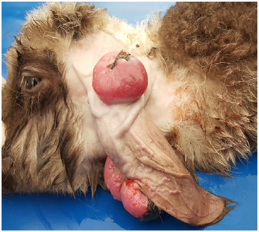
Gross appearance of congenital extraneural hemangioblastoma in a lamb consisting of 2 large solitary, brown-red masses on the skin of the left ear.
The excised masses were fixed in 10% neutral-buffered formalin, processed routinely, sectioned at 5 µm, and stained with hematoxylin and eosin, Masson trichrome, toluidine blue, and periodic acid–Schiff (PAS). For immunohistochemistry, sections were processed using the streptavidin–peroxidase method (ABC). After deparaffinizing and rehydrating, sections were rinsed in distilled water. After quenching endogenous peroxidase activity with 3% H2O2 (v/v) for 20 min, slides were washed twice in 0.01 M phosphate-buffered saline (PBS) for 5 min. Before adding primary antibody, slides were incubated with blocking serum (Histostain Plus bulk kit, Zymed, South San Francisco, CA) for 15 min to block nonspecific binding. Sections were then incubated with the following primary antibodies: CD45, neuron-specific enolase (NSE), glial fibrillary acidic protein (GFAP), factor VIII–related antigen (VIIIR:Ag), CD31, CD34, and S100, and kept at 4ºC overnight in a humidified chamber (Table 1). Slides were washed 4 times in 0.01 M PBS for 5 min, incubated with biotinylated secondary antibody (Histostain Plus bulk kit, Zymed) for 20 min at room temperature, and then washed 4 times in 0.01 M PBS for 5 min. After incubation with secondary antibody, sections were incubated with streptavidin–peroxidase conjugate (Histostain Plus bulk kit, Thermo Fisher Scientific, Waltham, MA) for 20 min, and then washed 4 times in 0.01 M PBS for 5 min, following enzymatic incubation. To visualize reactions, sections were kept in a solution of diaminobenzidine (DAB) for 1–2 min. After development of DAB reactions, sections were counterstained with Gill hematoxylin. Sections were then passed through alcohol and xylene and mounted directly with Entellan mounting medium (Merck, MilliporeSigma, St. Louis, MO). Appropriate negative and positive controls were used to verify staining. Slides were incubated in PBS, instead of the primary antibodies, as negative controls. Slides were examined and photographed using a light microscope (E-400, Nikon, Tokyo, Japan) equipped with a DS-RI1 video camera (DS-U3, Nikon).
Primary antibodies used to characterize an extraneural hemangioblastoma.
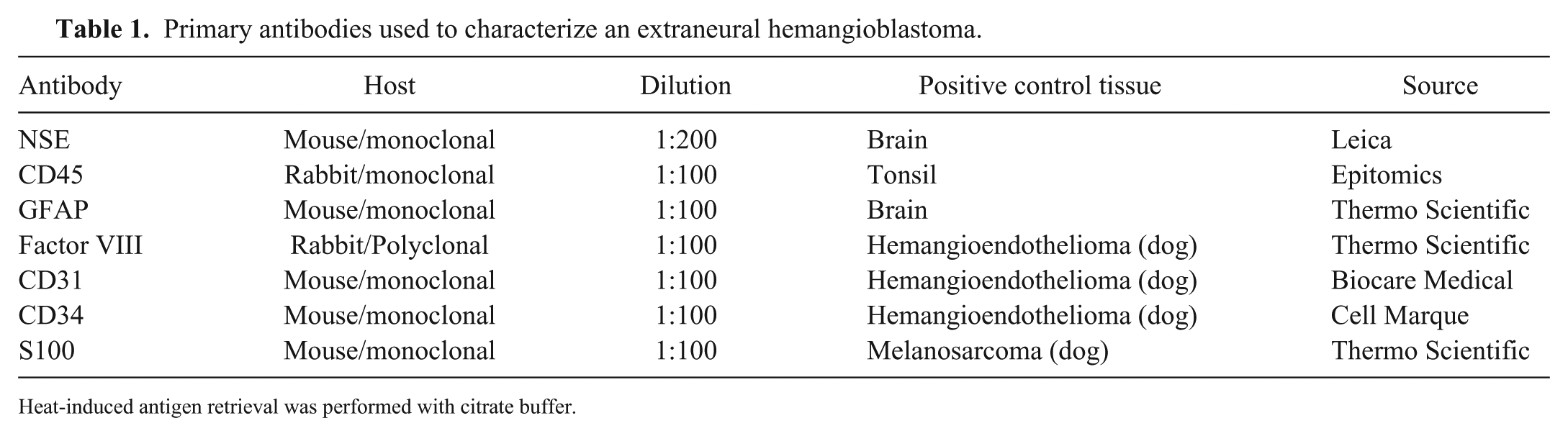
Heat-induced antigen retrieval was performed with citrate buffer.
Microscopically, the masses were encapsulated, cellular, and highly vascular, and consisted of closely packed, randomly oriented, capillary networks lined by plump endothelial cells. Capillaries were separated by pleomorphic stromal cells with indistinct cell borders, sometimes containing vacuoles within eosinophilic cytoplasm, and irregular, large, round-to-oval, vesicular nuclei with prominent nucleoli. Some stromal cells were binucleate. Moderate anisocytosis and anisokaryosis were occasionally present. Mitotic figures were rarely observed (<2 mitoses per 10 high-power fields). The stromal network of reticulin fibers contained groups of stromal cells and extensive fibrosis and hyalinization. Focal necrotic areas were present within the tumor. Extramedullary hematopoiesis was also present (Fig. 2A).
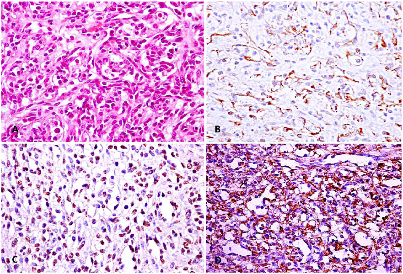
Congenital extraneural hemangioblastoma in a lamb.
Immunohistochemically, endothelial cells were strongly and diffusely positive for CD45, whereas stromal cells were negative (Fig. 2B). Stromal cells stained strongly positive for NSE (Fig. 2C). Some cells, interpreted as reactive astrocytes, stained with GFAP (Fig. 2D). Both the endothelial and stromal cells were negative for VIIIR:Ag, CD34, CD31, and S100. Based on these results, the tumor was diagnosed as a congenital extraneural HB.
HB cases are extremely rare in animals, but several intracranial HBs have been reported in dogs. In these cases, tumors were reported to have intramedullary, 5 intradural-extramedullary, 2 and intracranial localization.1,12 Extraneural HB cases have not been reported previously in animals, to our knowledge. Microscopic findings in our case conformed closely to the characteristics of human HB 26 and the description of the cases in dogs. 5 Histologically, HB is a capillary-rich neoplasm characterized by the presence of 2 main components, namely interstitial cells (stromal cells) and abundant and randomly directed capillary networks lined by endothelial cells, which can range from normal to plump. 13 Stromal cells are considered to be neoplastic components of the tumor, because aggregations of a similar cell type are not found in the developing brain or in any other adult tissue. 26 HB is rich in reticulin fibers, which provide separation of vascular cells from stromal cells. 5 Further, we detected vacuoles in stromal cells similar to those found in human HB. 8 We also noted extramedullary hematopoiesis. It has been reported that HBs might form blood islands containing potential extramedullary hematopoiesis in a similar manner to embryonic vasculogenesis. 15
If HBs arise in a typical location in the CNS and have a typical appearance, they are comparatively easy to diagnose. However, differential diagnosis can be difficult if HB occurs outside of the CNS. 27 Also, although the stromal cells in the tumor are usually bland, they often show significant nuclear pleomorphism suggestive of a more malignant nature. 27 In humans, immunohistochemistry is routinely used to assist in the diagnosis in both CNS 4 and extraneural 14 cases, especially where stromal cells show a positive reaction to NSE antibody. 14 In HB in dogs, stromal cells have been reported to be positive for NSE.2,5,6 In our case, stromal cells were positive for NSE.
CD45 is a marker for hematopoietic-derived cells, and as such is defined as a hematopoietic and/or vascular progenitor marker. 15 CD45 expression in HB indicates the onset of neovascularization and gradual development of the angioformative period. In our case, we speculate that the positive immunoreaction in endothelial cells with CD45 indicates that they are of hematopoietic origin. It has been shown that CD45 expression in endothelial cells is at both the gene and protein levels in sporadic and hereditary HB cases in humans. 15 Additionally, in human HB cases, endothelial cells have been reported to be generally positive with VIIIR:Ag and anti-CD31 and -CD34 antibodies. 3 However, in our case, no reaction was obtained with these markers. It has been reported that normal or neoplastic endothelial cells of vascular origin in cutaneous hemangiosarcoma do not produce positive results with VIIIR:Ag and anti-CD31 antibodies in sheep. 21 Additionally, a positive immunoreaction for CD34 could not be obtained in a dog with HB. 5
In our case, some stromal cells were positive for GFAP. In HB cases outside the CNS, it is uncertain whether these GFAP-positive cells are reactive astrocytes, stromal cells with differentiation of glial cells, or interstitial cells with phagocytic activity and cytoplasmic expression of GFAP. 27
Differential diagnosis in our case included other cutaneous vascular neoplasms in sheep. A single report of cutaneous hemangiosarcoma has been described in a sheep and consisted of pleomorphic spindle cells. 21 Hemangiomas have been reported rarely in sheep, characterized by blood-filled vascular spaces lined by a single layer of well-differentiated flattened endothelial cells. 24 Stromal cells detected by NSE antibody are the main microscopic findings that clearly distinguish HBs from other tumors.
Extensive discussion in the literature on HB reflects the difficulty in assigning an origin to the possible cell lines. 27 Suggestions include vascular, glial, neural, myofibroblastic, 19 endothelial cells; embryo choroid, neural, endocrine cells; and neuroectodermal-derived cells as possible origins of the stromal cells. 8
Footnotes
Declaration of conflicting interests
The authors declared no potential conflicts of interest with respect to the research, authorship, and/or publication of this article.
Funding
The authors received no financial support for the research, authorship, and/or publication of this article.