
Abstract
An ovarian choriocarcinoma was found in a 13-year-old cynomolgus monkey (Macaca fascicularis). The tumor was accompanied by a mature teratoma in the contralateral ovary. Histologically, the choriocarcinoma was characterized by nests of cells where cytotrophoblasts occupied the periphery with syncytiotrophoblasts at the center. Immunohistochemical staining for anti-human chorionic gonadotropin was positive in the syncytiotrophoblasts. The teratoma consisted of well-differentiated epidermal cells, sebaceous glands, hair follicles, cartilage, bone, and teeth. Choriocarcinoma metastases were in multiple organs. The concomitant development of choriocarcinoma and teratoma in the ovary is a consistent finding with the human counterparts of these lesions.
Ovarian choriocarcinoma is a malignant tumor arising from germ cells. In humans, the majority of these tumors are combined in varying proportions with other forms of malignant germ cell tumors, including teratoma, and therefore are regarded as belonging in the category of mixed germ cell tumors. 7 In nonhuman primates, ovarian teratomas alone have been reported; 1 , 4–6 however, ovarian choriocarcinoma has never been found. Here, we describe a rare case of ovarian choriocarcinoma accompanied by mature teratoma in the contralateral ovary of a cynomolgus monkey (Macaca fascicularis).
The monkey was born in our facility and physical appearance, body weight, hematology, and blood chemistry have been monitored periodically. Blood samples were taken several times to obtain control plasma, but otherwise this monkey was untreated and never mated. At 13 years of age, it developed prolonged vaginal bleeding. Because the monkey became severely anemic, it was euthanatized by exsanguination under ketamine anesthesia and then necropsied. Representative tissue samples were fixed in 10% neutral buffered formalin, and paraffin-embedded tissue was sectioned at 2 µm and stained with hematoxylin and eosin (HE) using standard procedures for light microscopic examination. Immunohistochemical technique was applied to the paraffin-embedded sections with anti-human chorionic gonadotropin (anti-HCG, rabbit polyclonal antibody, DAKO Japan Co., Kyoto, Japan) using the labeled streptavidin–biotin method (LSAB™ Kit, DAKO Japan Co.) without any antigen retrieval procedures.
At necropsy, the left ovary had a single white nodule that occupied 25% of the ovary. On cut surface, small multiple cysts and trabecular hard tissues were evident. The right ovary was enlarged to 7 × 6 × 6 cm and replaced by three large nodules. Each nodule was composed of mixtures of cystic, white solid, and dark-red soft regions. The uterine wall was thickened, and the lumen contained a large amount of dark-red mucinous material. The liver, lung, diaphragmatic pleura, omentum, and stomach contained many white to yellow nodules of various sizes.
Microscopically, the nodule in the left ovary consisted of tissues of different germ cell layers. Cysts were lined by keratinized epidermal cells and filled with keratin, and sebaceous glands were associated with hair follicles (Fig. 1). Discrete cartilage, bone, and teeth also were noted. These tissues were all well differentiated and showed no cellular or structural atypia. The histologic characteristics are consistent with a mature ovarian teratoma.
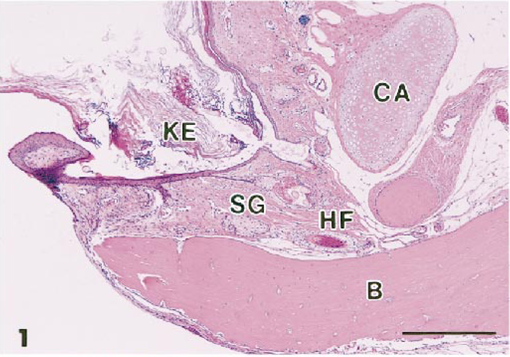
Left ovary; cynomolgus monkey. Tumor is a mature teratoma. CA = cartilage; KE = keratinized epithelium; SG = sebaceous gland; HF = hair follicle; B = bone. HE. Bar = 500 µm.
In the right ovary, the histologic features were entirely different from those described in the left ovary. The nodules were primarily composed of nests of highly cellular regions separated from each other by fine fibrous tissue (Fig. 2). Each nest consisted of two distinct cell types, one predominantly located along the periphery of the nest and the other at the center. Those cells seen in the periphery had indistinct cell borders and one large vesicular nucleus with one prominent nucleolus and were arranged in a solid sheet. In contrast, those that occupied the center had one small hyperchromatic nucleus, which occasionally formed multinucleated giant cells (Fig. 3). Immunohistochemical staining for anti-HCG was exclusively positive in these multinucleated giant cells (Fig. 4). This result and the morphologic findings strongly indicated that the cells with vesicular nuclei are equivalent to cytotrophoblasts and the multinucleated cells are syncytiotrophoblasts of the chorion. Areas of hemorrhage and necrosis were scattered over the large nodules; however, examination of multiple sections did not reveal any similar structure to the teratoma. None of the nodules found in the other organs of the monkey showed any morphologic similarities to the teratoma in the left ovary, but their histologic features were all consistent with those in the choriocarcinoma of the right ovary. The uterus also had multifocal lesions resembling the right ovarian neoplasm, though no grossly visible nodules were seen at necropsy.
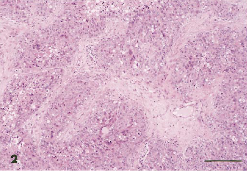
Right ovary; cynomolgus monkey. Tumor is a choriocarcinoma. HE. Bar = 200 µm.
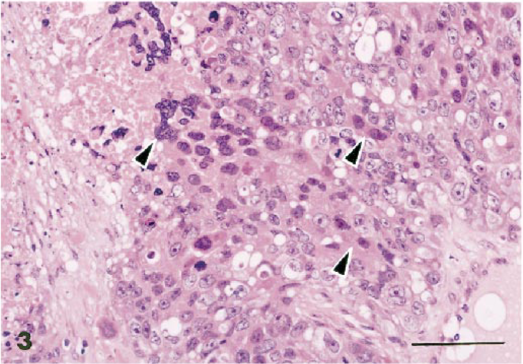
Choriocarcinoma; cynomolgus monkey. Note syncytiotrophoblasts (arrows) and cytotrophoblasts. HE. Bar = 100 µm.
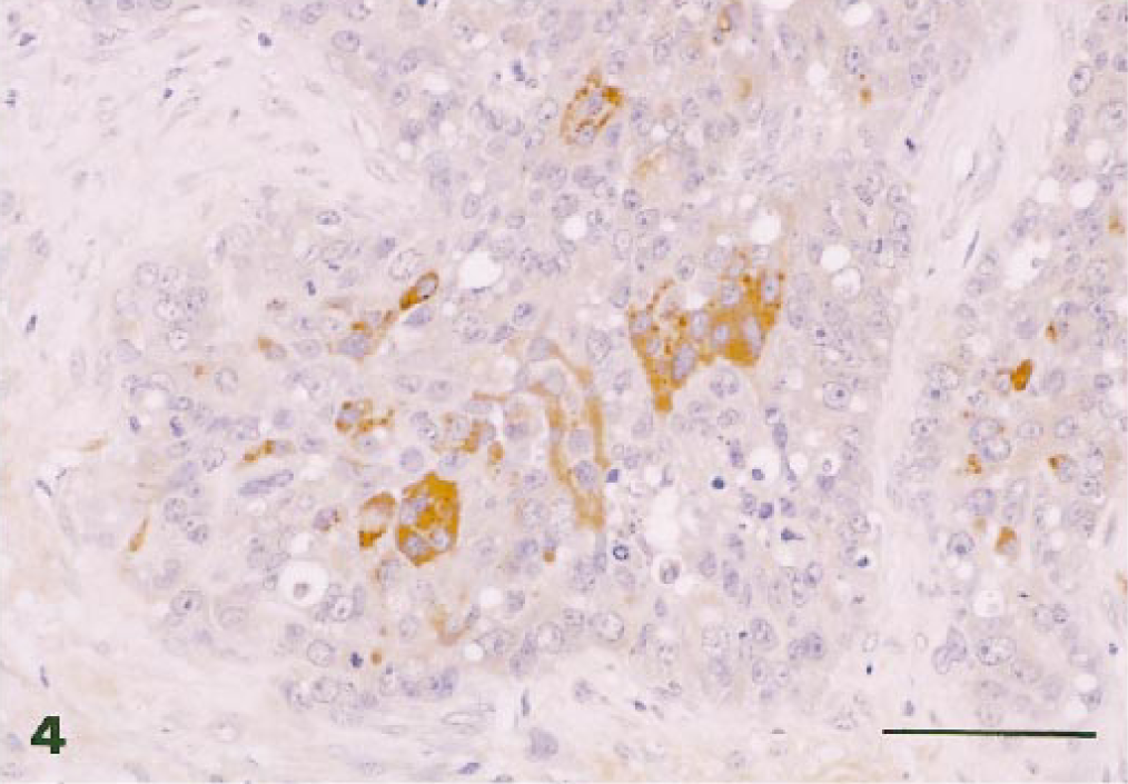
Choriocarcinoma; cynomolgus monkey. Syncytiotrophoblasts were positive with anti-human chorionic gonadotropin. Avidin–biotin–peroxidase complex method, Mayer's hematoxylin counterstain. Bar = 100 µm.
The incidence of spontaneous tumors in nonhuman primates is low in comparison with other animal species. This low incidence is due in part to the lack of studies of tumor incidence involving large groups of monkeys old enough when euthanatized to have developed tumors. The monkey in this report was bred and raised in our facility until it was 13 years of age.
Choriocarcinoma can develop either from the uterus or from the ovary and thereby can be gestational or of germ cell origin, respectively. 7 An intrauterine choriocarcinoma was associated with pregnancy in a rhesus monkey (Macaca mulatta). 3 The choriocarcinoma found in the ovary here could be a metastasis from the uterus; however, this is unlikely because the monkey had never been pregnant.
A close association between ovarian choriocarcinoma and other tumors of germ cell origin in humans has been reported. 2 Among eight patients with ovarian choriocarcinoma, four had dysgerminoma or teratocarcinoma in the same ovary. Although two distinct types of tumors of germ cell origin were also found in the present case, they were in the contralateral ovary, suggesting that each tumor developed independently.
Footnotes
Acknowledgements
We gratefully acknowledge animal care by Mr. S. Fukagawa, the technical assistance of Ms. K. Kunitoh, Ms. S. Ohi, and Ms. R. Yasui, and review of the manuscript by Dr. K. Tanaka.