
Abstract
Pulmonary myxoma is an uncommon neoplasm. A pale tan, lobulated, and well-circumscribed mass was discovered at slaughter in the left lung of a 5-year-old sheep. Histologically, the tumor was composed of spindloid to stellate cells in a myxoid matrix. Neoplastic cells were immunohistochemically positive for vimentin but did not express cytokeratins, S-100 protein, smooth muscle actin, desmin, or p53. On the basis of the histologic and immunohistochemical findings, this tumor was diagnosed as a myxoma.
Primary pulmonary myxomas are uncommon in humans 8, 11 or domestic animals. 9, 10 A pulmonary myxoma was reported in a sheep, but that tumor was not evaluated immunohistochemically. 10 In this communication, we describe macroscopic, histologic, and immunohistochemical findings of a pulmonary myxoma found in a sheep during postmortem examination at an abattoir.
As part of a survey to detect ovine pulmonary adenocarcinoma (Jaagsiekte), the carcasses of 3,864 sheep were examined at an abattoir in Van province in eastern Turkey from January 2004 to June 2007. The myxoma appeared as a mass 4 × 3.7 × 2 cm in the caudal lobe of the left lung of a 5-year-old Akkaraman ewe. The mass was well-circumscribed, soft, smooth-surfaced, white to tan, and lobulated with a gelatinous appearance on sectioning (Fig. 1). No other macroscopic lesions were observed.
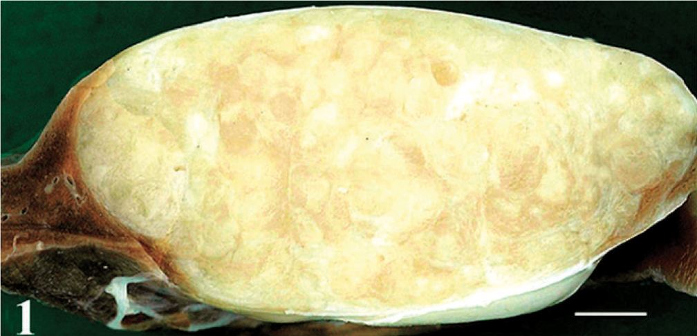
Lung; sheep. The pulmonary myxoma is a well-circumscribed, lobulated, pale tan to white mass. Bar = 5 mm.
Tissue specimens from major organs were fixed in 10% neutral buffered formalin and embedded in paraffin. Sections 4 μm thick were stained with HE, alcian blue (pH 2.5), toluidine blue, reticulin stain, and Masson's trichrome. Immunohistochemistry for vimentin, smooth muscle actin (SMA), desmin, cytokeratin (CK), p53, and S100 proteins was performed with the avidin-biotin immunoperoxidase complex (ABC) method (Universal LSAB 2 kit, Dako, Carpinteria, CA) (Table 1). Cervical leiomyoma (for SMA and desmin), canine mammary tumor (for vimentin and p53), and mucinous cholangiocarcinoma (for S100 and CK) were used for positive controls; nonimmunized mouse or rabbit serum was used for negative controls.
Characteristics of antibodies used for immunohistochemistry.
∗ Heat-induced antigen retrieval was performed with citrate buffer.
Histologically, the tumor was composed of spindloid to stellate cells in an abundant myxomatous matrix (Figs. 2,3). Lobules of neoplastic tissue were separated by fibrous septa. No pleomorphism, mitotic figures, atypia, necrosis, or multinucleation was detected. A few thin-walled vessels and hemosiderin-laden macrophages were in the tumor. The myxomatous matrix was alcianophilic at pH 2.5 and metachromatic with toluidine blue, which is characteristic of acidic glycosaminoglycans. Collagen and reticulin fibers of myxoid stroma were stained with Masson's trichrome and reticulin fiber stains, respectively. Immunohistochemically, neoplastic cells were positive for vimentin (Fig. 4) but negative for CK, SMA, desmin, S100 protein, and p53. The tumor was classified as a pulmonary myxoma. No metastases were detected.
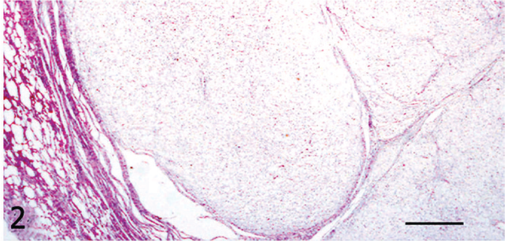
Pulmonary myxoma; sheep. Lobules of neoplastic tissue compress the adjacent pulmonary parenchyma. HE. Bar = 200 μm.
Pulmonary myxoma; sheep. Spindloid to stellate neoplastic cells in an abundant myxomatous matrix are divided into lobules by delicate fibrous septa. HE. Bar = 100 μm.
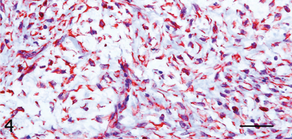
Pulmonary myxoma; sheep. Neoplastic cells are immunohistochemically positive for vimentin. ABC method, Mayer's hematoxylin counterstain. Bar = 50 μm.
Myxoma is a rare tumor with a few reports of its occurrence in the jaws of horses and cattle, 12 the heart 7 and synovia 3 of dogs, the heart of a cat, 2 and bronchi of a mare. 9 To the best of our knowledge, pulmonary myxoma has been reported only once before in a sheep. 10
The frequency of p53 immunoreactivity is generally higher in malignant tumors than in benign lesions. 5 In this ewe, with no cytologic features of malignancy and the lack of p53 immunoreactivity supported the diagnosis of a benign tumor.
Myxomas must be distinguished from other tumors that can have a myxomatous matrix, such as chondrosarcoma, chordoma, myxoid liposarcoma, peripheral nerve sheath tumor, embryonal rhabdomyosarcoma, leiomyosarcoma, myxosarcoma, and pulmonary hamartoma, as well as from nonneoplastic disorders, such as focal mucinous degeneration of soft tissues, nodular fasciitis, mucinous cyst, localized myxedema, and focal mucinosis. 1, 2, 4, 11, 12 In this case, neoplastic cells were immunohistochemically positive for vimentin and negative for CK, which confirmed their mesenchymal origin. The lack of nuclear atypia, mitotic figures, or p53 immunoreactivity were not consistent with a malignant neoplasm. Histologic features and the lack of immunohistochemical expression of S100 protein helped to rule out chondrosarcoma, chordoma, liposarcoma, and peripheral nerve sheath tumor. 1, 2, 4, 11 No histologic features of pulmonary hamartoma were observed in this case. The lack of expression of myogenic markers such as SMA and desmin helped to rule out rhabdomyosarcoma and leiomyosarcoma. 4 Pleomorphic or anaplastic sarcoma (also known as malignant fibrous histiocytoma) has variable immunohistochemical reactivity 4, 13 but is characterized by pleomorphism, frequent mitotic figures, hypercellularity, and hypervascularity, which were not features of the tumor in this sheep. The presence of reticulin fibers in the myxomatous matrix helped to rule out the possibility of nonneoplastic accumulations of mucin. 13 The gross, histopathologic, and immunohistochemical findings in this case are consistent with the results of previously reported myxomas in domestic animals and humans. 3, 7, 11
Myxoma is a rare tumor of various organs or tissues in humans 1, 8, 11 and domestic animals. 2, 3, 10, 12 All myxomas express vimentin immunohistochemically, 1– 3, 7, 11 but the expression of other markers, such as S100, SMA, and CK, depends on the anatomical location of the tumor. S100 immunoreactivity was negative in soft-tissue myxomas, intramuscular myxoma, normal dental follicle, and focal oral mucinosis; 6 however, S100 protein was expressed in 3 of 7 odontogenic myxomas and 7 of 12 cardiac myxomas. 6, 7 Odontogenic myxomas 6 and some cardiac myxomas 6, 7 expressed SMA, whereas human pulmonary myxoma was negative for this marker. 11 CK staining was positive in some soft-tissue myxomas and odontogenic myxomas, 6 but negative in a cardiac myxoma. 7
Myxomas are found most frequently in the human heart. 6 In veterinary medicine, cardiac myxoma has been reported in a cat 2 and a dog 7 as multilobulated, polypoid tumors. Tumor embolization is a major complication of cardiac myxomas. The right atrial myxoma can dislodge and cause pulmonary embolism with resultant infarction or hemorrhage. 11 In a canine cardiac myxoma, myxomatous emboli were widely disseminated in intrapulmonary arteries. 7 No atrial lesions were present in this sheep.
In the sheep of this communication, pulmonary myxoma appeared as a solitary mass, as described in human cases, 8, 11 whereas the only previously reported ovine pulmonary myxoma 10 was described as multinodular mass. Although rare, myxoma should be included in the differential diagnosis for pulmonary masses in sheep.