
Abstract
In an experimental study, we evaluated acoustic immittance in rabbits in order to use these data as normative values for further experimental investigations. This study is the first experimental evaluation of both conventional 226 Hz and multifrequency tympanometry (MFT) in rabbits. For the investigation, we used 33 female New Zealand rabbits weighing 3.2–4.4 kg and aged six months. Bilateral measurements using conventional 226 Hz and MFT were performed under general anaesthetic. A 226 Hz tympanogram was recorded for all animals by conducting an air pressure sweep from +200 to −400 daPa at a rate of 50 daPa/s. Subsequent tympanograms were recorded over a wide frequency range from 250 to 2000 Hz. The acoustic impedance device used in this study provided reproducible and evaluable tympanograms. The applied tone frequency of 226 Hz proved to be especially suitable for determining compliance. Normative data obtained from our study reveal the resonance frequency to be 1368 ± 205 standard deviation (SD) for the right side and 1413 ± 216 SD for the left side. The values for physiological acoustic immittance of the middle ear in the rabbit obtained here can serve as normative data in subsequent experimental animal studies.
Tympanometry at 226 Hz has become a routine clinical measure for evaluating the acoustic immittance of the middle ear. Tympanometry is suitable for detecting middle ear disorders, such as otitis media, an interrupted ossicular chain and defects of the tympanic membrane. Multifrequency tympanometry (MFT) is a technique for evaluating the immittance of the middle ear over a wide frequency range.
In the field of experimental evaluation, MFT has been shown to be useful for monitoring diseases in certain animal studies (Margolis 1995, Cole & Kwochka 2000, Hsu et al. 2000). Margolis (1995, 1998) performed MFT on normal chinchillas and Mongolian gerbils in order to obtain normative data against which to compare results obtained from animals with middle ear pathology. They induced different diseases and measured the resonance frequency; their interventions included artificially extending the ear canal, inducing serous otitis media and disrupting the ossicular chain. The results are similar to those in healthy human subjects and patients with middle ear diseases.
In this study, we intended to define normative values serving as the basis for further experimental studies on the middle ear of the rabbit. Particularly in experimental otology, the middle ear of the rabbit has become the standard model for the development of new prostheses replacing the ossicular chain. While normative values of inner ear function do exist, findings concerning middle ear function are insufficient.
This study is the first experimental evaluation of both conventional 226 Hz and MFT in rabbits. Based on the results of the experimental examinations, reference values were established for both physiological tympanograms and resonance frequency.
Methods
Animals
This experimental animal study involved a total of 33 approximately six-month-old New Zealand white female rabbits originating from the Charles River animal breeding farm (Sulzfeld, Germany). The selection of the species was based on its similarity to human middle ear anatomy and the standardized, microsurgical approach to the tympanic cavity that is favoured (Steinbach 1973).
The animals had a body weight of between 3.2 and 4.4 kg. They were housed in individual cages in a climate-controlled room at the Medical University of Hannover's Central Animal Laboratory. The cages measured around 50 × 60 × 45 cm in size, with a floor consisting of a perforated metal plate. They were equipped with a nipple drinking system providing the animals with water ad libitum. The rabbits received 100 g of a pelleted, all-in-one food (SSNIFF, Soest, Germany) and 150 g of autoclaved straw daily. The temperature in the room was around 20 ± 2°C, with humidity at 55 ± 5%. The animals were kept under an artificially controlled, 12 h light/dark cycle.
Anaesthetic management
All measurements were performed under inhalation anaesthetic. Anaesthesia by intubation has crucial advantages over anaesthesia by barbiturates. The depth of anaesthesia can be better controlled, enabling mortality to be considerably reduced (Lipman et al. 1997).
Prior to anaesthesia, each animal was examined as to its general condition in order to eliminate the risk of anaesthetic complications. Body weight was determined in order to allow precise individual dosage of the medication. Two premedication drugs were applied intramuscularly: 25 mg/kg of ketamine (Ketanest®; Albrecht GmbH and Co KG, Aulendorf/Württemberg, Germany) and 5 mg/kg of midazolam hydrochloride (Midazolam®; CuraMed Pharma GmbH, Hameln, Germany). Anaesthesia was induced by applying the short-acting anaesthetic agent Propofol-Lipuro 1% (Propofol®; B Braun Melsungen AG, Melsungen, Germany) at a dosage of 1 mg/kg, into the auricular vein of the left ear via an indwelling venous catheter (0.9 mm × 25 mm; B Braun Melsungen AG). Glycopyrrolate 5 mg (Robinul®; Riemser Arzneimittel AG, Riems Island, Germany) was then administered intramuscularly, with 5 mg of buprenorphine hydrochloride (Temgesic®; Essex Pharma GmbH, Munich, Germany) administered subcutaneously. Following endotracheal intubation, narcosis was maintained with 1.5% isoflurane (Forene®; Abbot GmbH, Wiesbaden, Germany). An infusion of 10 mL/kg/h of Sterofundin-HEG-5 (B Braun Melsungen AG) was used to stabilize the circulatory system of the animals. In order to ensure that decreasing body temperature did not influence the derivates, the body temperature of the animals (taken rectally) was stabilized at 39 ± 0.5°C using a heating mat on which the rabbits were placed in prone position. To avoid temperature variations, an additional red-light heating lamp was activated when the temperature fell by >0.5°C. Temperature readings were taken continuously throughout the experimental period. Anaesthesia was performed using a semi-open anaesthetic machine (Dräger Medical AG and Co KGaA, Lübeck, Germany) and maintained using a mix of isoflurane (Forene®; Abbot GmbH) and oxygen (1.5%/1.5 L/min on average). Once the measurements had been completed, the animals were immediately euthanized using 3 mL of pentobarbital administered intravenously (Eutha 77®; Pitmann-Moore, Inc, Washington, USA) while still under anaesthetic.
Auditory assessments
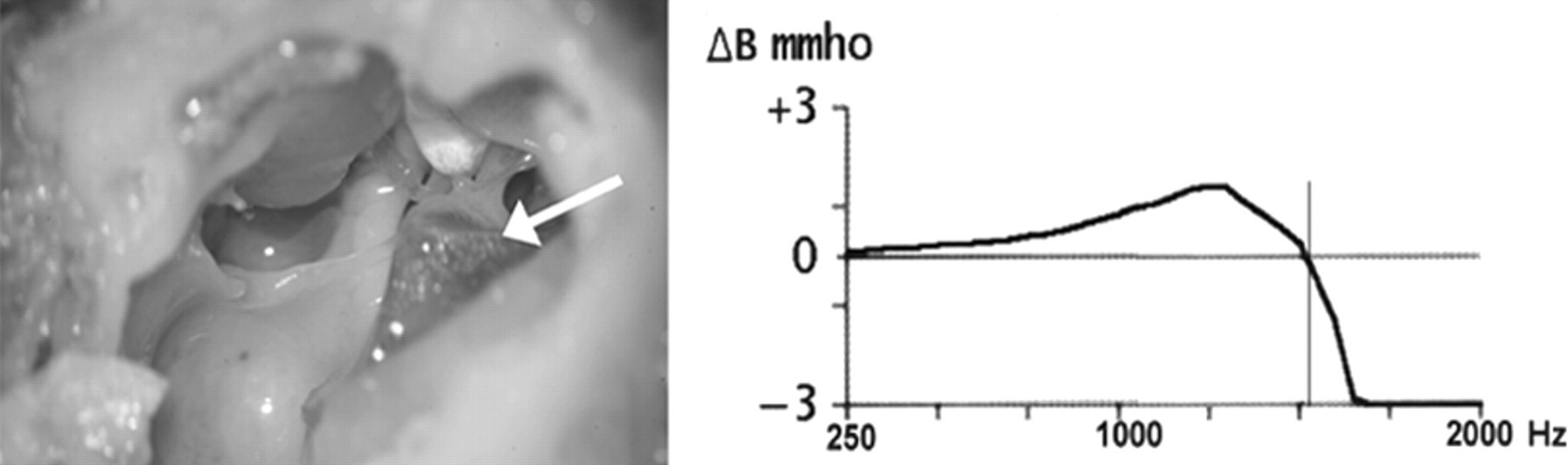
Tympanometry was performed using the GSI 33 Version 2 tympanometer (Grason-Stadler, Inc, Littleton, USA). The system is capable of recording tympanograms from 226 to 2000 Hz at 1/6 octave intervals. The complexity of the ear canal acoustics does not permit accurate calibration above 2000 Hz. The probe was hermetically sealed in the ear canal close to the osseous border, a standard clinical probe being used. A 226 Hz tympanogram was recorded for all animals by conducting an air pressure sweep from +200 to −400 daPa at a rate of 50 daPa/s. The following tympanograms were recorded over a wide frequency range from 250 to 2000 Hz. The immittance values thus recorded were computer-processed to provide standard MFT curves. The computer analysed resonance frequency, generating a diagram in which ΔB is 0 (Figure 1).
In order to establish the ideal probe-tone frequency for recording the tympanograms, we took repeat measurements at different frequencies (226, 678 and 1000 Hz).
Microscopic examination
A microscopic examination of the middle ear was carried out in order to ensure that no fluid (or any other pathological change) had led to a distortion of the measured data. The most reliable method is therefore the direct removal of the temporal bones and subsequent microscopic examination.
Assessment under the surgical microscope was carried out immediately following euthanasia and preparation of the temporal bones (with opening of the tympanic bulla). The ossicular chain and surrounding tissue were microscopically examined for increased vascularization and new tissue formation. Tweezers were used to move the eardrum slightly so that, in the functional test, the coupling of the ossicular chain to the surrounding structures could be assessed.
Statistical analysis
For all recorded data, the arithmetic mean, standard deviation (SD), median and minimum and maximum values were calculated. Data were tested for normal distribution using the Kolgomorov-Smirnov and Anderson-Darling goodness-of-fit tests. The Wilcoxon signed-rank test was used to calculate the significance levels (P) of the measured data obtained. The statistical program used was Statistical Analysis System (SAS Institute, Inc, Cary, USA).
Results
Overall, performing tympanograms using the GSI 33 Version 2 tympanometer proved straightforward. The probe-tone frequencies can be used in the rabbit in the same way as with the human middle ear, so that tympanograms could be recorded in 100 % of cases. One drawback was the lack of a means of electronically storing the data, hence a written record had to be maintained.
226 Hz tympanometry
Using probe-tone frequencies of 226, 678 and 1000 Hz for the repeat measurements, adequate tympanograms were obtained, with the highest degree of reliability achieved at 226 Hz. Tables 1 and 2 present the values for compliance (mean and SD) and amplitude pressure for both ears. None of the statistical analyses performed revealed any significant differences (P > 0.05) for the compliance values determined from the tympanogram. Trends in the median values revealed almost no dependence on the time of measurement for either the left or the right ear.
Average physiological compliance (mL) for both ears, as measured by conventional tympanometry using a 226 Hz probe tone
Average physiological amplitude pressure (daPa) for both ears, as measured by conventional tympanometry using a 226 Hz probe tone
Multifrequency tympanometry
In our studies, the success rate in determining the resonance point was 100%. Normative data obtained from our study reveal the resonance frequency to be 1368 ± 205 for the right side and 1413 ± 216 for the left side (Table 3). The maximum value recorded for the right ear was 1750 and the mimimum was 1050. On the left side, the maximum was 1800 and the minimum was 1000.
Average physiological resonance frequency (Hz) for both ears, as measured by multifrequency tympanometry
Microscopic examination
Following euthanasia and removal of the temporal bones, the bulla was opened and the tympanic cavity was examined under the microscope. In all the test animals, the site showed no sign of irritation, fluid accumulation or inflammation. In two animals, accumulations of viscous fluid were observed, indicating seromucous otitis media. These animals were removed from the study.
Anaesthetic
Four rabbits died in the initial phase during the anaesthesia procedures carried out as part of the preparatory work. Autopsies performed on the deceased animals suggested vagal cardiac arrest. As a result, the anaesthesia regimen was augmented by the anticholinergic agent Robinul®, dosed at 5 mg/animal in the form of an intramuscular injection.
Discussion
Whereas, in the field of animal experimentation, normative values for MFT exist for species such as the chinchilla and Mongolian gerbil (Margolis 1995, 1998), no data on the resonance behaviour of the rabbit middle ear can currently be found in the literature. However, the anatomical situation is described in detail and surgical access routes exist, which allow experimental implantation of the newly developed ossicular prostheses.
Tympanometry is a non-intrusive method and can be performed under sedation. Intubation anaesthesia, as used in this study, has the advantage of allowing better control while registering the vital parameters.
The tympanometer employed in this study (GSI 33, Version 2) has already been successfully used by Cole and Kwochka (2000) to obtain tympanograms in dogs. It has the advantage of allowing both conventional and MFT to be performed. In the present study, the machine was successfully tested for use in rabbits. We recorded 100% diagnostically usable tympanograms. Now that the device has been refined for use in human medicine, the scale of the tympanogram is tailored to humans. For use in rabbits, it would be helpful to be able to individualize this scale, so that lower compliance values could be shown more clearly.
226 Hz tympanometry
Various probe-tone frequencies (226, 678 and 1000 Hz) were tested in the present study. In their investigations, Counter et al. (1989) report that they failed to obtain adequate tympanograms in rabbits at a probe-tone frequency of 226 Hz. In the present authors' own study, however, tympanograms with comparable curves were obtained at all three of the frequencies used. In the present investigation, therefore, following the recommendation by Kiessling (1982), a probe-tone frequency of 226 Hz was used in order to minimize the influence of the factors of friction and mass and thus determine the compliance as precisely as possible.
Amplitude pressure
In our studies, amplitude pressure was between 0 and −100 daPa, thus falling within the normal range for data in human medicine (Shahnaz & Polka 1997, Miani et al. 2000, Lehnhardt & Laszig 2001). The pressure fluctuations observed in the rabbit are, however, still within the tolerance range of 100 daPa. A comparison of both ears produced no statistically significant differences. A possible explanation for the negative amplitude pressure measured in several rabbits could be seen in the animals' failure to swallow while under anaesthetic. This may lead to the formation of negative pressure in the middle ear, as the air is reabsorbed via the mucous membranes of the middle ear and no air can subsequently flow in via the closed tube. Cole and Kwochka (2000) also report changes to the tympanogram during lengthy anaesthesia in dogs.
In order to ensure that the inhalation anaesthetic agent was unable to influence the amplitude pressure, we used isoflurane. Both halothane and nitrous oxide (laughing gas) are reported in the literature as influencing amplitude pressure, whereas no such effects are observed when isoflurane is used (Kuschnir et al. 1981, Lawrence et al. 1994). The increase in amplitude pressure is attributed to the accumulation of laughing gas in closed cavities through direct exchange with the nitrogen in the air.
Compliance
Compliance values are considerably lower than those for the human middle ear, which supports the findings of Rosowski (1994). The reason for this is the greater stiffness of the rabbit middle ear. As compliance is the inverse of stiffness, a stiff eardrum has a lower compliance, whereas a flaccid eardrum leads to a higher compliance (Lehnhardt & Laszig 2001).
Multifrequency tympanometry
The physiological resonance point for the rabbit ear was found to average 1368.1 Hz (preoperative, right ear), which is higher than in humans (974 Hz). The significant differences are mainly due to the different size and the corresponding functional effects. According to Schönfelder et al. (1990), the surface area of the tympanic membrane and the base of the stapes, as well as the length of the incus, are nearly twice as great as in the rabbit. This results in different leveraging interactions which affect the biomechanics of the tympanic membrane and ossicles. The different surface ratios of the tympanic membrane and stapes, as well as the leveraging effect of the manubrium of the malleus and the incus, result in a total amplification factor of 48.6 as opposed to 21.4 in humans (Lehnhardt & Laszig 2001). The increased rigidity of the system results in the resonance point shifting to a higher part of the frequency range.
MFT is suitable for evaluating the immittance of the middle ear. The results will serve as normative values for future experimental studies.
Footnotes
Acknowledgement
This project is supported by the German Research Foundation (DFG) within the collaborative research programme SFB 599/D1.