
Abstract
A systemic framework for teaching vascular sonography that is centered on hemodynamics is useful to unify the initial test-by-test approach that developed with the pioneering and expansion of the field. Vascular sonography education is most powerful when taught with a system-level view of the circulation that connects how vascular anatomy (form) and physiology (function) are inextricably linked with hemodynamics (flow). Furthermore, understanding hemodynamic principles is key to using waveform morphology as a diagnostic tool. Teaching a system-based, hemodynamic-centered perspective aligns with the objectives of the recent Consensus Statement on Doppler Waveforms by the Society for Vascular Medicine and the Society for Vascular Ultrasound and equips the vascular sonography student to recognize, understand, and appropriately document vascular findings.
In the early 1950s, physicians who sought a non-invasive alternative to angiography began developing novel techniques to assess their patients. Groundbreaking studies catalyzed an interest in the application of ultrasound for vascular testing, resulting in tools such as continuous wave (CW) Doppler, duplex ultrasound imaging with color, and spectral Doppler.1,2 These techniques, were developed one at a time, initially focused on application to a single vascular bed. In each instance, technology deepened hemodynamic understanding (see Table 1).
Vascular Ultrasound Application Development by Year.
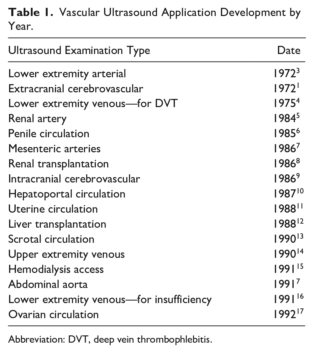
Abbreviation: DVT, deep vein thrombophlebitis.
Development of this array of technology provided both detailed renderings of a patient’s anatomy and previously unobservable physiologic characteristics of real-time human blood flow. Doppler waveforms offered insightful maps of hemodynamic properties that were unobtainable by surgical or cadaveric exploration of vessels and impossible to simulate in laboratory settings.18-20 These tools were capable of acquiring information that was beyond the contemporary understanding in the field at the time and fueled the modern era of clinical hemodynamics, with the waveform as its centerpiece, providing a written language for the translation of vascular physiology. 20
Proposed Systemic Approach: Vascular Form and Function Are Inextricably Linked to Flow
Since the advent of vascular testing, the modern field of clinical hemodynamics has solidified principles of arterial and venous flow patterns, offering explanations for the underlying phenomena that govern vascular flow. Vascular sonography education is most powerful when it places these hemodynamic principles at its core, providing a cohesive framework for understanding flow within the vascular system as a whole, as well as the significance of its individual parts. A foundational, system-level view of the circulation that connects how vascular anatomy (form) and physiology (function) are inextricably linked with hemodynamics (flow) provides a fundamental approach to explaining normal and pathologic states, rather than just describing them. Furthermore, this perspective can be used to synthesize how these elements are captured in duplex ultrasound and indirect vascular testing modalities. This approach provides a framework that supports critical thinking and skillful imaging of the vascular system.
Foundations: Anatomy, Physiology and Hemodynamics (Form, Function, and Flow)
The vascular system is not a disconnected collection of unique vessels that operate in isolation from one another. Rather, it is a complex interconnected system whose parts work in balance, with each vessel’s effectiveness arising from its position and structure, as well as the health of the other mechanisms of circulation (heart, other blood vessels, organs, and blood18,21). A systemic view recognizes that all these attributes contribute to hemodynamic patterns and, subsequently, normal and abnormal waveform morphology within any given vessel.
Because the vascular system is a closed pressurized circuit, flow at any given location is a product of conditions both up and downstream from the point of interest. Specifically, all flow characteristics are derived from the interplay of
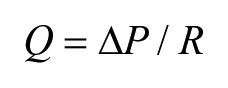
From this formula, it is noted that the main driver of flow is pressure gradient and the main detractor is resistance. The interplay between pressure and resistance is reflected in fluctuation of flow velocities, which are mapped over time to yield waveforms.20,21
Arterial Hemodynamics
For the arteries, the primary source of flow is the dynamic pressure of the cardiac cycle, while resistance arises largely from the distal arteriolar beds.
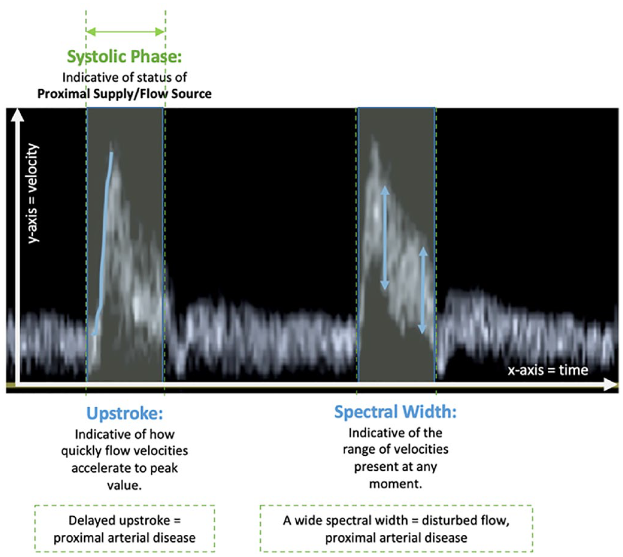
Systolic phase components of arterial waveforms and their hemodynamic significance.
Assessment of the systolic phase of a waveform reveals the efficacy of source pressure and provides information on the health of the upstream components of circulation: the more proximal arteries and the heart.
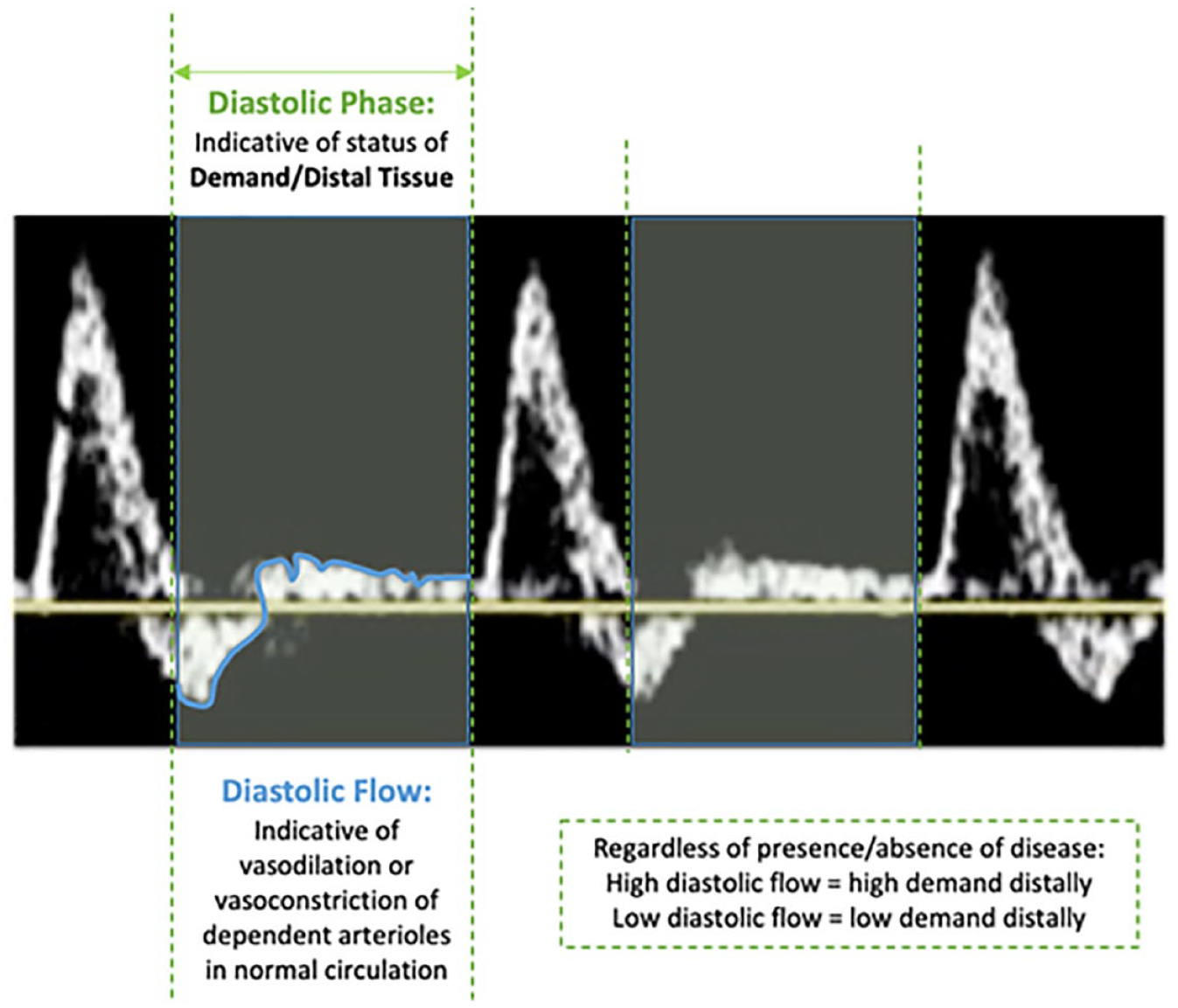
Diastolic phase components of arterial waveforms and their hemodynamic significance.
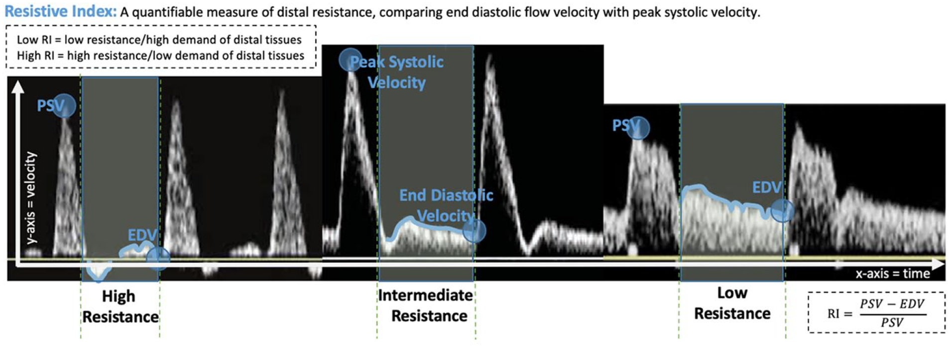
Varying resistivity levels of arterial waveforms.
Assessment of the diastolic phase of a waveform provides information on the level of resistance created by the physiologic state of downstream arteriolar beds.
Venous Hemodynamics
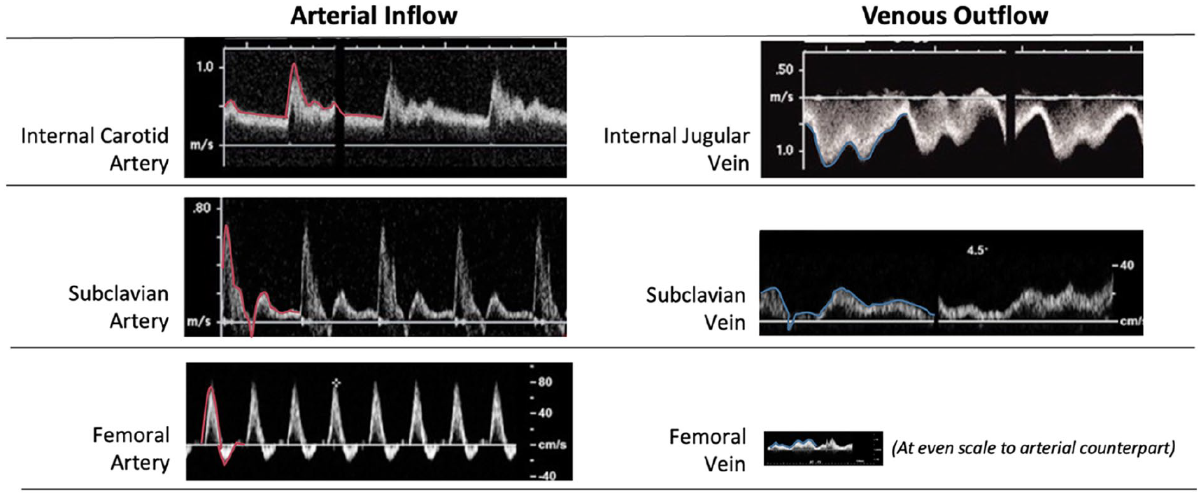
Arterial inflows as compared with their venous outflows.
Much different than its high-pressure arterial counterpart, the low-pressure venous system is a delicate balance between source pressure and downstream resistance, with direction and velocity of flow indicating which is the dominant force in any given moment. When source pressure is dominant over downstream resistance, centripetal (toward the heart/antegrade) flow ensues. When downstream resistance is dominant over source pressure, centripetal flow ceases and centrifugal (away from the heart/retrograde) flow may occur.
Venous waveforms are meaningful maps of the pressure balance within the venous system, demonstrating normal flow volumes and responsiveness to patterns of cardiac and respiratory motion based on their position within the vascular system (see Figures 5 and 6).
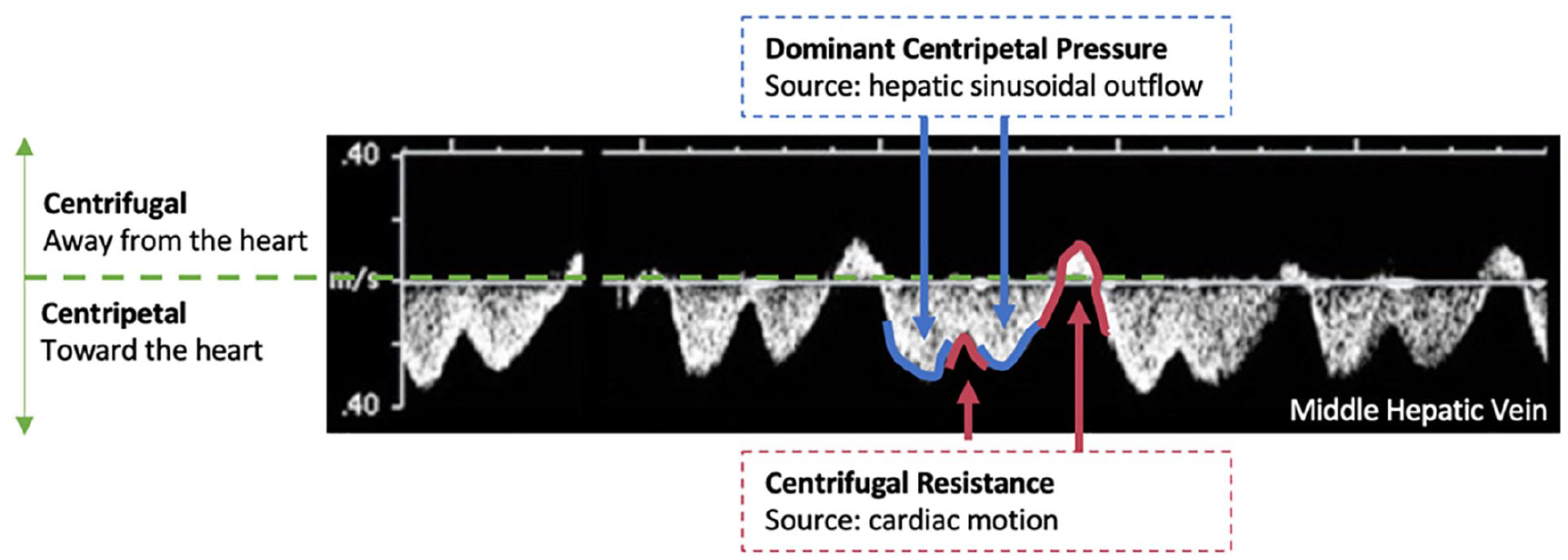
Influences of venous waveform flow velocity and direction in a middle hepatic vein waveform.
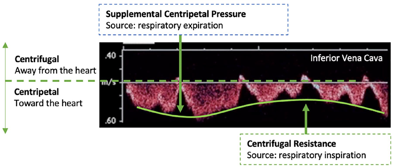
Effects of respiration on a venous waveform of inferior vena cava flow.
A hemodynamic-centered approach to teaching vascular ultrasound offers, by nature, a systemic perspective of the vascular system because it recognizes that the blood within the circulatory system forms a single continuous body of fluid, with flow at any given point being dependent on the conditions both up and downstream, namely, at the source of blood flow and its destination.
Discussion
A hemodynamic-centered approach to vascular sonography education, as introduced in the previous section, extends beyond teaching traditional anatomic and physiologic characteristics of the vascular system and seeks to integrate how particular waveform appearances result from flow dynamics created by these. It defines and describes the varying elements that characterize normal flow and constructs an explanation of what underlying anatomic and physiologic characteristics support them: both at the point of interest and systemically. Vascular educators can achieve a hemodynamic-centered approach by (1) creating an initial foundation of knowledge that demonstrates how the three key content areas of anatomy, physiology, and hemodynamics are interdependent and broadly responsible for subsequent waveforms and (2) referencing this hemodynamic foundation when teaching individual vascular tests using a standardized framework for explaining specific flow characteristics within the vessels of any vascular subsystem.
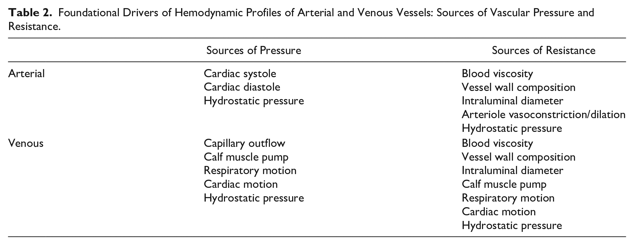
The hemodynamic foundation initially taught should provide a cohesive framework to explain both normal and pathologic flow states. Using Ohm’s law, all normal anatomic and physiologic attributes can be explained as contributing to either the pressure that contributes to antegrade flow or the resistance that opposes it. To emphasize the dependent nature of flow at any point within a given vessel, students should be taught to think in terms of the source of flow and its destination. For arteries, this can be framed in terms of supply and demand, whereas for veins, the terms (centripetal) source pressure and (centrifugal) resistance are more useful. Sources of pressure and resistance that should be explored are noted in Table 2.
Foundational Drivers of Hemodynamic Profiles of Arterial and Venous Vessels: Sources of Vascular Pressure and Resistance.
The difference between pressure and resistance at any given point is demonstrated in velocity fluctuations over time, which are mapped by waveforms. A thorough comprehension of how anatomy and physiology contribute to hemodynamics paves the way for a firm understanding of waveform morphology. After normal states are explained, all vascular pathology, by contrast, should then be framed as processes that disrupt anatomy and physiology and subsequently alter the normal balance of flow and resistance, which is reflected in abnormal waveforms (See Figure 7).
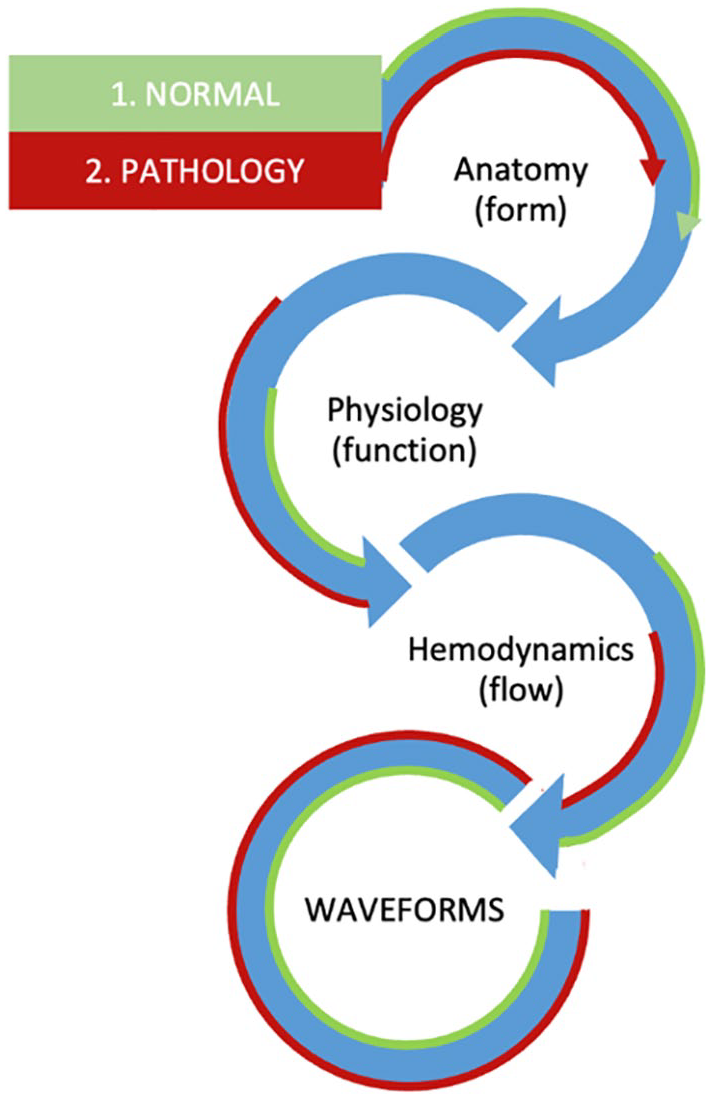
Pedagogical approach to hemodynamic-centered vascular education.
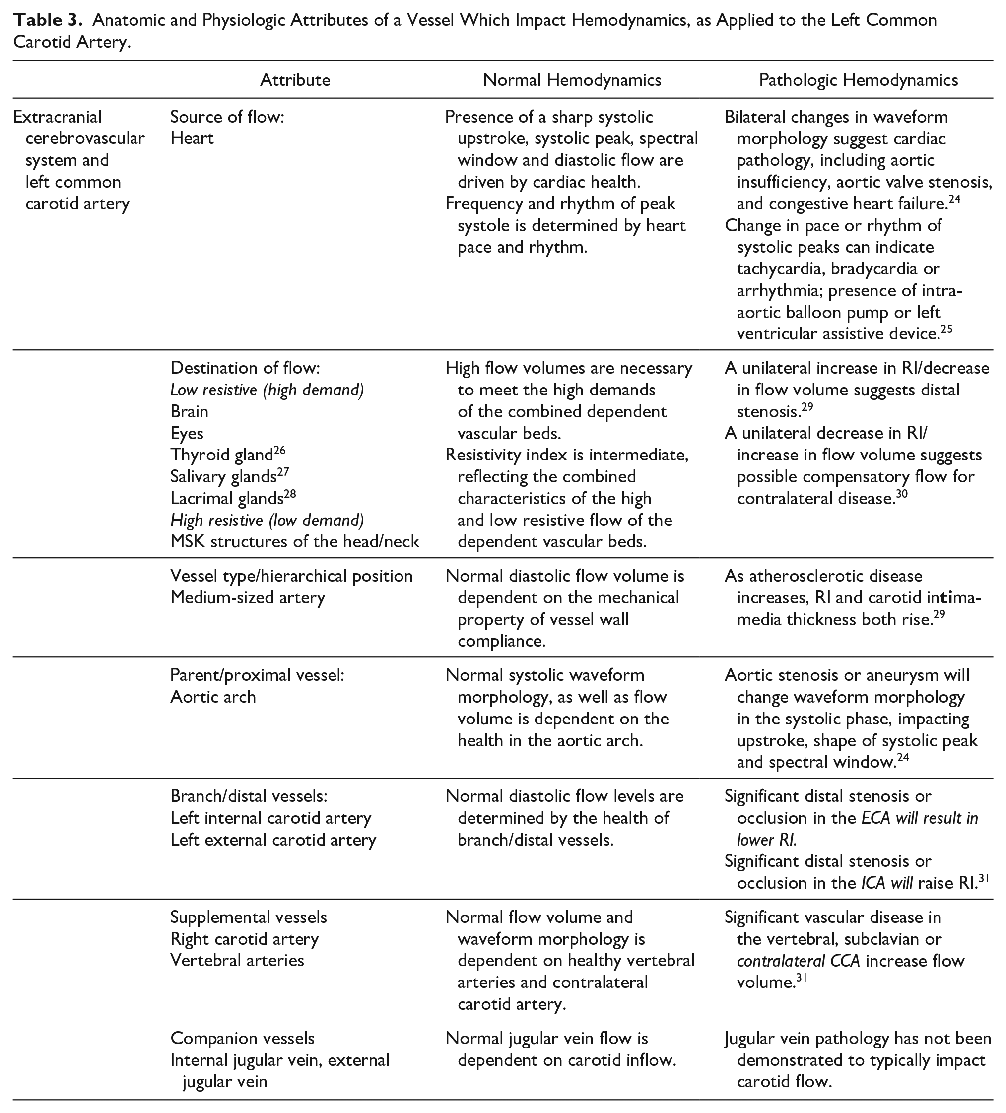
Once this initial foundation has been laid, the same approach should also be applied in standardized fashion to teaching vascular testing of specific structures. This involves identifying all vessels that collectively serve a dependent vascular bed and then identifying the sources of pressure and resistance among them. This methodology captures the many factors that may influence flow and waveforms in any individual vessel, which can range from localized pathology (like stenosis in the vessel of interest), proximal pathology, distal pathology, or pathology in an associated vessel (such as renal vein thrombosis impacting renal artery waveform; subclavian occlusion impacting vertebral artery waveform). Table 3 uses the example of the left common carotid artery to demonstrate how this systemic, hemodynamic perspective can be applied to map fundamental anatomic and physiologic attributes of vascular structures that are traditionally taught, alongside an explanation of how pathologic change impacts waveform morphology.
Anatomic and Physiologic Attributes of a Vessel Which Impact Hemodynamics, as Applied to the Left Common Carotid Artery.
Recently, a consensus document by the Society for Vascular Medicine and the Society for Vascular Ultrasound has clearly defined and standardized descriptive terms for normal and pathologic Doppler waveforms in both the peripheral arterial and venous systems. This important achievement not only codifies a shared language for discussing waveform morphology but also identifies the necessary skill level for vascular sonographers to possess. The authors state, All interpreting physicians and sonographers should be able to describe Doppler waveforms, be able to identify the changes which occur with physiologic and disease states, and effectively communicate these waveform characteristics to interpreting physicians so that consistent information is given to referring providers.
32
It should be the goal of vascular educators to provide necessary instruction in waveforms and the hemodynamic principles underlying them so that students can achieve competence in these areas. The list of major descriptors and modifying terms for waveform morphology that were defined in the consensus document serve to establish the scope of necessary knowledge for vascular sonographers. Vascular students, therefore, will need foundational instruction in anatomy, physiology, and hemodynamics to support an understanding of the significance of these key terms, as listed in Table 4.
Key Major Descriptors and Modifier Terms Used as Scope of Necessary Information for Hemodynamic-Centered Vascular Education.
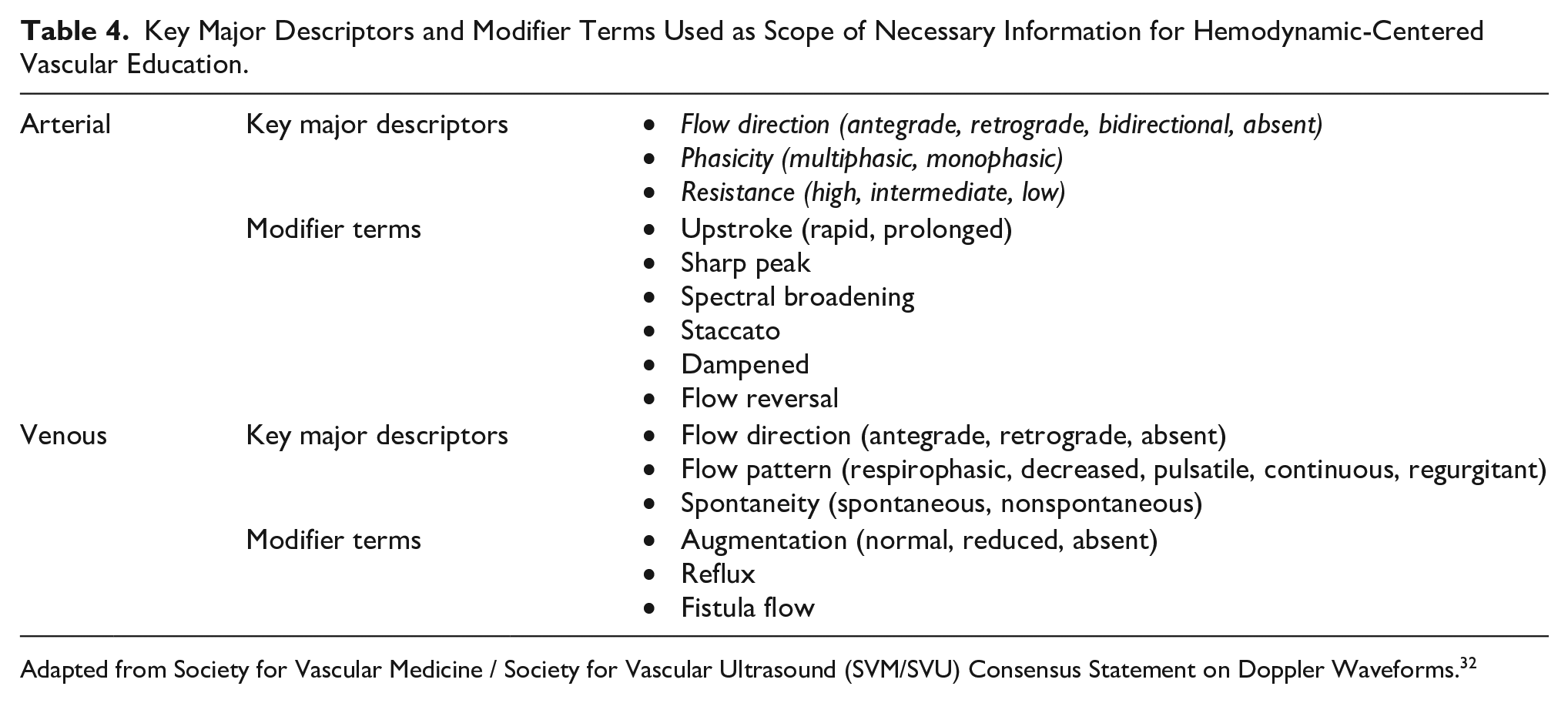
Adapted from Society for Vascular Medicine / Society for Vascular Ultrasound (SVM/SVU) Consensus Statement on Doppler Waveforms. 32
The systemic, hemodynamic-centered framework offered above provides foundational reasoning about the significance of varying anatomic and physiologic attributes of vascular structures, as well as patterns of waveform morphology. This is a useful educational tool because it offers a cohesive perspective for teaching students to understand the causes of normal and abnormal findings of vascular testing, beyond recognizing their appearances. Although key hemodynamic principles were only applied to basic normal waveform morphology in this article, they can also be readily applied to additional normal and pathologic characteristics.
While the normal appearance of vascular structures and waveforms vary from vessel to vessel, findings for each are in line with the principles of hemodynamics. When combined with the well-documented sonographic appearances of waveforms for many circulatory subsystems, these explanations provide students with a central framework that is meaningful for understanding the significance of the waveforms they are recording.
Footnotes
Declaration of Conflicting Interests
The author declared no potential conflicts of interest with respect to the research, authorship, and/or publication of this article.
Funding
The author received no financial support for the research, authorship, and/or publication of this article.