
Abstract
Caudal duplication is a rare condition that results in varying degrees of duplication of organs within the gastrointestinal, genitourinary, reproductive, spinal, and vertebral systems. Polymelia is a rare congenital defect that presents as supernumerary limbs. This case report describes a fetus with characteristics of both conditions but does not present with the classic scenario of either condition.
Caudal duplication is a rare condition that includes a wide range of gastrointestinal, genitourinary, vertebral, and spinal anomalies. 1 The phrase caudal duplication syndrome was first introduced in 1993 by Dominguez in an effort to describe the association of gastrointestinal, genitourinary, and distal neural tube malformations. 2 Case reports have documented varying degrees of duplication of the external genitalia, cervix and uterus, bladder, colon, anus, distal spine, and limbs. In addition to duplication anomalies, myelomeningocele, hemivertebrae, omphalocele, umbilical hernia, and imperforate anus have also been reported. The syndrome is rare, occurring in less than 1 per 100 000 births. 3 It appears to be more common in females 2:1 without racial or familial predilection. 4
Polymelia is an uncommon congenital anomaly that includes the presence of accessory or supernumerary limbs. 5 It is rarely reported in humans 6 and is usually associated with chromosomal defects and/or environmental factors. 5 The accessory limb(s) can be small or deformed and attached to various regions of the body. 6 Limb malformations occur in approximately 6 per 10 000 live births and usually affect the upper limbs more than the lower limbs 3:1. 6
Limb development is a very complex process and involves several factors. The exact cause of caudal duplication is unknown. Although a few theories have been proposed, such as an insult during limb development or incomplete separation of monozygotic twins, 7 polymelia is thought to be caused by genetic factors or incomplete twinning, often including the presence of a parasitic twin. 8 We present a unique case of a fetus with characteristics of both caudal duplication and polymelia.
Case Report
A 21-year-old, non-diabetic female (gravida 2, para 1) was referred from an outside 3D sonography center for possible lack of fetal heart tones. Estimated gestational age was 13 weeks 3 days. A sonogram performed at 9 weeks was within normal limits and the fetus had a heart rate of 176 beats per minute. Maternal history included daily tobacco use and recent onset of abdominal cramping.
The second sonogram confirmed fetal demise with an estimated gestational age of 12 weeks 6 days. The fetus was in the dependent portion of the uterus, making evaluation somewhat difficult. The head and thorax appeared normal (Figure 1). Inferiorly, there appeared to be several echogenic linear structures suggesting multiple femurs (Figure 2). After the onset of preterm labor, a nonviable fetus was delivered intact. The head and thorax appeared grossly normal; however, there were six complete lower extremities present (Figure 3).
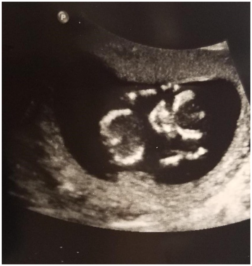
Sonographically normal appearing fetal head and torso.
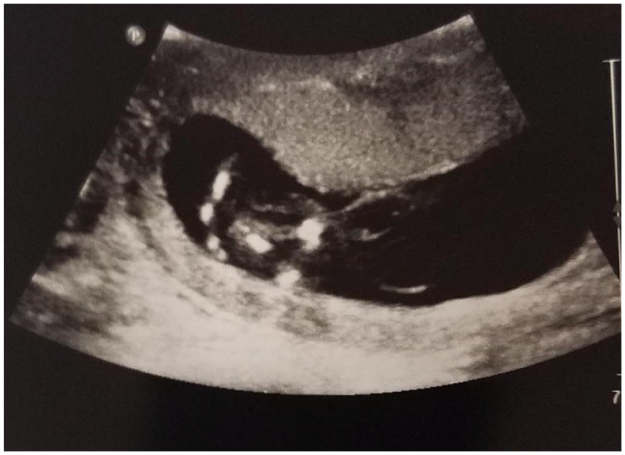
Sonogram of the inferior portion of the fetus demonstrating multiple linear echogenic structures.
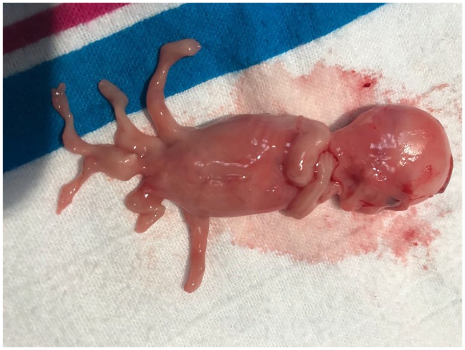
Gross specimen of the fetus confirming the presence of six lower extremities.
An initial diagnosis of polymelia with possible caudal duplication syndrome was made. The fetal tissues failed to grow in culture, and chromosome analysis could not be performed. Therefore, a microarray analysis was performed on DNA extracted from snap frozen tissue. No parental tissue was requested at the time of the analysis. The parents chose not to seek genetic counseling.
Perinatal autopsy confirmed a male fetus with a normal karyotype and no regions of homozygosity. Six complete lower extremities, including feet and toes were identified. No polydactyly or syndactyly was present. The limbs were arranged in a rostral to caudal pattern. The most normal extremities were located anteriorly and the most rudimentary pair more posteriorly. The three pairs of extremities were each associated with a variably formed pelvis.
Upon further evaluation, there were no cardiac, respiratory, urinary, or reproductive anomalies noted. The only other finding was intestinal malrotation. Polymelia was ruled out as there was no indication of a parasitic twin or abnormal karyotype. Despite the lack of anomalies within other organ systems, the final diagnosis of caudal duplication syndrome was made, presumably due to the presence of multiple pelvises and lower extremities.
Discussion
Caudal duplication is a rare syndrome that encompasses a wide variety of organ duplication anomalies. The organs affected are typically of the genitourinary, reproductive, gastrointestinal, spinal, and vertebral systems. Duplication of the external genitalia1,2,4,9–14 and bladder1–4,9–13,15,16 have been reported within several case studies and appear to be common presentations of the syndrome. Additional reported anomalies include duplication of the anus,1–4,9,12 colon,1,2,4,9–14 urethra,2,4,7,10,11,16 and uterus.3,4,10,17 Associated anomalies such as imperforate anus,10,14,17 omphalocele,2,10 lipomeningocele, 12 myelomeningocele, 2 and lipomyomeningocele 18 have also been documented. This would appear to be the first reported case of caudal duplication that did not include organ duplication or other associated anomalies.
Interestingly, only a few cases included an accessory or duplicated extremity.2,14–18 One of the cases reviewed by Dominguez et al included a patient with a third hypoplastic leg and iliac bone that arose from the area of a meningocele. 2 Arnone et al described a case that included a foot-like structure that arose from the patient’s right labia majora. 17 Zhao et al described a case of congenital limb duplication in which the child had an extra pelvis and lower extremity that arose from the sacral area. Although the child had other associated anomalies similar to those of caudal duplication, that specific diagnosis was not made. 18
Polymelia was one of the initial diagnoses considered for this case. This condition is also very rare and results in supernumerary limbs. There are four classifications based on where the extra limbs are attached: the head, spine, thorax, or pelvis. The subcategory of pyromelia is when the limbs are attached to the native pelvis. 5 This could be considered as a differential diagnosis, but the extra limbs in our case each had their own variably formed pelvis. Polymelia is also associated with genetic and environmental factors, 5 neither of which were reported by the patient or identified postnatally in our case. In addition, a parasitic twin is often present with polymelia. 8 Perinatal autopsy of the fetus did not show evidence twinning. Therefore, the diagnosis of polymelia was no longer considered for this fetus.
Through an in-depth review of the literature, another possible differential diagnosis presented itself, dipygus. Dipygus is a severe and complete form of caudal duplication. 14 The classic presentation is complete lumbosacral duplication that includes two pelvises and four lower extremities. 15 Similar to caudal duplication, dipygus usually includes anomalies of the genitourinary and reproductive organs, such as duplication of the external genitalia and bladder. 15 Ulman et al reported a case of an infant with duplication of the pelvis, two extra extremities, and duplication of the ileum, external genitalia, and bladder. 15 A case report by Al Alayet et al described an infant with two pelvises, four lower legs, bilateral imperforate ani, duplicated external genitalia, and gastroschisis. 14 The cause of dipygus is unknown but is thought to be a result of incomplete monozygotic twinning. 16 Our case is similar to dipygus by having multiple extremities, but the fetus had three pelvises and six lower extremities, no duplication anomalies, or evidence of twinning.
The etiology and embryonic pathophysiology of caudal duplication remains unclear. 14 Some authors theorize it is the result of incomplete division of monozygotic twins.7,10,16 Pang et al suggested that all duplication disorders of the spinal cord result from an abnormal adherence of the ectoderm and endoderm. 19 In contrast, Domingeuz et al suggested that caudal duplication results from damage to the caudal cell mass during 23rd and 25th day of gestation. Due to the vast variety of anomalies included within caudal duplication, it may be possible all theories are correct, but apply differently to each case. Limb development occurs during the four and fifth week of gestation. Due to this very small window when deformities can occur, it is understandable why limb anomalies are indeed so rare. 18
Treatment for caudal duplication, polymelia, and dipygus should be evaluated on an individual basis. Treatment plans should always be customized according to the extent and severity of the duplication and the functionality of the organs involved. 12 Considerations should include the number and severity of anomalies present in each case. Surgical management must be adapted to each case and aim to achieve the best functional results rather than restore typical anatomy. 11
Conclusion
Caudal duplication is a very rare condition and although an exact diagnosis was elusive for this case, it is undeniable that sonography played an instrumental role in the identification of the limb anomalies. Prenatal sonography is an invaluable tool in the assessment of gross congenital anomalies. As sonographers, we are trained to evaluate fetal anatomy and rule out many common anomalies and syndromes during a routine anatomy scan. At times, the sonographic findings do not fit into the prescribed list of known abnormalities. Sonographers must use their judgment, knowledge, and professionalism to form a conclusion to assist in the diagnostic process. Lesson learned from this case relates to the adage of “if you hear hooves, you typically look for horses, not zebras.” Sometimes even if you cannot believe what you think you see, in rare cases, you might have just found the zebra.
Footnotes
Declaration of Conflicting Interests
The authors declared no potential conflicts of interest with respect to the research, authorship, and/or publication of this article.
Funding
The authors received no financial support for the research, authorship, and/or publication of this article.