
Abstract
Cystic lesions of the liver may include simple cysts, multiple cysts arising in the setting of polycystic liver disease, parasitic or hydatid (echinococcal) cysts, cystic tumors, and abscesses. Sonography is the diagnostic imaging procedure of choice for hepatic cysts, so knowledge of the sonographic features of the most common types of cystic hepatic lesions should enable an accurate diagnosis to be made directly or in conjunction with clinical and laboratory data. This study of 183 patients was done to identify and evaluate focal hepatic cystic lesions in Sudanese adults using ultrasonography (US) and to determine the diagnostic role of ultrasound-guided fine-needle aspiration (FNA). Most cystic lesions identified were solitary lesions (74.3%). The most common site for lesions was the right lobe of the liver (66.1%). Simple hepatic cysts were seen in 20.2% of patients; the most common type of lesion was hepatic abscess (41.5%). Autosomal dominant polycystic kidney disease accounted for only 2.2% of lesions. There was a statistical association between focal hepatic cystic lesions and patient age (P < .001). US-guided FNA was done in 48.6% of patients, and these findings were in agreement with sonographic features, with the FNA aspirate color and consistency correlating with lesion histology.
Cystic lesions of the liver include simple cysts, multiple cysts arising in the setting of polycystic liver disease, parasitic or hydatid (echinococcal) cysts, cystic tumors, and abscesses. These conditions can usually be distinguished on the basis of the patient’s symptoms, clinical history, and the radiographic appearance of the lesion.1–4 Simple cysts are the most common type of cystic liver lesion and are seen in approximately 2.5% of the population. 4
Cystic liver lesions in adults can have a number of etiologies, including congenital, neoplastic, and infectious conditions, as well as trauma and miscellaneous entities.1,4 These lesions may be encountered on radiologic examinations, often unexpectedly, and a number of cystic lesions have grave outcomes. Sonography is the diagnostic imaging procedure of choice for hepatic cysts, so the knowledge of the sonographic features of the most common types of cystic hepatic lesions should enable an accurate diagnosis to be made directly or in conjunction with clinical and laboratory data.4,5 Sonographically, cystic lesions of the liver may present in several forms and as solitary or multiple lesions. Autosomal dominant polycystic kidney disease is the most common congenital disorder that shows hepatic involvement by cysts.5,6 The purpose of this study was to identify and evaluate focal hepatic cystic lesions in Sudanese adults using ultrasonography (US) and to determine the diagnostic role of US-guided fine-needle aspiration (FNA).
Materials and Methods
All Sudanese patients with hepatic cystic lesions who were seen in the National Centre for Gastrointestinal Tract and Liver Disease were candidates for the study. Ultrasonography identified 183 patients; 105 were male (57%) and 78 were female (43%). Sonographic examinations were all done using a Fukuda ultrasound machine (Fukuda Denshi, Tokyo, Japan) with a 3.5-MHz curvilinear transducer. The scanning technique used in this study followed the protocol determined by Hagen-Ansert 7 and Schafer et al. 8 Patients were scanned in the supine position; US measurements of the anteroposterior and transverse diameters (in millimeters) were done for each lesion. For aspiration procedures, needles with a 0.4- to 0.45-mm external diameter (26–27 gauge) on a “binder valve” were used to allow better sampling of very small lesions. Aspirations were done based on a suspicious finding on sonography and under sonographic guidance. During the FNA procedures, local anesthesia was used, and the needle was inserted and advanced at an acute angle underneath the probe, which was held perpendicular to the skin. Approximately 2 cc of aspirated fluid from each participant was sent for cytology.
To collate all the clinical data and US findings, we used a data collection sheet for all participants, which included age, sex, clinical diagnosis, and US and FNA findings. Statistical analysis was done using SPSS version 16 (SPSS, Inc, an IBM Company, Chicago, Illinois). Descriptive statistics, nonparametric correlations, and χ2 testing were applied as appropriate to determine statistical significance, taken to be P < .05.
Results
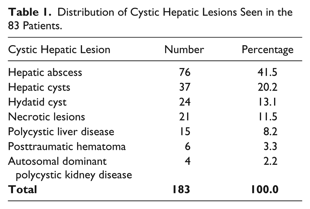
Mean age of the 183 patients included in the study was 50 ± 16.5 years. One hundred five patients (57%) were male and 78 were female (43%). Table 1 summarizes the sonographic findings of hepatic cystic lesions. Most lesions were solitary lesions (136 of 183, 74%). The most common site of the lesions was within the right lobe of the liver (121 lesions, 66%). The most prevalent lesion found, in 76 patients (41.5%), was hepatic abscess (Figure 1); 55 of these were found in patients younger than 60 years (73%). Autosomal dominant polycystic kidney disease was the least common finding, seen in only four patients (2.2%). Simple hepatic cysts were seen in 37 patients (20.2%) (Figure 2), hydatid cysts in 24 patients (13.1%) (Figures 3–5), necrotic lesions in 21 patients (11.5%), congenital polycystic liver disease in 15 patients (8.2%), and hematoma/biloma in 6 patients (3.3%). Table 2 summarizes the nonparametric correlations that were applied, and χ2 testing showed statistical association between the focal hepatic cystic lesions and increasing age, the presence of gallstones, and diabetic status, P < .01.
Distribution of Cystic Hepatic Lesions Seen in the 83 Patients.
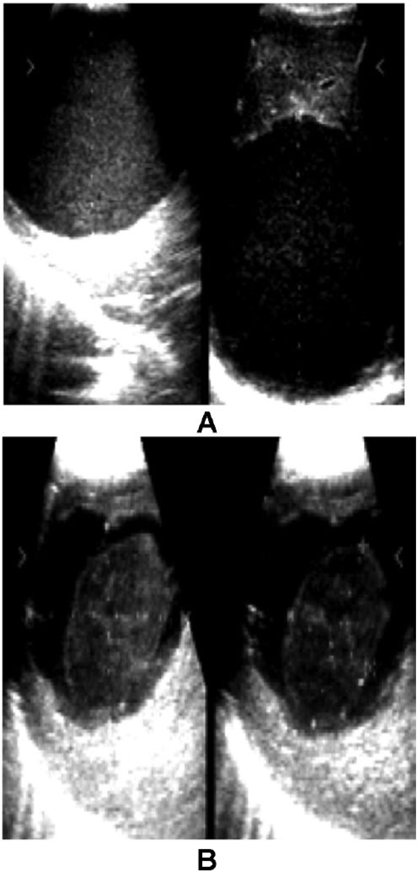
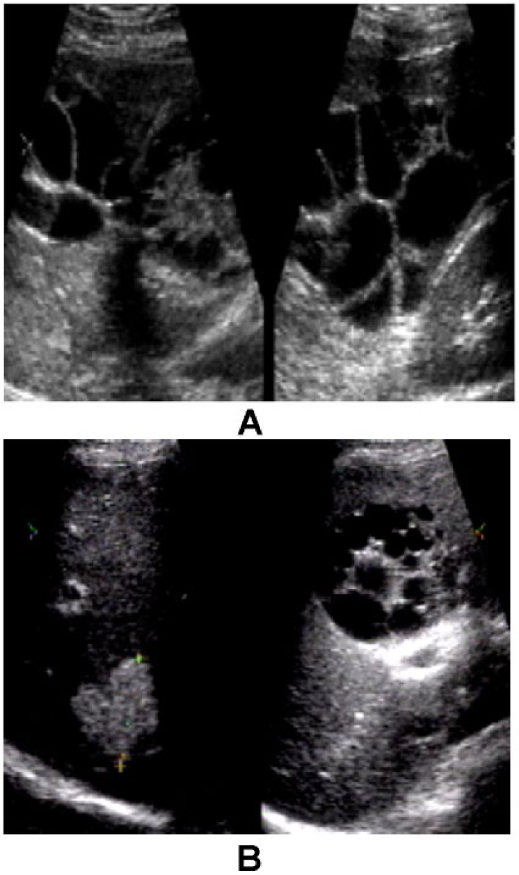
Gray-scale images of the liver of a 35-year-old man showing a partially cystic lesion with echogenic material consistent with hepatic abscess. (A) Before fine-needle aspiration. (B) One week after aspiration.
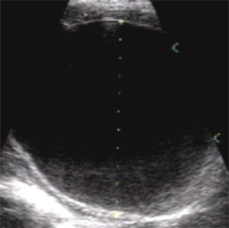
Gray-scale image of the liver of a 40-year-old man showing a simple hepatic cyst.
Gray-scale image of a complex hepatic lesion consistent with hydatid cyst.
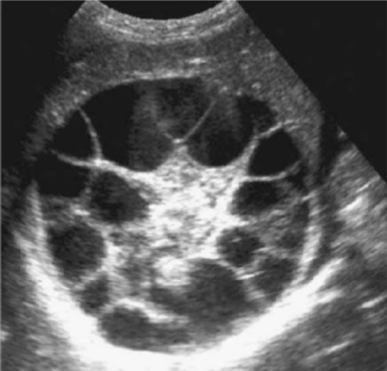
Gray-scale image of a large multicystic hepatic lesion with a honeycomb appearance in the right lobe of the liver consistent with hydatid cyst.
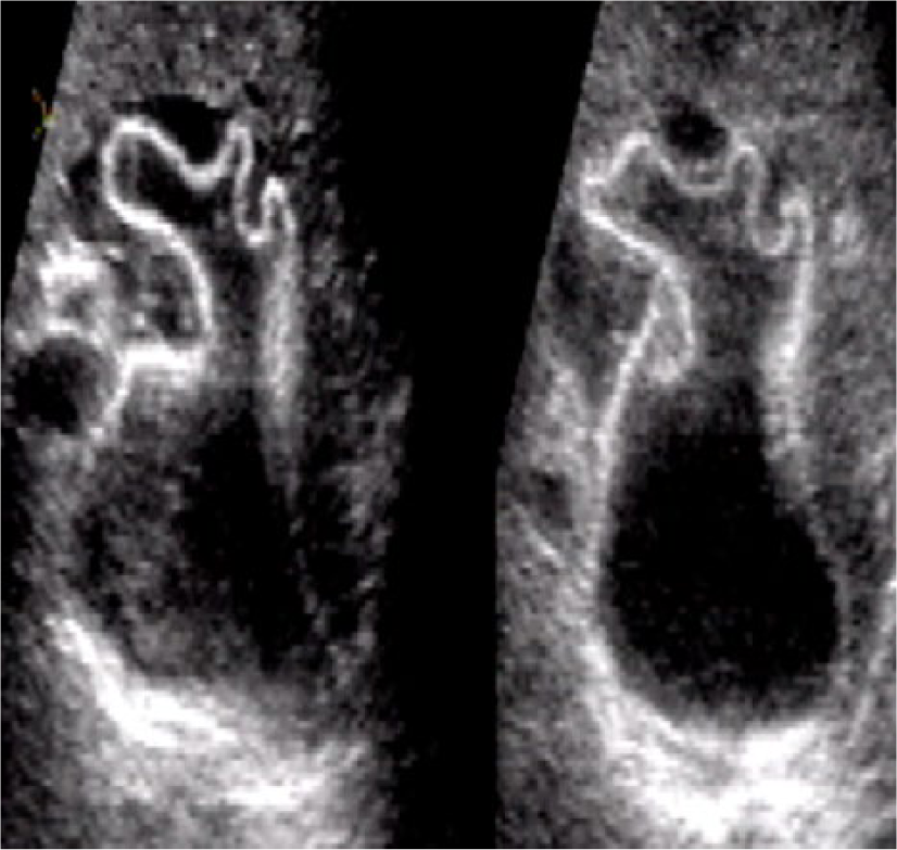
Gray-scale images of a multiloculated cyst with daughter vesicles containing echogenic material consistent with hydatid cyst. (A) Before fine-needle aspiration. (B) After aspiration.
Nonparametric Correlations for the Study Variables.

Figure 6 summarizes the correlation between US features of the cystic lesions of the liver and the US-guided FNA findings; 44.5% of aspirates were purulent/colored, 23.6% were clear, 13.5% had color confirmed hepatic amoebiasis, and 4.5% were bloody and/or with bile content.
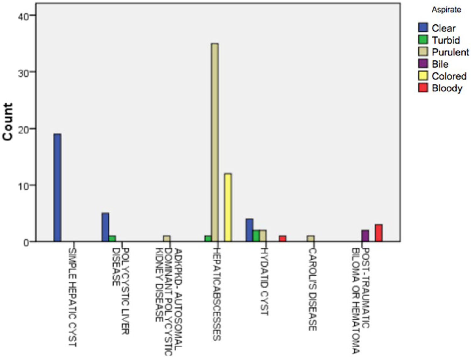
Correlation between the findings of ultrasound-guided aspiration with sonographic features of the hepatic cystic lesions.
Discussion
Solitary lesions were found in 74.3% of the patients, and lesions were most commonly found in the right lobe of the liver. Hepatic abscesses accounted for the greatest percentage of lesions, and cystic hepatic lesions became increasingly more common with age, the strongest independent predictor for the occurrence of a cystic lesion of the liver. The results of this study are in agreement with a study by Halvorsen et al 9 that also showed the sensitivity of sonography in detecting liver abscesses was 79% compared with computed tomography (CT) imaging. The present study is also in agreement with the findings of Sakijan, 10 who showed that 84% of liver abscesses occupied the right lobe. While some authors have proposed that the demographics of liver abscess has shifted toward the sixth and seventh decades of life, 11 this study showed that 72.7% of liver abscesses found were in patients younger than 60 years.
A simple cyst was seen in 37 patients (20.2%), which to some extent correlates with the findings of Power et al 4 ; one difference was that in their study, simple cysts were the most common type of cystic liver lesion. The finding of a simple cyst in this study exceeded the prevalence found by Gaines and Sampson, 5 who showed that simple hepatic cysts occur in only 2.5% of the population. The findings of the present study lie between the results shown by these two authors. The present study also showed a strong correlation between the sonographic image features and the findings of ultrasonographically guided fine-needle aspiration in characterizing cystic lesions of the liver. This is in agreement with the results of Parajuli et al, 12 who showed that ultrasonography and CT-guided (FNA) cytology are sensitive methods in diagnosing deep-seated cystic hepatic lesions. However, a limitation of the present study is that FNA cytology was available for only slightly less than half of the patients, and this may diminish the strength of its overall value in diagnosing cystic lesions of the liver.
Conclusion
The results of this study showed that the right lobe of the liver was the most common site of hepatic cystic lesions in Sudanese patients and that hepatic abscesses were the most commonly seen cystic lesion. There was a strong statistical association between the presence of focal hepatic cystic lesions and patient age. Furthermore, ultrasonographically guided fine-needle aspiration specimens correlated well with sonographic image features, with good correlation between aspirate consistency, clarity, and color with the images of the hepatic cystic lesions.
Footnotes
Acknowledgements
The researchers thank IbinSina Hospital-Khartoum-Sudan (National Centre for Gastrointestinal Tract and Liver Diseases) for allowing this study.
Declaration of Conflicting Interests
The authors declared no potential conflicts of interest with respect to the research, authorship, and/or publication of this article.
Funding
The authors received no financial support for the research, authorship, and/or publication of this article.