
Abstract
Background
Finite element analysis (FEA) is a computational method used to evaluate stress, strain, and deformation in complex structures. In orthodontics, understanding biomechanical responses within teeth, periodontal ligament, and supporting bone is difficult through direct clinical measurement. Therefore, FEA has emerged as a valuable research tool to simulate orthodontic force systems and predict biological responses.
Objective
To review the principles of finite element analysis and discuss its applications, advantages, limitations, and clinical implications in orthodontics.
Methods
A narrative review of the literature was conducted focusing on the fundamental steps involved in FEA, including model generation, meshing, assignment of material properties, application of boundary conditions, and computational analysis of stress and strain within orthodontic structures.
Results
FEA provides a three-dimensional simulation of orthodontic force systems and enables detailed evaluation of stress distribution in teeth, periodontal ligament, alveolar bone, and orthodontic appliances. It has been widely applied in studying tooth movement biomechanics, mini-implant stability, bracket–archwire interactions, orthognathic surgery planning, and aligner therapy. The technique allows non-invasive visualization of biomechanical responses and supports optimization of appliance design and treatment mechanics.
Conclusion
Finite element analysis is a powerful tool that enhances the understanding of orthodontic biomechanics and assists in improving treatment planning and appliance design. With advancements in digital imaging, artificial intelligence, and patient-specific modeling, FEA is expected to play an increasingly important role in evidence-based orthodontic practice.
Keywords
Introduction
Orthodontic tooth movement is caused by the application of regulated pressures to the alveolar bone, which induces remodeling through stress and strain in the periodontal ligament (PDL). Because direct in vivo measurement of these stresses is challenging, computational approaches like finite element analysis (FEA) give a non-invasive and precise way to observe internal biomechanical reactions. 1
Initially developed in engineering, FEA was applied to dentistry in the 1970s to investigate stress distributions in dental and craniofacial structures. With developments in 3D imaging, digital modeling, and computer power, it has become an indispensable research tool in orthodontics.
Principles of the Finite Element Method (FEM) in Orthodontics
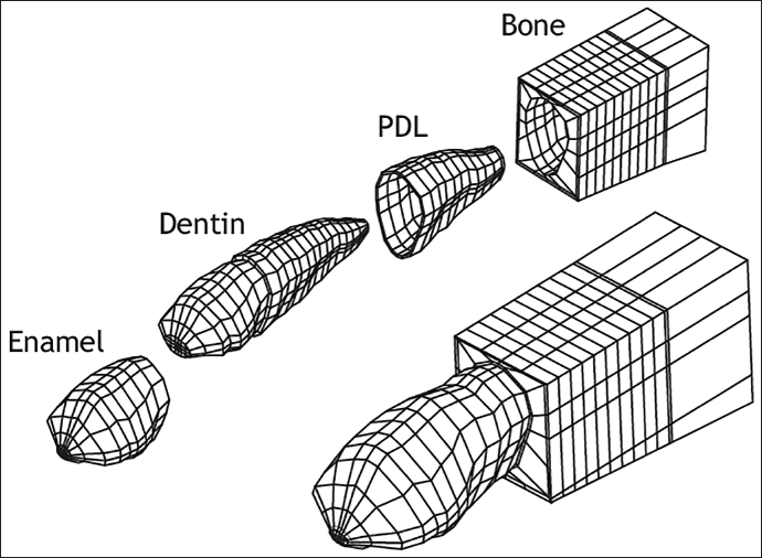
Discretization (meshing) of the structure (in Figure 1)
The complex geometry of the tooth–PDL–bone system is divided into small, finite elements (e.g., tetrahedral or hexahedral).
Each element is connected at discrete points called nodes.
This allows localized stress–strain analysis. 1
Assignment of material properties
Application of boundary conditions
The model is constrained to simulate anatomical fixation points (e.g., the cranial base or the alveolar bone).
Forces or moments representing orthodontic loads (from brackets, wires, or temporary anchorage devices (TADs)) are applied. 3
Formulation of element equations
For each element, mathematical equations are formulated that relate nodal displacements to applied loads using stiffness matrices.
These equations obey the laws of mechanics (Hooke’s law, equilibrium, and compatibility). 4
Assembly of the global stiffness matrix
All individual element equations are combined into a global system representing the entire structure.
This system reflects how all elements interact under applied forces. 5
Solution of equations
The global stiffness matrix is solved (usually numerically) to obtain nodal displacements.
From these, strain and stress values within each element are computed. 6
Visualization and interpretation
The computed stresses, strains, and displacements are visualized using color-coded 3D models.
Clinically, these results help interpret how orthodontic forces act on teeth, bone, and supporting structures. 7
Indications of FEA in orthodontics
FEA is indicated for evaluating orthodontic force systems, predicting tooth movement patterns, analyzing stress distribution in the PDL and alveolar bone, optimizing appliance design, assessing anchorage requirements, and comparing different treatment mechanics prior to clinical application.
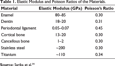
Elastic Modulus and Poisson Ratios of the Materials.
Finite Element Mesh of Tooth, Periodontal Ligament, and Alveolar Bone Used to Simulate Orthodontic Forces.
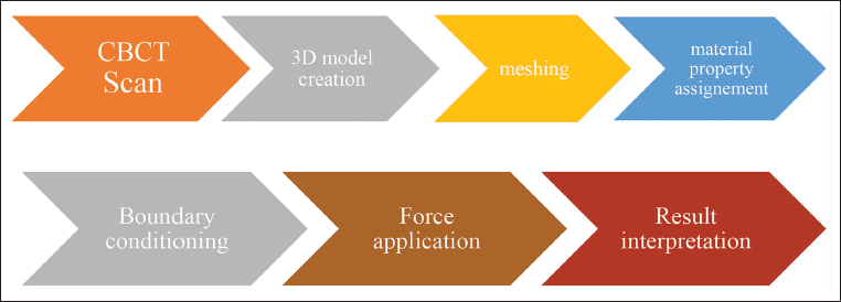
Steps Involving in Finite Element Analysis
The workflow of finite element analysis (FEA) in orthodontics. It begins with a CBCT scan to obtain accurate three-dimensional anatomical data of the patient, which is then used to create a digital 3D model of teeth, bone, and surrounding structures. This model is subdivided into small elements through meshing to enable mathematical analysis. Subsequently, appropriate material properties such as elasticity and stiffness are assigned to each tissue to simulate real biological behavior. Boundary conditions are then defined to restrict unwanted movements and stabilize the model, followed by the application of orthodontic forces with specified magnitude and direction. Finally, the results are interpreted in terms of stress distribution, strain patterns, and tooth displacement, providing insights into the biomechanical response of dental structures under applied forces (Figure 2).
Sequential Steps Involved in Finite Element Analysis of Orthodontic Systems, from Image Acquisition to Stress Visualization.
Applications of FEA in Orthodontics
Tooth Movement Biomechanics
FEA helps understand how forces and moments act on teeth and the PDL. 1 Studies have demonstrated stress concentration in the cervical and apical areas during tipping and bodily tooth movement.
How different bracket-slot sizes and archwire dimensions influence tooth displacement and stress distribution was further analyzed to determine their biomechanical effects. 7
Clinically, these findings help orthodontists predict areas of high stress during tooth movement and determine the optimal force magnitude for controlled and efficient orthodontic treatment.
Mini-implants and Anchorage Mechanics
FEA models are used to evaluate stress patterns around TADs. It also analyzes the effect of implant diameter and cortical bone thickness on implant stability. 8 Optimal placement angles can also be predicted using FEA. 9
Mini-implant stability improves when the implant is placed perpendicularly (90°) with a larger diameter (1.6–2 mm) and greater length (6–10 mm). Increasing these parameters reduces stress on both bone and implant. Angulated insertion increases stress due to a lever-arm effect. Since this was an in vitro study, more in vivo research is needed to understand biological responses.
These findings assist clinicians in selecting appropriate mini-implant dimensions and insertion angles to enhance primary stability.
Orthodontic Brackets and Archwires
Stress and frictional forces between different bracket–archwire systems have been simulated using FEA to optimize force transmission. 10
Comparisons between self-ligating and conventional brackets have also been made to study frictional resistance.
Such simulations guide clinicians in selecting bracket–archwire systems that minimize friction and optimize biomechanical efficiency.
Orthognathic Surgery and Skeletal Anchorage
FEA assists in evaluating postoperative stress distribution on fixation plates and screws, predicting the risk of failure. 11 It helps simulate skeletal displacement in orthognathic procedures for precise planning.
Clear Aligners and Custom Appliances
Recent studies have applied FEA to analyze the predictability of aligner-induced tooth movement. 12 Patient-specific aligner designs and force optimization can be achieved through iterative simulation.
This improves the predictability of aligner-induced movements and aids in attachment design customization.
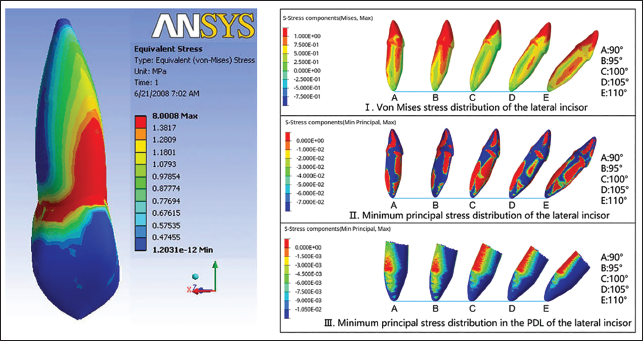
Biomechanical Evacuation of Orthodontic Force Effects Using Finite Element Analysis
Finite element analysis can be used to study the biomechanical response of a tooth and its supporting structures under different orthodontic force directions. By simulating forces at various angulations, the model helps visualize how stress is distributed within the tooth and periodontal ligament, allowing clinicians to understand how changes in force application influence tooth movement and tissue response. This approach provides a scientific basis for optimizing force systems to achieve efficient and safe orthodontic treatment (Figure 3).
Stress Distribution Pattern in the Periodontal Ligament During Orthodontic Tooth Movement Obtained Using Finite Element Analysis (FEA).
Advantages of FEA
A non-invasive and repeatable method.
Enables three-dimensional visualization of stress and strain patterns.
Allows parametric analysis of force magnitude, direction, and appliance design.
Reduces ethical and practical limitations associated with animal or human experimentation.
Limitations of FEA
Biological tissues are often modeled as linearly elastic, which does not fully represent the viscoelastic and anisotropic properties of the PDL and bone. 13 Simplified geometries and boundary conditions may not accurately reflect in vivo environments. Therefore, validation of FEA results with clinical and experimental studies remains essential.
Future Directions
Future advancements in FEA include the development of patient-specific models using cone-beam computed tomography (CBCT) and intraoral scans, integration with artificial intelligence and machine learning for predictive modeling, time-dependent simulations incorporating bone remodeling and PDL viscoelasticity, and the use of digital twin systems for orthodontic treatment planning and monitoring.
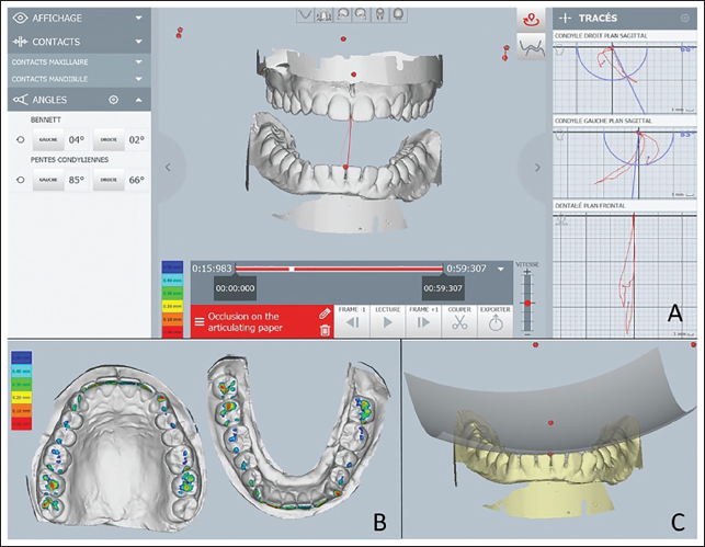
Advanced FEA studies analyzing how jaw movements and model detail influence stress and tooth behavior in dental structures (Figure 4).
Frontiers Finite Element Analysis of Dental Structures: The Role of Mandibular Kinematics and Model Complexity. (A) Digital Analysis of Mandibular Movement and Condylar Paths; (B) Color-mapped Occlusal Contact Distribution on Dental Arches; (C) Virtual Articulation Showing the Maxillomandibular Relationship.
Software Used in FEA in Orthodontics
Imaging and model generation software
Mimics
Dolphin Imaging
3Shape Dental System
Blue Sky Plan
FEA software
ANSYS
ABAQUS
COMSOL Multiphysics
HyperMesh
Orthodontic treatment planning software
Dolphin Orthodontics
OnyxCeph3D
ClinCheck
Conclusion
FEA is an advanced computational tool that bridges theoretical biomechanics and clinical orthodontic practice. By accurately simulating orthodontic forces and their effects on teeth and supporting structures, FEA aids in optimizing appliance design, improving treatment mechanics, and enhancing precision in orthodontic care. With ongoing advancements in digital imaging and computational technologies, FEA is expected to play an increasingly significant role in evidence-based and individualized orthodontic treatment.
Footnotes
Declaration of Conflicting Interests
The authors declared no potential conflicts of interest with respect to the research, authorship, and/or publication of this article.
Ethical Approval
Not applicable.
Funding
The authors received no financial support for the research, authorship, and/or publication of this article.
Informed Consent
Not applicable.