
Abstract
Inositol hexakisphosphate kinases (IP6Ks) regulate a myriad of cellular processes, not only through their catalytic activity (which synthesizes InsP7, a multifunctional inositol pyrophosphate signaling molecule) but also through protein–protein interactions. To further study the enzymatic function and distinguish between these different mechanisms, specific inhibitors that target IP6K catalytic activity are required. Only one IP6K inhibitor is commonly used: N2-(m-(trifluoromethyl)benzyl) N6-(p-nitrobenzyl)purine (TNP). TNP is, however, compromised by weak potency, inability to distinguish between IP6K isoenzymes, off-target activities, and poor pharmacokinetic properties. Herein, we describe a new inhibitor discovery strategy, based on the high degree of structural conservation of the nucleotide-binding sites of IP6Ks and protein kinases; we screened for novel IP6K2 inhibitors using a focused set of compounds with features known, or computationally predicted, to target nucleotide binding by protein kinases. We developed a time-resolved fluorescence resonance energy transfer (TR-FRET) assay of adenosine diphosphate (ADP) formation from adenosine triphosphate (ATP). Novel hit compounds for IP6K2 were identified and validated with dose–response curves and an orthogonal assay. None of these inhibitors affected another inositol pyrophosphate kinase, PPIP5K. Our screening strategy offers multiple IP6K2 inhibitors for future development and optimization. This approach will be applicable to inhibitor discovery campaigns for other inositol phosphate kinases.
Keywords
Introduction
Inositol pyrophosphates, such as 5-diphosphoinositol pentakisphosphate (InsP7), regulate many cellular processes, although most attention is given to their actions at the interface of cell signaling and bioenergetic metabolism. 1 InsP7 is synthesized by inositol hexakisphosphate kinases (IP6Ks); mammalian cells express three of these: types 1, 2, and 3. IP6K1 and IP6K2 are ubiquitously expressed, whereas IP6K3 expression is mainly restricted to the cerebellum and skeletal muscle. 2 Genetic experiments in mice have revealed that IP6Ks have several non-overlapping functions. For example, only the IP6K1 knockout displays low body weight, low insulin levels, male sterility, 3 protection from thrombotic challenge, 4 and lower weight gain on a high-fat diet. 5 These studies, and other work,6,7 suggest that inhibition of IP6K1 could be of therapeutic benefit in treating diabetes, obesity, and thrombosis. It has also been reported that IP6K2 promotes cancer cell migration, invasion, and tumor metastasis via inactivation of the tumor suppressor liver kinase B1 (LKB1). 8 Therefore, inhibitors of IP6K2 offer promise as new cancer therapeutics.
Chemical probes that inhibit IP6Ks could also be used as research tools for functional characterization of their kinase activities, and also to distinguish this kinase-directed mechanism from separate, noncatalytic roles mediated by protein–protein interactions. Currently, only one IP6K inhibitor is in routine use: N2-(m-(trifluoromethyl)benzyl) N6-(p-nitrobenzyl)purine (TNP). This compound is, however, compromised by weak (low-micromolar) potency, inability to distinguish between different IP6K isoenzymes, and off-target liabilities. 6
A recent study by Wormald et al. 9 described how IP6K assays that monitor adenosine triphosphate (ATP) consumption can be developed and used for compound-screening campaigns. This group derived proof-of-principle of their approach using an annotated set of 1280 compounds, the Library of Pharmacologically Active Compounds (LOPAC). At least one pharmacological activity is known for each of these compounds, and the main purpose of this library is to examine the performance of a high-throughput assay, rather than to identify tractable inhibitors. 9 A successful screening exercise, however, depends on identification of chemically tractable hit molecules. One approach to reach this goal, in an efficient manner, lies in the curation and application of smaller, focused libraries with functionally and/or chemically related properties. 10
To support our selection of focused compound sets, we describe here how we first compared the conserved core structure of an IP6K (from Entamoeba histolytica; PDB: 4O4F) with that of protein kinase A (PDB: 1L3R), and we found the nucleotide-binding sites to exhibit a substantial degree of similarity. Therefore, we reasoned that a focused screen using molecules known to have features of protein kinase inhibitors would be a potentially successful approach. Thus, we screened human IP6K2 with two focused compound sets: a 5000-kinase library (“5K library”) from the Center for Integrative Chemical Biology and Drug Discovery, University of North Carolina (UNC CICBDD), 11 and the GSK Published Kinase Inhibitor Set (PKIS). 12 We identified several novel hits for IP6K2, which showed specificity over PPIP5K, another inositol pyrophosphate kinase.
Materials and Methods
Protein Expression and Purification
The catalytic domains of recombinant human PPIP5K2 13 and full-length recombinant human IP6K2 14 were prepared as described previously. The purity of these proteins was estimated to be >90% as judged by SDS-PAGE (sodium dodecyl sulfate–polyacrylamide gel electrophoresis). The purified proteins were concentrated to between 1 and 10 mg/ml and stored at −80 °C.
IP6K2 and PPIP5K TR-FRET Assays for Adenosine Diphosphate (ADP) Detection
ATP-driven kinase activity was measured by detecting ADP formation from substrate phosphorylation using the Adapta Universal Kinase Assay (Thermo Fisher Scientific, Waltham, MA, USA). The enzyme reaction was performed using optimized final conditions of 400 nM IP6K2, 10 μM ATP, and 10 μM InsP6; and 500 nM PPIP5K, 10 μM ATP, and 10 μM 5-InsP7. All assays were performed in 384-well plates in kinase reaction buffer: 50 mM HEPES pH 7.5, 0.01% Brij-35, 10 mM MgCl2, and 1 mM EGTA. Reaction conditions (additional details below) were selected such that displacement of the ADP tracer from the antibody was 70–80% (according to the manufacturer’s guidelines).
Compound Screening
Compounds from a kinase-focused library of 4727 molecules (the “5K library”) and from the PKIS of 843 molecules were screened at 10 µM and 1 µM, respectively, using a Mosquito (TTP Labtech, Melbourn, UK) to dispense 50 nL compound (in DMSO) from the stock plates (1 mM for the 5K library and 0.1 mM for PKIS) in 384-well assay plates. A Multidrop Combi Reagent Dispenser (Thermo Fisher Scientific) was used to dispense 2.5 µL of (2×) kinase to the assay plates, then incubated for 20 min at room temperature, and this was followed by addition of 2.5 µL of (2×) ATP and substrate to a final concentration of 10 µM, as stated above. The enzymatic reaction was performed for 30 min, and the amount of ADP produced was detected by adding 2.5 µL of detection solution Adapta Eu-anti-ADP antibody, Alexa Fluor 647 ADP tracer, and EDTA for a final concentration of 2 nM, 12 nM, and 10 mM, respectively. After an additional 30 min equilibration period, the plate was read on an EnVision (Perkin Elmer, Waltham, MA, USA) plate reader (excitation, 320 nm; emission, 665 nm and 615 nm). The HTRF signal was calculated as a ratio of the signals from the 665 nm (acceptor) and 615 nm (donor) channels.
IC50 Determination
Inhibitors (threefold serial dilutions, 10 points) were incubated for 10–20 min at room temperature with each kinase prior to addition of substrates at the concentrations, and the enzymatic reaction was performed as described above. Percent inhibition was calculated on a scale of 0% (i.e., activity with DMSO vehicle only) to 100% (no enzyme added) using full column controls on each plate. The interquartile mean of 16 wells of each control was used per plate. ADP titration curves were routinely run on all compound-screening and IC50 plates to assure that the enzyme assays produced near 20% maximum ATP turnover.
TNP Synthesis
A suspension of 6-chloro-2-fluoro-9H-purine (200 mg, 1.0 mmol) in DMF (2.0 mL) was added to 4-nitro benzylamine (300 mg, 1.0 mmol) and triethylamine (200 mg, 2.0 mmol). The reaction mixture was heated at 90 °C under microwave irradiation for 20 min, quenched with water, and extracted with ethyl acetate. The organic layer was concentrated. The crude product was washed with 10 mL MeOH and filtered to yield the desired product, 2-fluoro-N-(4-nitrobenzyl)-9H-purin-6-amine (150 mg, 50%), which was used without further purification. A suspension of 2-fluoro-N-(4-nitrobenzyl)-9H-purin-6-amine (350 mg, 1.2 mmol) in (3-(trifluoromethyl)phenyl)methanamine (1.5 mL) was heated at 155 °C for 2 h. Silica was added to the reaction mixture. The mixture was loaded directly to an ISCO column and purified using a mixture of hexanes and ethyl acetate, followed by another purification on pre-HPLC (high-performance liquid chromatography) to provide the desired product, TNP (150 mg, 28%). 1 H NMR (400 MHz, CD3OD) δ 8.11 (s, 1H), 8.08–7.98 (m, 2H), 7.55–7.34 (m, 6H), 4.84 (s, 2H), and 4.68 (s, 2H).
HPLC Assay of IP6K2 Activity
The base buffer for assaying IP6K2 activity was 100 mM KCl, 16.5 mM NaCl, 20 mM HEPES pH 7.2, 8 mM MgCl2, 1 mM EDTA, and 10 μM [3H]InsP6 (15,000 dpm). Two alternative concentrations of ATP were added, either 5 mM or 50 μM, with the quantity of IP6K2 adjusted to 30 nM or 100 nM, respectively. Assays were run for 15 min at 37 °C, and then quenched, neutralized, and assayed by strong anion exchange HPLC, as previously described. 14
Results and Discussion
A recent report describes the development of a high-throughput bioluminescence assay (ADP-Glo; Promega, Madison, WI, USA) for IP6K activity. 9 Here, we report the development of an alternative enzymatic assay for IP6K2 and PPIP5K. We used a TR-FRET readout (Adapta; Thermo Fisher Scientific) that detects ADP resulting from phosphate transfer to substrate from ATP; ADP is detected with a europium-labeled anti-ADP antibody, which displaces an acceptor-labeled ADP ( Figure 1A ). The TR-FRET format protects against false positives due to the time-resolved nature of the fluorescence readout. In addition, the ratiometric readout for TR-FRET (acceptor/donor fluorescence) enhances assay robustness. Therefore, the TR-FRET assay is particularly well suited for screening campaigns and compound characterization. 15
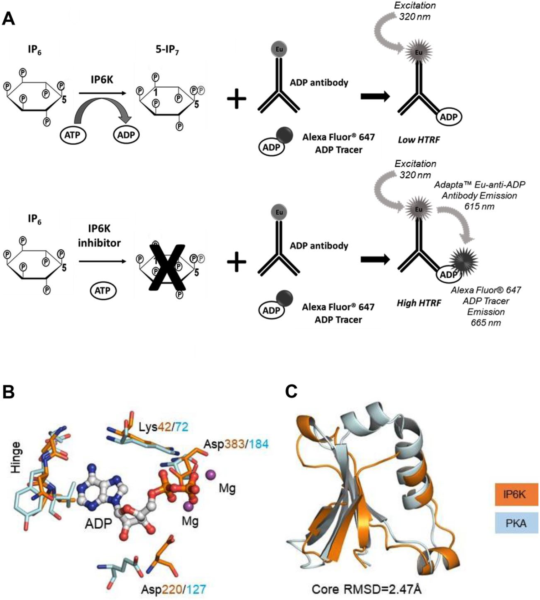
The inositol hexakisphosphate kinase type 2 (IP6K2) screening strategy. (
We paid particular attention to the choice of a compound library for screening. Our decision was based on the following structural analysis: Using the structure of EhIP6K as a template, we constructed a homology model
14
of the ATP-binding site of human IP6K2, which we compared with the corresponding region of a prototypical protein kinase [protein kinase A (PKA);
Our screening assay was designed to preferentially identify substrate competitive inhibitors by using substrate concentrations significantly lower than the Km value. Whereas previous studies suggest the IPK Km for ATP is near 1 mM, 17 we chose an ATP concentration of 10 µM ATP. This low concentration also enabled us to reliably quantitate the amount of ADP formed in the enzyme reaction, given the affinity of the anti-ADP antibody and its degree of selectivity over ATP. We chose conditions that would allow for a maximum of approximately 20% conversion of ATP to ADP during the time course of the reaction. This low turnover assured that the reaction remained in the linear, initial velocity phase (data not shown).
We screened both IP6K2 and PPIP5K with the protein kinase–focused sets, and overall the screen performed well with an average signal-to-background ratio of ~2 and average Z-prime value >0.8 ( Fig. 2 ). No hits were identified for PPIP5K. For IP6K2, of the 5570 compounds screened, 96 compounds yielded >50% inhibition (1.7% hit rate) ( Fig. 2 ). We discarded 22 of these compounds because they altered europium donor emission by more than two standard deviations from the mean. That is, approximately 0.4% of the hits in the primary screen were removed due to quenching of donor fluorescence alone. The remaining 74 hits were retested with dose–response curves (see below).
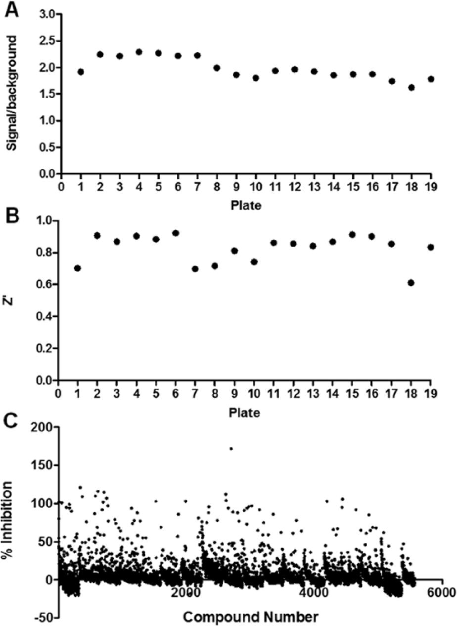
Assay performance for the inositol hexakisphosphate kinase type 2 (IP6K2) screening campaign. The kinase-focused library from the Center for Integrative Chemical Biology and Drug Discovery, University of North Carolina (UNC CICBDD) (5K) (plates 1–16), and the GSK Published Kinase Inhibitor Set (PKIS) (plates 17–19) were screened using the Adapta Universal Kinase Assay (Thermo Fisher Scientific). (
We also evaluated the IP6K inhibitor, TNP, as a positive control during screening and dose–response follow-up. Notably, we found TNP to have an IC50 value of 7.36 µM for IP6K2 ( Fig. 3A ). This is significantly weaker than the IC50 value of 0.43 µM (Ki value of 0.24 µM) previously reported for IP6K1. 18 To confirm our result, we resynthesized TNP to produce a highly pure compound and obtained the same IC50 value as with commercially obtained material. Note that our IC50 value for TNP is close to that previously reported using the ADP-Glo Max assay (Promega). 9
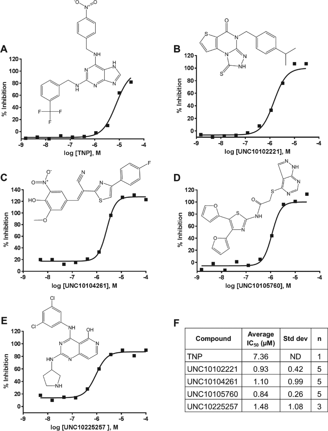
Representative dose–response curves for inhibition of inositol hexakisphosphate kinase type 2 (IP6K2) by (
We found that 46 of our initial hits were validated in secondary screening (i.e., IC50 < 5 µM). The dose–response curves for four illustrative hit compounds, plus TNP, are shown in Figure 3 . One of these, UNC10225257 from the PKIS compound library, is a quite promiscuous inhibitor that targets >80 kinases with >90% inhibition at 1 µM. 12 Thus, it is unlikely this would be an optimum compound for further development. Reassuringly, none of the confirmed hits for IP6K2 inhibited PPIP5K. Generally, we did not observe significant structural similarities among the top 46 hits (other than noting they contained the kinase “hinge-binding” motif). 16 Having a number of distinct chemotypes for future follow-up work is an encouraging outcome from our screening efforts.
To further validate our hits for IP6K2, we either repurified the compound (UNC10102221) or remade fresh solutions from solid stocks (UNC10104261 and UNC10105760), and performed an orthogonal assay; we used HPLC to monitor metabolism of radiolabeled InsP6 substrate. This is a laborious and time-intensive assay, so we only provide data with two inhibitor concentrations, 1 µM and 10 µM, and two ATP concentrations, 50 μM and 5 mM. As shown in
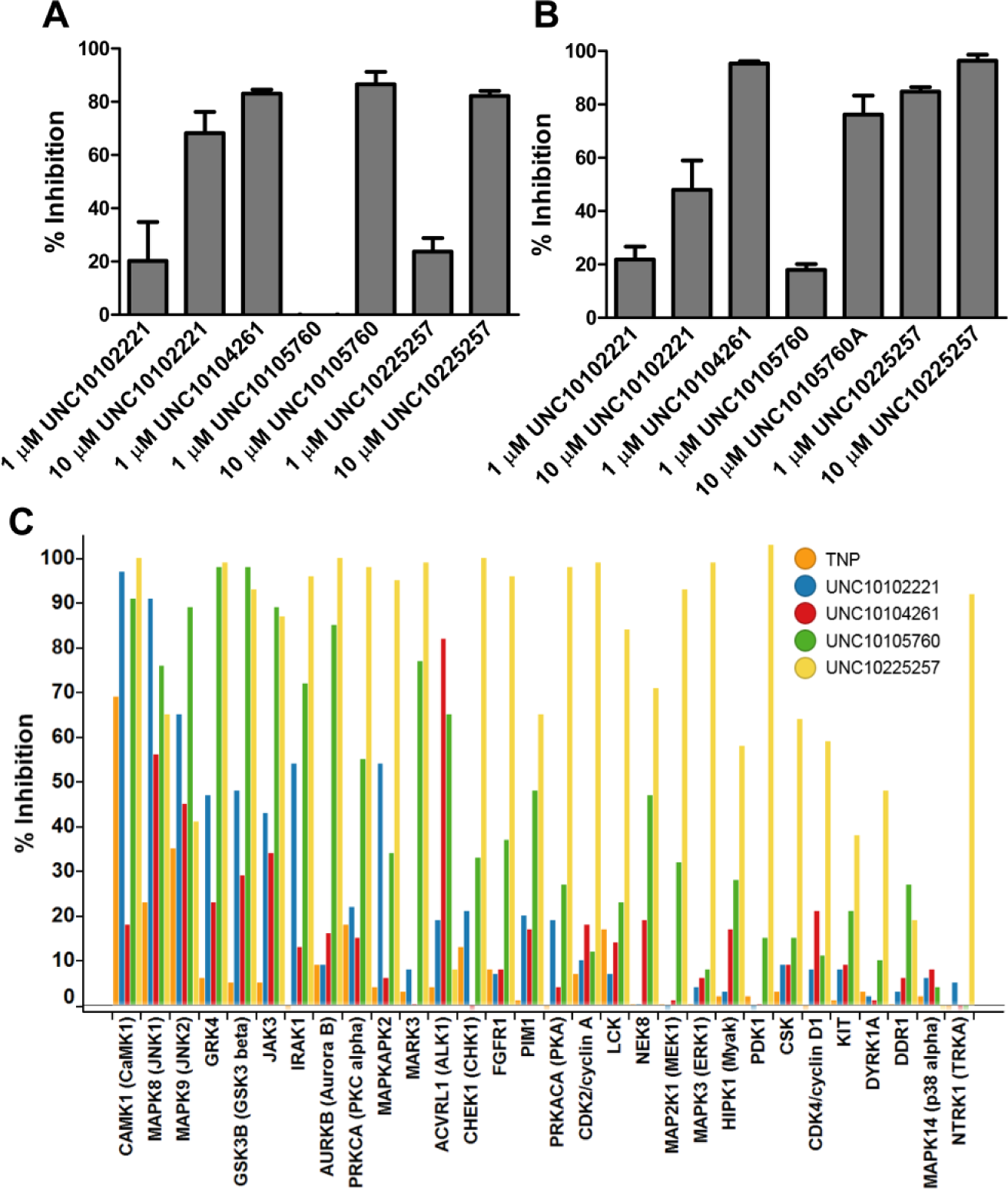
Evaluation of selected inositol hexakisphosphate kinase type 2 (IP6K2) inhibitors by high-performance liquid chromatography (HPLC) and protein kinase selectivity assays. Assays were performed as described in the Materials and Methods section using either (
A key development in this study has been our structure-based evidence that IP6K2 may be targeted by compounds resembling protein kinase inhibitors ( Fig. 1 ). This has allowed us to conduct an efficient screening campaign; we have used relatively small, focused libraries containing molecules that have features that are either known or computationally predicted to target a protein kinase nucleotide-binding site. This new approach is validated by our identification of several novel inhibitors of IP6K2 with IC50 values in the micromolar range. These results also suggest that commercially available kinase-profiling panels, such as those supplied by CarnaBio, DiscoveRx, Eurofins, ReactionBiology, and Thermo SelectScreen, should include IP6Ks and perhaps other inositol phosphate kinases. Although most of these kinase panel screening services do contain inositol lipid kinases (e.g., PI3K, PI4K, and PIP5K1), these are a functionally and structurally different class of proteins from the inositol phosphate kinases. Overall, our targeted library approach could also prove useful for inhibitor discovery campaigns against other inositol phosphate kinases.
Footnotes
Acknowledgements
We thank Daowei Huang for help with confirming TNP via nuclear magnetic resonance (NMR).
Declaration of Conflicting Interests
The authors declared no potential conflicts of interest with respect to the research, authorship, and/or publication of this article.
Funding
The authors disclosed receipt of the following financial support for the research, authorship, and/or publication of this article: This research was funded by the National Institutes of Health under grant R01 DK101645.