
Abstract
Trichobezoar is a rare disorder of the young girl with a psychiatric profile. It is suspected when there is a notion of trichophagia, although its clinical manifestations are non-specific. Imaging through CT scan, plays a fundamental role in the diagnosis of extensive forms.
Keywords
Introduction
Trichobezoar is the presence of hair clumps in the gastrointestinal tract. It is the most common type of bezoar. It is a rare disease usually found in female subjects with a psychiatric background. The clinical presentation of this condition is polymorphic.
We illustrate through this case a type of presentation of trichobezoars.
Observation
A 16-year-old girl was admitted to the emergency department for epigastralgia of progressive onset associated with nausea and vomiting.
The history revealed a mention of trichophagia and no psychiatric follow-up. The physical examination showed abdominal distension in a hemodynamically and respiratory stable patient with discolored conjunctiva. However, there was no fever or jaundice.
The rest of the somatic examination was unremarkable.
Biologically, there was a moderate hyperleukocytosis due to neutrophils. There was also a hypochromic microcytic anemia and elevation of CRP, while the rest of the biological test was normal. An abdominal CT scan was performed (Figure 1).

Axial slice abdominal CT scan at portal venous phase showing an intra-gastric mass (star) distinct from the gastric wall, heterogeneous iso-dense by the presence of air bubbles and not enhanced by iodinated contrast. The diagnosis of trichobezoars was retained and the patient was scheduled for surgery.
Intraoperative exploration confirmed this diagnosis (Figure 2).
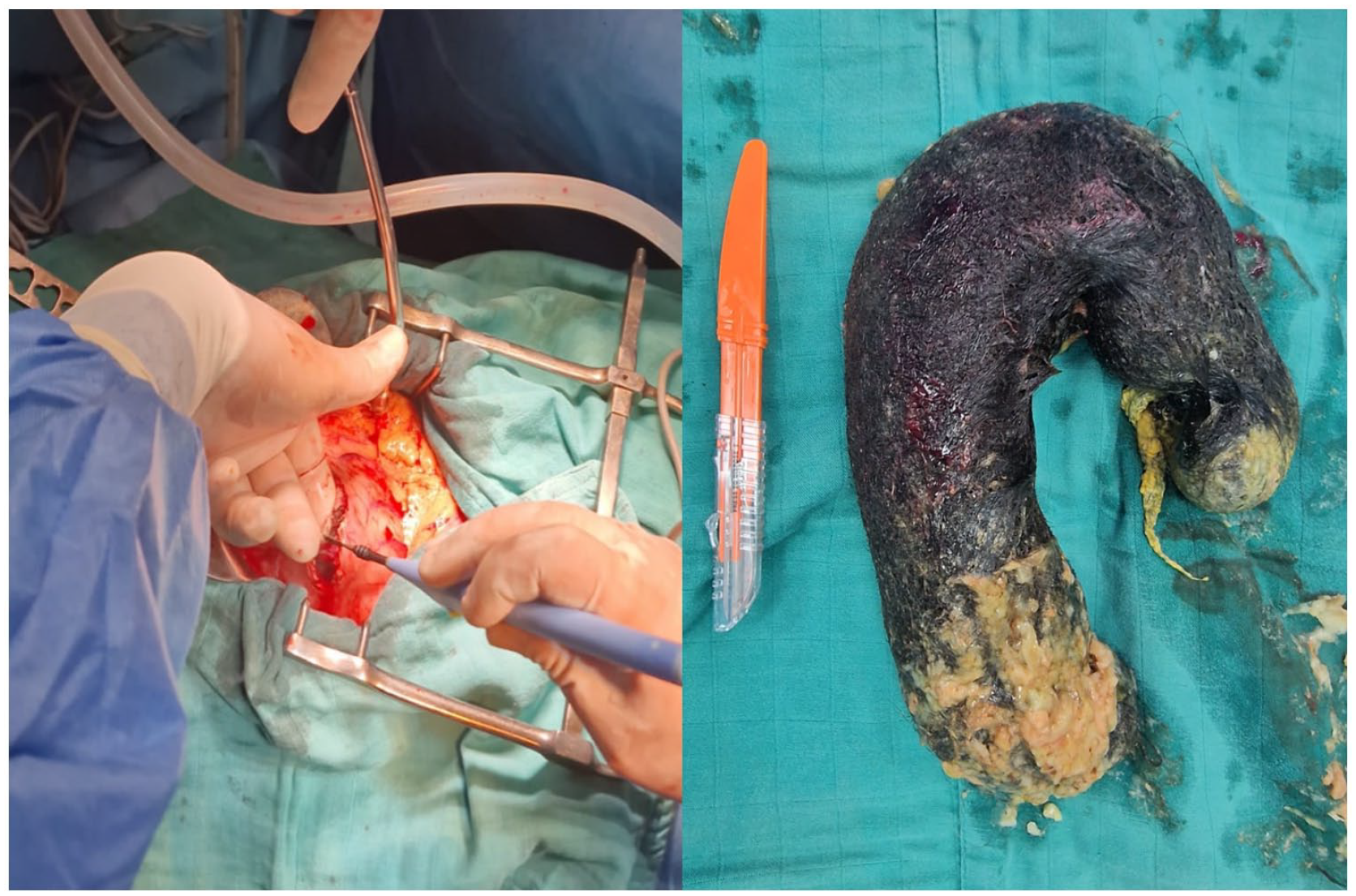
Operating part for a trichobezoar.
Discussion
Bezoar is a condition described in young girls (90%), often between 10 and 19 years of age. 1 It corresponds to the formation of a concretion or mass in the digestive tract as a result of an accumulation of various substances. Based on the type of aggregated substance, a distinction is made between: trichobezoar (hair or wool or paper mache), phytobezoar (vegetable fibers), lactobezoar (curdled milk), and pharmaco-bezoar (drugs). 2
Hair is found in more than half of the cases of bezoar with a predilection for the stomach and can extend to the duodenum and even beyond the angle of treitz, thus creating the classic “Rapunzel syndrome.” 3
Several disorders intervening at different levels would favor the occurrence of this condition. They can occur as a result of poor dietary selection (diet rich in fiber, ingestion of food difficult or not digestible), the presence of psychiatric disorders (depression, intellectual disability, behavioral disorders), digestive abnormalities (insufficiency of mastication, disorder of gastric and duodenal motility, gastric hypo acidity due to an antisecretory treatment), or partial gastrectomy. 4 This disease is manifested by various and non-specific symptoms such as: abdominal pain, nausea, vomiting, skin, and mucous membrane pallor which were found in our patient. Mechanical alopecia and weight loss can also be found.
In some cases, complications such as digestive hemorrhage, occlusion, and perforation reveal the disease.
Moreover, these clumps alter digestion, intake, and absorption of nutrients responsible for a malabsorption syndrome translated essentially on the biological level by a moderate microcytic hypochromic anemia. 5
Endoscopy is the first examination carried out when there is a suspicion of localized bezoar and has the advantage of being diagnostic and therapeutic, while for extensive forms, imaging through abdominal CT is of great help in assessing the extension. It may reveal a gastric mass more or less extended to the rest of the digestive tract without heterogeneous parietal attachment due to the presence of air bubbles within it and not enhanced after injection of iodinated contrast. On magnetic resonance imaging, the trichobezoar appears as a mass with variable signal in T1 and T2 weighting and not enhanced after injection of gadolinium. 6
The management of the patient aims at extracting the foreign body by endoscopy (minimal form) or surgery (extensive form) and also at correcting the carinal syndrome without forgetting the psychiatric aspect (referral to a psychiatric consultation to manage a possible depression or an eating disorder).
In this case, the patient presented with a bezoar taking all the stomach, which was extracted by surgery through a median supra umbilical gastrotomy under general anesthesia. The postoperative care was normal and the patient was referred to a psychiatrist.
Conclusion
Trichobezoar is a rare disease whose diagnosis is suspected in a young patient in the presence of a report of trichophagia. It is confirmed by endoscopy in the minimal forms and imaging through abdominal CT especially in the advanced forms.
Footnotes
Acknowledgements
I would like to express my gratitude to my professors and all the colleagues who participated in the completion of this work.
Author Contributions
ID: conception of the work, design of the work, and acquisition of data; W-YT: acquisition of data; AZ: acquisition of data; EM: acquisition of data; NB: revising the work critically for important intellectual content; IN: revising the work critically for important intellectual content.
Declaration of Conflicting Interests
The author(s) declared no potential conflicts of interest with respect to the research, authorship, and/or publication of this article.
Funding
The author(s) received no financial support for the research, authorship, and/or publication of this article.
Ethics Approval
Our institution does not require ethical approval for reporting individual cases or case series.
Informed Consent
Written informed consent was obtained from a legally authorized representative for anonymized patient information to be published in this article.