
Abstract

In new drug discovery research, pharmacokinetic study is very important to rationalize in vivo efficacy and to evaluate the most suitable dose. We usually analyze the drug concentration in blood or homogenates of animals by manual liquid phase extraction methods. However, it is necessary to routinely handle many samples and the work is troublesome. Thus, we planned the automation of the analysis process.
We identified four key requirements for the development of a new apparatus as follows;
Compatible with upper and lower layer extraction
Fully automated from sampling to HPLC analysis
User-friendly setting and monitoring
Compact and extensible system
We designed the automated processes for upper and lower layer extraction, as shown in Figure 1 and 2, based upon the corresponding manual treatments.

Automated process in liquid phase extraction methods for plasms concentration analysis (upper layer extraction).
In the case of upper layer extraction (Figure 1), sampling is performed with disposable pipettes in order to prevent contamination, buffer solution and organic solvent are added to the sample containing the target drug, the mixture is vibrated and then centrifuged to separate the organic phase and the aqueous phase completely, and the isolation of the organic phase is carried out with a disposable pipette. The obtained extract is evaporated with a nitrogen gas flow and heating to give a residue which is then dissolved in a small amount of solvent for HPLC. This solution is then injected to the HPLC system. Finally, the concentration of the target drug in the sample is calculated by the host computer.
On the other hand, lower layer extraction can be accomplished with an invented extraction device which consists of a cylinder cap and a glass tube which has internal tube (Figure 2). When the cylinder cap is inserted to the glass tube in which has been added the sample, buffer solution, and an organic solvent such as chloroform, the liquid mixture in the internal tube will be removed. After vibration and centrifugation, the cylinder cap is raised up gradually, so that the only lower organic layer will rise up in the internal tube. Thus, a disposable pipette can remove the organic layer without contamination by the upper layer. The obtained organic layer is then will be treated in a similar manner to that described for the upper layer extraction. The invented analyzer is shown in Figure 3. It consists of three units, a liquid extraction device (the upper pan of the apparatus), a HPLC system (the lower part of the apparatus) and a host computer which is operated by Windows 3.1 and can control 4 machines at once. The extraction device can be set with up to 48 sample tubes. The analysis conditions (number of samples, sampling volume, extraction solvents, etc.) can be inputed from the host computer or a touch-panel in the apparatus. Monitoring of the operation can achieved by the host computer.

Automation process in the case of lower layer extraction.
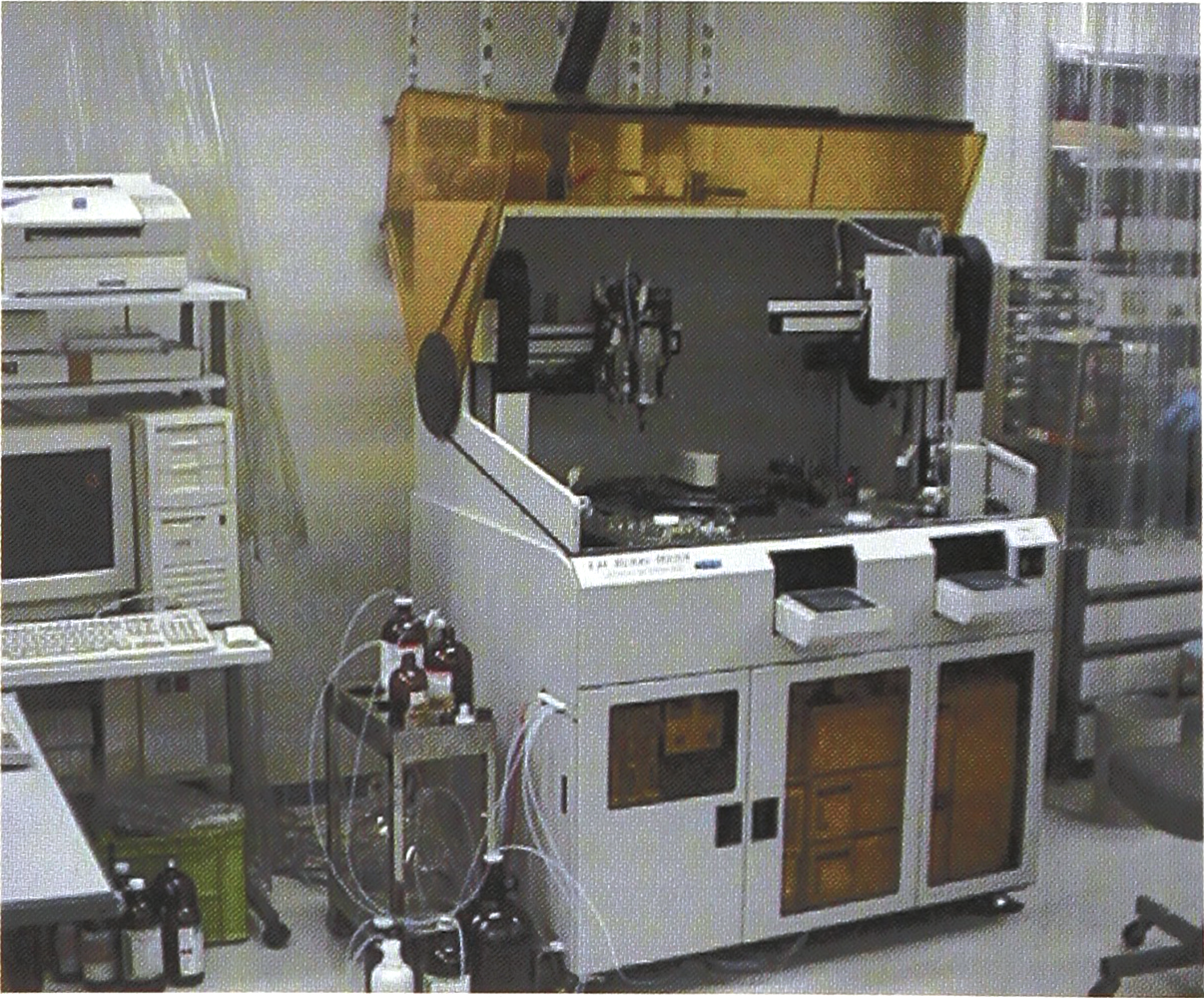
The invented analyzer consists of three units, a liquid extraction device (the upper part of the apparatus), a HPLC system (the lower part of the apparatus) and a host computer
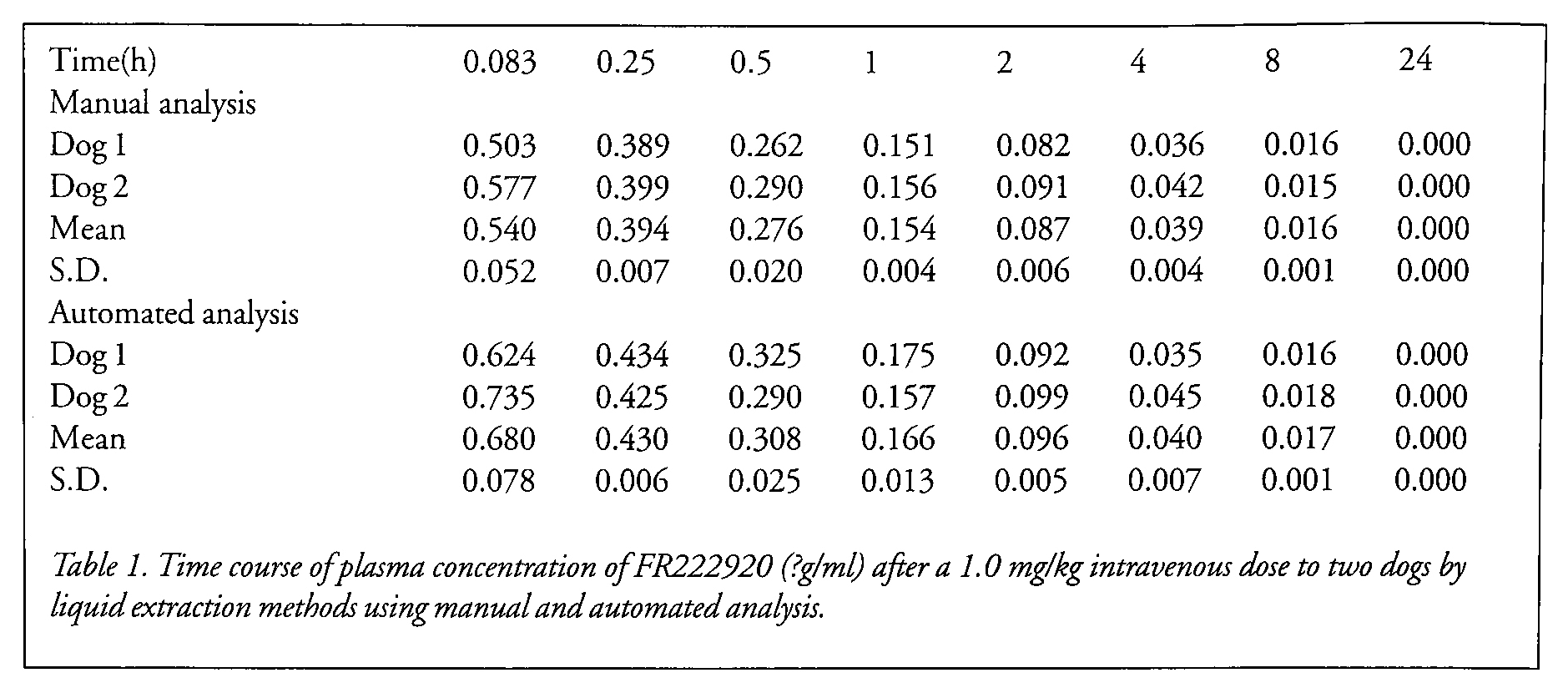
As a demonstration, we compared automated analysis with manual analysis by using the same dog plasma samples which contained an investigative drug, FR222920 (internal research code number). As a result, we could confirm that the developed apparatus showed good performance, as shown in Table 1.
Time course of plasma concentration of FR222920 (?g/ml) after a 1.0 mg/kg intravenous dose to two dogs by liquid extraction methods using manual and automated analysis.
This developed analyzer may become the first automated apparatus on sale in Japan, and makes it possible to release pharmacological researchers from monotonous and repetitious work and brings accurate pharmacokinetics data to the researchers in a timely fashion.
EXPERIMENTAL
PREPARATION OF PLASMA SAMPLES OF FR222920 FROM TWO DOGS:
Blood samples (2.5 ml) were collected from two dogs (beagle, male, 12 months) after a 1.0 mg/kg intravenous dose of FR222920 at the times of 0.083, 0.25, 0.5, 1, 2, 4, 8, and 24 hours after injection. 25μl of heparin sodium (1000 units/ml) was added to each obtained blood sample as an anticoagulants. The mixtures were centrifuged at 3000 r.p.m. for 5 minutes to obtain the plasma samples (1.0 ml).
MANUAL ANALYSIS OF PLASMA CONCENTRATIONS OF FR222920 IN TWO DOGS:
Each dog plasma sample (0.1 ml) was added to a mixture of mobile solvent (0.1 ml, acetonitrile: 20 mM aqueous potassium biphosphate = 65: 35), 0.1 M aqueous sodium hydroxide (0.5 ml), and extraction solvent (4.0 ml, isoarnyl alcohol: n-hexane = 2; 98), The mixture was shaken for 5 minutes, and centrifuged at 3000 r.p.m. (3000 G) for 5 minutes. 3.0 ml of the upper organic layer was separated with a pipette and evaporated by nitrogen gas flow to give a residue which was then shaken for 10 seconds to dissolve in 0.1 ml of the mobile phase. The mixture was centrifuged at 3000 r.p.m. and 80μl of the mixture was injected to the HPLC system. The conditions of analysis was as follows;

Each concentration of FR222920 in the plasma was calculated for the obtained data and a calibration curve.
AUTOMATED ANALYSIS OF PLASMA CONCENTRATIONS OF FR222920 IN TWO DOGS:
Each dog plasma sample (0.1 ml) was added to a mixture of a mobile solvent (0.1 ml, acetonitrile: 20 mM aqueous potassium biphosphate = 65: 35), 0.1 M aqueous sodium hydroxide (0.5 ml), and extraction solvent (3–5 ml, isoamyl alcohol: n-hexane = 2: 98). The mixture was vibrated at 3500 r.p.m. for 5 minutes, and centrifuged at 2500 r.p.m. (3000 G) for 5 minutes. 2.5 ml of the upper organic layer was separated and evaporated by nitrogen gas flow to give a residue which was then shaken for 10 seconds to dissolve in 0.1 ml of the mobile phase. The mixture was centrifuged at 3000 r.p.m. and 80μl of the mixture was injected to the HPLC system. The analysis was performed using the same conditions as the manual analysis.
Each concentration of FR222920 in the plasma was calculated for the obtained data and a calibration curve which could be obtained by the automated apparatus.
Footnotes
ACKNOWLEDGMENT
The authors wish to express thanks to Mr. Naotaka Sawada, Akiyoshi Banba, and Katsuhiko Saito of Dai-Nippon Seiki Co. Ltd. for helpful assistance in design and programming of the automated apparatus, and to Mr. Hiroyoshi Sakai for the drug concentration analysis under both manual and automated conditions.
1)
Yoshihiro Murakami, Shintaro Nishimura*, Akihiro Noda, Norihiro Harada, and Hideo Tsukada, Automated synthesis of 18F labeled FK960 (N-[4-acetyl-1-piperazinyl)-p-fluorobenzamide monohydrate): Journal of Labelled Compounds and Radiopharmaceuticals, Vol. 40, No.12, pp.1–3, 1997
2)
Shintaro Nishimura, Kazuyoshi Yajima, Norihiro Harada, Yasuhiro Ogawa and Nobuyoshi Hayashi, Automated synthesis of radiopharmaceuticals for PET: an apparatus for [1-11C]labelled aldoses, Journal of Automatic Chemistry, Vol. 16, No.6, pp. 195–204, 1994