
Abstract
There is a growing demand in the biopharmaceutical industry for high-throughput, large-scale N-glycosylation profiling of therapeutic antibodies in all phases of product development, but especially during clone selection when hundreds of samples should be analyzed in a short period of time to assure their glycosylation-based biological activity. Our group has recently developed a magnetic bead–based protocol for N-glycosylation analysis of glycoproteins to alleviate the hard-to-automate centrifugation and vacuum–centrifugation steps of the currently used protocols. Glycan release, fluorophore labeling, and cleanup were all optimized, resulting in a <4 h magnetic bead–based process with excellent yield and good repeatability. This article demonstrates the next level of this work by automating all steps of the optimized magnetic bead–based protocol from endoglycosidase digestion, through fluorophore labeling and cleanup with high-throughput sample processing in 96-well plate format, using an automated laboratory workstation. Capillary electrophoresis analysis of the fluorophore-labeled glycans was also optimized for rapid (<3 min) separation to accommodate the high-throughput processing of the automated sample preparation workflow. Ultrafast N-glycosylation analyses of several commercially relevant antibody therapeutics are also shown and compared to their biosimilar counterparts, addressing the biological significance of the differences.
Introduction
Since the discovery of the importance of protein glycosylation, the needs for novel sample preparation and glycoanalytical tools are rapidly emerging. 1 During the development of therapeutic proteins, the analysis of glycosylation is a key. Currently in the biopharmaceutical field, almost 400 of the more than 900 biotherapeutics are monoclonal antibodies (mAbs) with an estimated increase to $70 billion in business worldwide in 2015. 2 mAbs are highly complex molecules with a complicated higher order structure subject to important co- and posttranslational modifications, such as glycosylation. The therapeutic substance (the molecule itself) and drug product (the pharmaceutically formulated final product) are heterogeneous (i.e., a mixture of several slightly different structures, e.g., due to glycosylation variations). In the early stage of biopharmaceutical drug development, transfection allows many variants to be expressed, altogether representing a significant challenge for their identification. Therefore, there is a great demand for high-throughput sample characterization methods, especially glycosylation analysis. 3 Generally used techniques include nuclear magnetic resonance (NMR), mass spectrometry, chromatography, capillary electrophoresis (CE), and the combination of them. 4 Most of these methods require sample preparation and derivatization steps, including glycan release, derivatization, purification, and preconcentration. In addition, current protocols include numerous centrifugation and vacuum–centrifugation steps, making full automation of the process by liquid-handling robots a challenge. 5
One of the most commonly used enzymes to release the N-glycan moieties of glycoproteins is PNGase F [peptide-N4-(N-acetyl-beta-glucosaminyl) asparagine-amidase] digestion due to its reliable and specific cleavage capability under simple and mild conditions. 6 Traditionally, overnight digestion protocols are used at 37 °C, but efforts were taken to accelerate the enzymatic reaction by microwave irradiation, ultra-high-pressure cycling, or immobilized PNGase F in micro reactors. However, besides the significant gain in glycan release speed, these approaches are expensive and somewhat complicated, and by using harsh conditions, some sample degradation is inevitable. 7
In general, liquid phase carbohydrate analysis is performed by either high-pressure liquid chromatography (HPLC) or CE. Because sugars lack chromophore and fluorophore groups and, unless sialylated, acetylated, phosphorylated, or sulfated, do not possess charged moieties either, techniques like refractive index or electrochemical detection could be used, but their sensitivity and specificity are insufficient for low-level glycan analysis. Therefore, CE and LC both require chemical derivatization prior to separation. In this study, CE with laser-induced fluorescent detection was used for high-sensitivity glycan analysis. The released carbohydrates were tagged via reductive amination with 8-aminopyrene-1,3,6-trisulfonic acid (APTS). 8 The labeling reaction scheme is specific to the reductive end of the sugars; therefore, only one APTS molecule binds to one sugar molecule, allowing easy quantification ( Fig. 1 ).
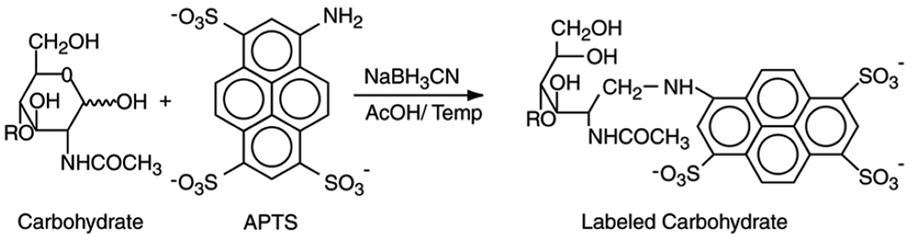
APTS labeling reaction of glycans. APTS, 8-aminopyrene-1,3,6-trisulfonic acid.
After the Schiff base formation, a reduction step is necessary to obtain a stable conjugate that is most commonly done by the addition of sodium-cyanoborohydride (NaBH3CN), but the use of 2-picoline-borane (pic-BH3) has also been suggested as an alternative. 9 Although the labeling reaction is well optimized and highly effective, in some instances silalic acid loss was observed 10 with a rate that was dependent on the reaction time and temperature. For sufficient labeling yield, the tagging reagent is used in great excess; therefore, highly efficient cleanup before CE analysis is of great importance. 11
Magnetic bead–mediated techniques have been widely used for decades in the fields of genomics and proteomics, mostly in cleanup and enrichment using special interaction-based capture mechanisms. 12 Carboxyl-coated magnetic microparticles, routinely used in nucleic acid purification protocols, can be used in a similar fashion for selective glycan capture via solid phase reversible immobilization (SPRI). Organic solvents, such as acetonitrile, force the carbohydrates to be crowded at the surface of the magnetic beads. In this way, both the remaining polypeptide chains after glycan release as well as the nonreacted excess ATPS can be readily removed from the reaction mixtures with high efficiency. 13 By the application of aqueous media, the captured glycans are released from the beads for further processing. 14
Applying this magnetic bead–based glycan sample preparation protocol with a laboratory automation workstation, a large number of samples was processed in 96-well plate format within a couple of hours, requiring no centrifugation or vacuum–centrifugation steps. Ultrafast separation (<3 min) of the resulting fluorophore-labeled glycans was accomplished by high-performance capillary electrophoresis with laser-induced fluorescent detection. The automated sample preparation and analysis method was applied to therapeutic antibodies of high biopharmaceutical importance.
Materials and Methods
Chemicals and Reagents
Water (HPLC grade), acetonitrile, immunoglobulin G, lithium acetate, acetic acid, and sodium-cyanoborohydride were obtained from Sigma-Aldrich (St. Louis, MO, USA). APTS and carbohydrate separation buffer (NCHO) were from SCIEX (Brea, CA, USA). The Agencourt CleanSeq magnetic beads were from Beckman Coulter (Brea, CA, USA), and the PNGase F enzyme and digestion reaction kit was from ProZyme (Hayward, CA, USA). The murine mAb standard was from Waters (Milford, MA, USA). The therapeutic antibody samples [innovator, biosimilar, and antibody drug conjugate (ADC)] were the kind gift of HLBS International (Seattle, WA, USA).
Capillary Electrophoresis
CE with laser-induced fluorescence detection (CE-LIF) was performed on a PA 800 Plus system (SCIEX). For rapid sample analysis, a 20 cm (effective length) NCHO capillary (30 cm total length, 50 µm ID) was filled with 1% PEO (MW 900,000; Sigma-Aldrich) solution in 25 mM lithium acetate buffer (pH 4.75). The separation voltage was 30 kV in reverse polarity mode (cathode at the injection side and anode at the detection side), resulting in E = 1000 V/cm electric field strength. The samples were pressure injected by 3 psi (20.68 kPa) for 6 s. The 32 Karat (version 9.1) software package (SCIEX) was used for data acquisition and analysis.
Automation for Large-Scale Sample Processing
Fully automated sample preparation was performed on a Biomek FXP Laboratory Automation Workstation (Beckman Coulter), which was set up with 96-well plate holders, a magnetic stand, 1000 µL and 25 µL pipette tip holders, a quarter reservoir, along with sample and reagent vials (
Table 1
Labware Used for Glycan Analysis Automation.
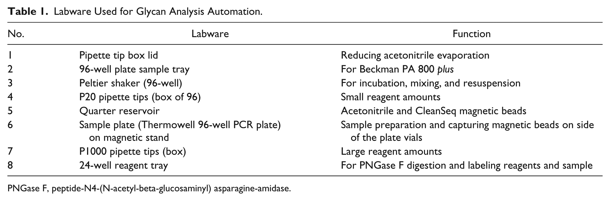
PNGase F, peptide-N4-(N-acetyl-beta-glucosaminyl) asparagine-amidase.
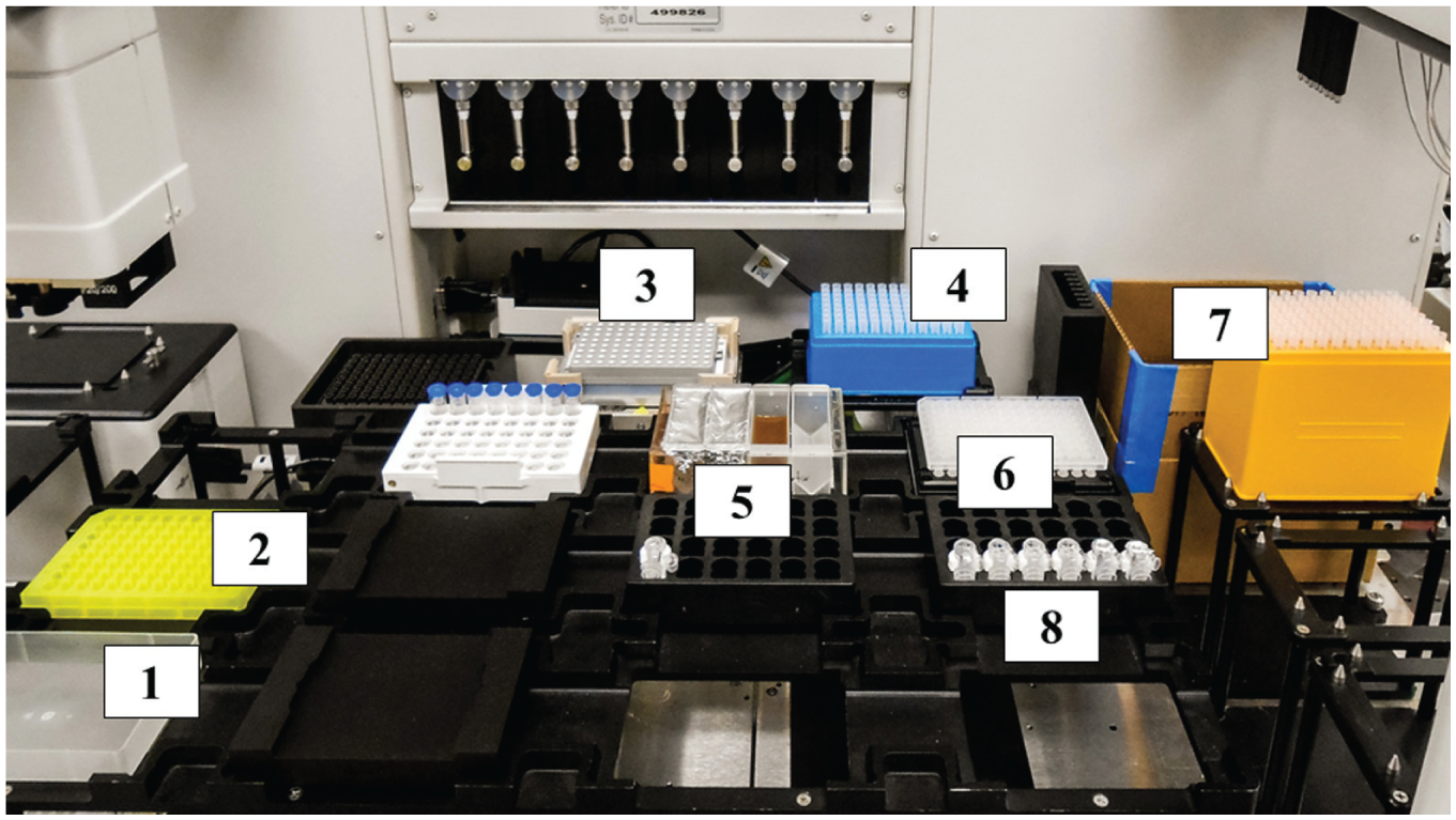
Experimental setup of the Biomek FXP Laboratory Automation Workstation.
Results and Discussion
The entire workflow of the rapid and automated glycan sample preparation protocol is shown in Figure 3 . The endoglycosidase digestion using PNGase F was performed at 50 °C for 1 h, followed by capture of the released glycans by the CleanSeq magnetic beads using 87.5% acetonitrile medium. 15 Reductive amination-based labeling of the captured carbohydrates was initiated in situ on the beads by the addition of the fluorophore (40 mM APTS in 20% acetic acid) and the reductive agent (1 M sodium cyanoborohydride in tetrahydrofurane) followed by incubation at 37 °C for 2 h. Then, acetonitrile was added to the reaction mixture to yield 87.5% final concentration, necessary to recapture the glycans by the beads. For high-efficiency APTS dye removal, the beads were washed three times in every step, first with water for easy and fast resuspension, followed by acetonitrile to yield 87.5% final concentration for glycan recapture. The fluorophore-labeled glycans were then eluted from the beads by the addition of 25 µL water and were ready for ultrafast CE-LIF analysis.
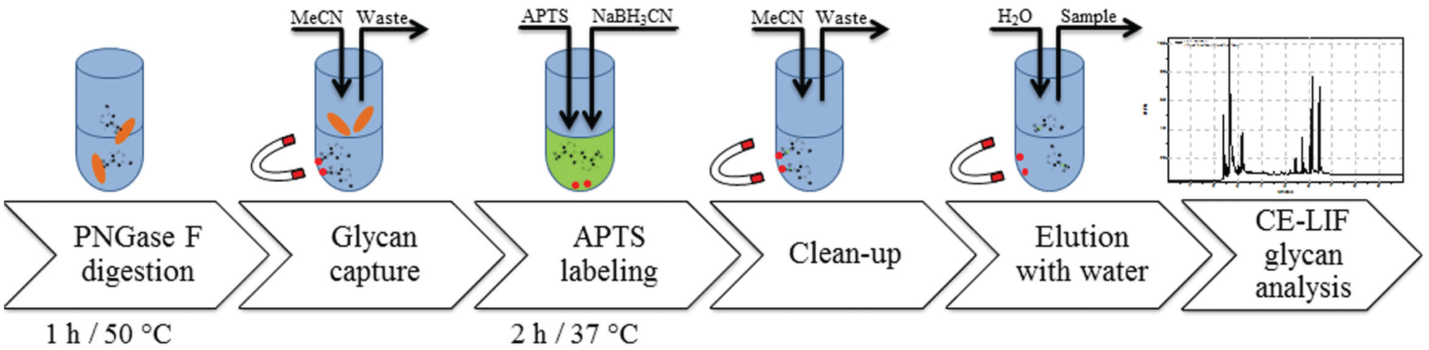
Laboratory automation flowchart for magnetic bead–based sample preparation and CE-LIF analysis of N-glycosylation of therapeutic antibodies. CE-LIF, capillary electrophoresis with laser-induced fluorescence detection.
Automation Steps
Magnetic bead suspension is added from the quarter reservoir (
Fig. 2
,
The sample solution (preferred concentration: 10 mg/ml) is added to each well from the sample rack (or reservoir) to the sample plate (
Fig. 2
,
The sample is denatured by the addition of the denaturation buffer at 65 °C for 10 min (
Fig. 2
,
In parallel with steps 1–3, the digestion mixture is prepared (
Fig. 2
,
After the denaturation step, the digestion mixture (
Fig. 2
,
The deglycosylation reaction is implemented on the vortex shaker (
Fig. 2
,
Acetonitrile is added to each sample to obtain 87.5% final acetonitrile concentration for glycan capture (
Fig. 2
,
In parallel with step 7, the labeling mixture is made containing APTS (fluorophore), sodium-cyanoborohydrate (reductive agent), and acetic acid (catalyst) (
Fig. 2
,
After step 7, the supernatant is removed, and the labeling mixture (
Fig. 2
,
The labeling reaction mixture is incubated at 37 °C for 2 h on the vortex shaker (
Fig. 2
,
The reaction is stopped by the addition of acetonitrile (
Fig. 2
,
Cleanup after the labeling reaction:
Elution of the APTS-labeled glycans with water (
Fig. 2
,
Sample transfer into a sample tray to be used in the CE instrument (
Fig. 2
,
Repeatability of the Automated Sample Preparation Process and CE-LIF Analysis
Glycan release, carbohydrate partitioning, APTS labeling, and sample cleanup were all optimized for full automation using the magnetic bead–based protocol, resulting in <4 h processing time with excellent yield and good repeatability. CE-LIF analysis of the fluorophore-labeled glycans was also optimized for ultrafast separation to accommodate the high throughput of the automated sample preparation process with the use of a gel-buffer system containing 1% high-molecular-mass polyethylene oxide. The electropherograms of the APTS-labeled immunoglobulin G (IgG) glycans injected from a 96-well plate after the automated sample preparation process are shown in Figure 4 . The actual measured repeatability was 2.6%, whereas the reproducibility was 4.1%, which could be significantly improved with the use of bracketing standards. 16 Please note that full separation of all the major IgG glycans was obtained in less than 3 min with excellent run-to-run repeatability. The glycan structures are shown in Figure 4 following the Oxford symbolic nomenclature.
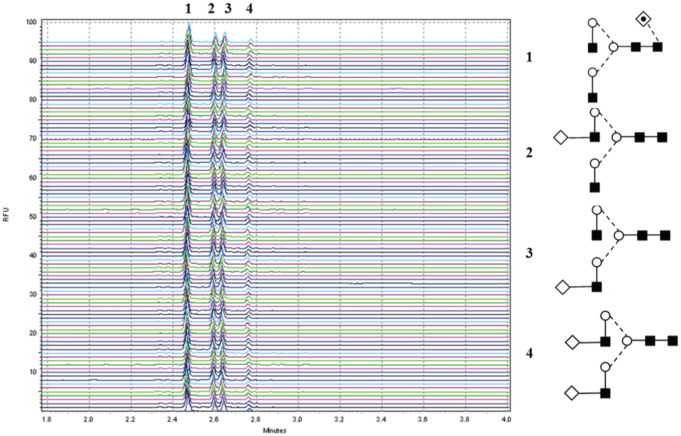
Repeatability study by CE-LIF analysis of APTS-labeled IgG glycans prepared by using the laboratory automation workstation in a 96-well plate format using the magnetic bead–based protocol: (
Application of the Automated Sample Preparation Workflow for the N-Glycosylation Analysis of Therapeutic Antibodies
Next, we applied our fully automated protocol to the analysis of therapeutic antibodies, including an innovator, a biosimilar, and a derivative in the form of antibody drug conjugate (ADC), to demonstrate the vastly improved speed of the method. Figure 5 depicts the resulting electropherograms. Trace (F) is the maltooligosaccharide ladder and also shows the degree of polymerization of the peaks as G3 for maltotriose, G5 for maltopentaose, G7 for maltoheptaose, and G9 for maltonanoose. These maltooligomers are used in glucose unit (GU) value calculation for database-based structural elucidation. 17
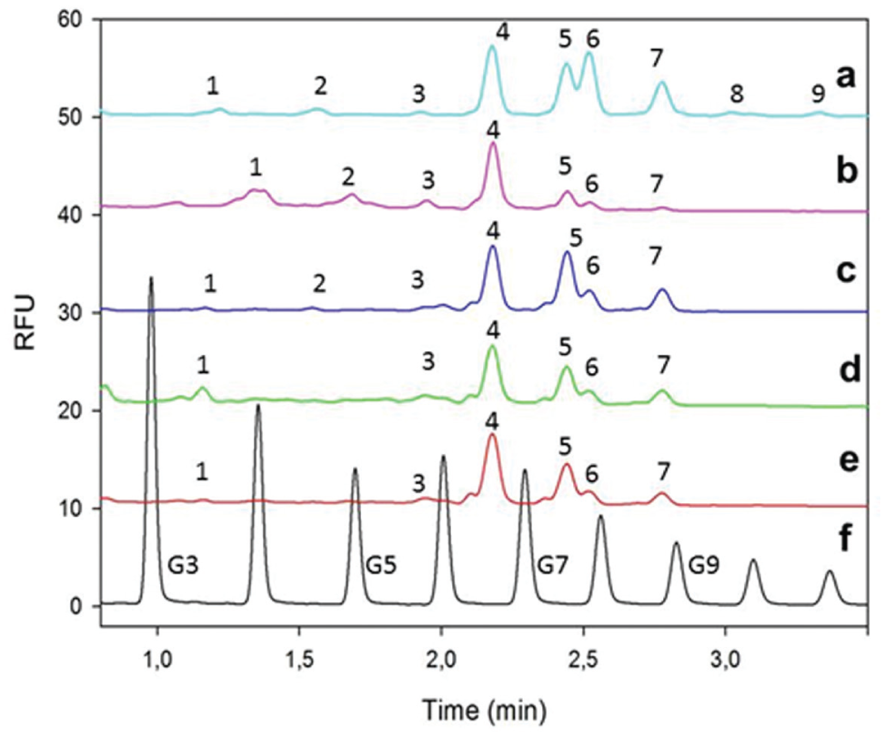
Ultrafast CE-LIF analysis of APTS-labeled N-glycans released from a murine mAb standard (
Trace (
Conclusion
In this article, we introduced a novel automated sample preparation scheme for an optimized magnetic bead–based protocol of N-glycosylation analysis of antibody therapeutics from endoglycosidase digestion, through optimized fluorophore labeling and cleanup, to ultrafast CE-LIF separation with high-throughput sample processing in a 96-well plate format. Programming of the laboratory automation workstation was simple, and the system was robust while also flexible to handle large numbers of samples. Automated liquid-handling processing offered fast and precise sample preparation, reduced flow-induced shear strain on native biological sample matrices, and minimized contamination risks. Due to the large amount of deck space available in the laboratory automation workstation used, background electrolyte preparation for the CE-LIF measurement was also done automatically. Fully automated sample preparation in this instance means no human intervention is needed from the beginning to the end of the sample preparation process. To attain higher accuracy liquid handling, or with the use of unknown sources or amounts of samples, conductive pipette tips can be used for high-precision liquid handling. In summary, the laboratory automation workstation used in these experiments was capable of large-scale sample processing to accommodate rapid glycan analysis of therapeutic antibodies for the biopharmaceutical industry. The usefulness of the automation protocol was demonstrated by the rapid N-glycosylation analyses of a few commercially relevant antibody therapeutics. In addition to the comparison of the glycosylation profile of an innovator drug to its biosimilar counterpart, the biological significance of the differences was also addressed.
Footnotes
Acknowledgements
The generous support of Don Arnold and Navaline Quach, as well as the help of Mike Kowalski and Bee Na Lee, is greatly appreciated. The authors also acknowledge the support of the MTA-PE Translation Glycomics project (#97101).
Abbreviations
PNGase F: peptide-N4-(N-acetyl-beta-glucosaminyl) asparagine-amidase; APTS: 8-aminopyrene-1,3,6-trisulfonic acid; CE-LIF: capillary electrophoresis with laser-induced fluorescence detection; HPLC: high-pressure liquid chromatography; SPRI: solid phase reversible immobilization technology; CTC: complement-dependent cellular cytotoxicity; GU: glucose unit.
Declaration of Conflicting Interests
The authors declared no potential conflicts of interest with respect to the research, authorship, and/or publication of this article.
Funding
The authors disclosed receipt of the following financial support for the research, authorship, and/or publication of this article: The generous support of Don Arnold and Navaline Quach, as well as the help of Mike Kowalski and Bee Na Lee, is greatly appreciated. The authors also acknowledge the support of the MTA-PE Translation Glycomics project (#97101).