
Abstract
Automated cell cultivation is an important tool for simplifying routine laboratory work. Automated methods are independent of skill levels and daily constitution of laboratory staff in combination with a constant quality and performance of the methods. The Biomek Cell Workstation was configured as a flexible and compatible system. The modified Biomek Cell Workstation enables the cultivation of adherent and suspension cells. Until now, no commercially available systems enabled the automated handling of both types of cells in one system. In particular, the automated cultivation of suspension cells in this form has not been published. The cell counts and viabilities were nonsignificantly decreased for cells cultivated in AutoFlasks in automated handling. The proliferation of manual and automated bioscreening by the WST-1 assay showed a nonsignificant lower proliferation of automatically disseminated cells associated with a mostly lower standard error. The disseminated suspension cell lines showed different pronounced proliferations in descending order, starting with Jurkat cells followed by SEM, Molt4, and RS4 cells having the lowest proliferation. In this respect, we successfully disseminated and screened suspension cells in an automated way. The automated cultivation and dissemination of a variety of suspension cells can replace the manual method.
Introduction
Research with cell cultures is a common method for investigation in regenerative medicine, 1 cancer research, and tissue engineering. 2 Therefore, regulatory and commercial cell culture methods are required for scalable and robust production processes.1,3 Cell cultivation is a general process in research and laboratory work to expand cells for bioscreening experiments. Traditionally, these are repetitive and long-lasting manual work steps with a risk of contamination and human errors associated with high costs. The automation of cell cultivation is required to simplify the processes in combination with increased quality, repeatability, precision, and stability of the individual batches under consistent sterile environmental conditions with efficient costs and time.4–8
Available studies on automated cell cultivation systems focus exclusively on monolayer cultures. Another cell type includes suspension cell lines, which are essential for research, especially for the investigation of leukemia. They require different methods for cell handling and devices for the automated cultivation and dissemination of cells compared with adherent cells (see Fig. 1 ).
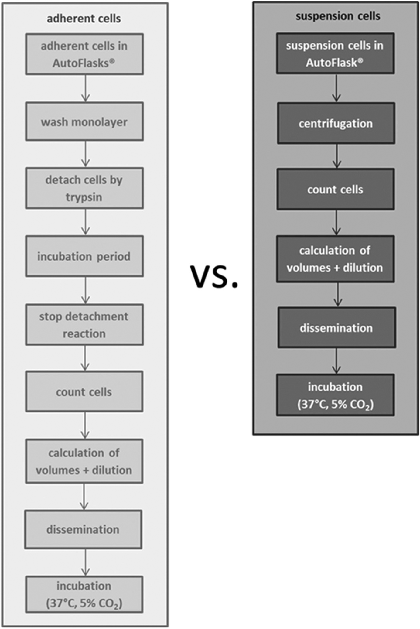
Schematic overview of the cell handling of adherent cells versus suspension cells. The general steps for the dissemination of adherent cells contrast with the same procedure for suspension cells.
We addressed this topic by integrating a fully automated system. The Biomek Cell Workstation (Beckman Coulter, Krefeld, Germany) is designed for the regular cell cultivation of different cell types (adherent cells and suspension cells). These cell cultures enable visualization and bioscreening using a high-throughput screening (HTS) system. We integrated the parallel cultivation and dissemination of different suspension cells (Jurkat, SEM, RS4, and Molt4) with automated bioscreening using a WST-1 proliferation assay.
Systems for Automated Cell Cultivation
Currently, there exists a variety of solutions for automated cell cultivation to improve laboratory work. 9 These systems require robotic arrangements for labware handling, hoods with filters for sterile conditions, liquid handling systems, cell count devices, and incubators (37 °C, 5% CO2). Other systems include devices for the detection of biological processes and cameras for the evaluation of the confluence. The arrangements have to be programmable and monitorable using specific software. Mainly, these systems are modular and flexible and related to hardware and software (see Table 1 ).9,10
Summary of the Automated Cell Culture Systems.

HCS, high-content screening; HIV, human immunodeficincy virus; PBS, phosphate-buffered saline.
Liu et al. 1 investigated the expansion of anchorage-dependent human Caucasian osteosarcoma (HOS) cell lines with the horizontally arranged CompacTSelecT (Automation Partnerchip-TAP Biosystems, Hertfordshire, UK) cell culture system. This system enables the regular steps for automated cell cultivation, including passaging, seeding, and cultivation. The CompacTSelecT includes a six-axis anthropomorphic robotic arm that enables access to 90 T175 barcoded flasks and a plate incubator (37 °C, 5% CO2). Furthermore, a cell counter (Cedex; Roche Innovatis AG, Bielefeld, Germany), a medium pump, and two flask decappers and holders are integrated. A HEPA filter supports the sterile condition. Jain et al. 11 investigated the AI.CELLHOST (Hamilton Robotics, Reno, NV) system, a robot for automated cell cultivation and fluorescence imager, for the cultivation of SH-SY5Y, BE(2)M17, and HEK293T cell lines. This system provides high-content and high-throughput screenings for genomic, proteomic, and transcriptomic analysis. The AI.CELLHOST includes a Hamilton Microlab STAR (Hamilton, Martinsried, Germany) for liquid handling, a Cellavista cell counter (Roche, Basel, Switzerland) for the evaluation of the cell count and confluence, and a Cytomat 24C incubator (Thermo Scientific, Waltham, MA) for up to 504 SBS plate stackers under sterile conditions, which is guaranteed by a flow chamber with the HEPA filter systems (Bigneat, Hampshire, UK). These devices are associated with a MICROLAB SWAP robot arm (Hamilton). Furthermore, a control system with a graphical interface is integrated as well as a storage solution for media.
TECAN (Männedorf, Switzerland) offers the Cellerity for automated cell culturing. The different options support many cell culture processes (passaging, isolation, suspension handling, counting, and incubation) for high-throughput bioscreening and transfections. The housing associated with a HEPA filter system supports sterile handling. The CellGEM (Cell Growth Expansion and Maintenance) software is intuitively controllable with process optimization by scheduling steps. Corning (Corning, NY) RoboFlask Cell Culture Vessels are used within the system. The cells are evaluated (counting, viability, size, morphology) based on the Trypan blue test using the CEDEX cell analyzer (Roche, Mannheim, Germany). The reusable steel tips enable slow-volume pipetting (20 µL, 100 µL), low-volume pipetting (5–50 mL), penetration through the septa of the Corning RoboFlask Cell Culture Vessels, and cleaning steps (interior, exterior).12,13 Schultz et al. 14 used the modified Cellerity system for the cultivation and transfection of cells to investigate the human immunodeficiency virus (HIV) pseudovirus high-quality production of 140 to 1000 mL. The footprint of this system is 5 × 3 m. The modular standard Freedom EVO 200 platform for cell handling is the basis of the system. Furthermore, the Flask Flipper, a flask transfer station, a liquid handling arm (LiHa), vessels for solutions, and the robotic manipulator arm (RoMa) are located and enclosed on the worktable. The Flask Flipper allows the shaking and knocking as well as turning of the four flasks horizontally and vertically in one step for liquid handling using the liquid handling arm (LiHa) with eight reusable steel needles. The flasks are stored at barcode-defined positions in a Storex incubator (Liconic, Montabaur, Germany) (37 °C, 5% CO2). The Cedex Cell Counter (Roche) is connected with the worktable and enables cell counting as well as viability testing. The proprietary Freedom Evoware Plus Software controls the devices (RoMa, the LiHa, the Flask Flipper, and the transfer station), and the CellGEM (Cell Growth, Expansion, Maintenance) software enables the time scheduling of the cell-handling process.
The BioCel system (Velocity 11, Palo Alto, CA) from Agilent Automation (Santa Clara, CA) enables high-throughput processes using a fast central radial robotic arm. The environmental conditions are supported by different solutions, for example, filter systems with ultra-low penetration air (ULPA) filters, unidirectional airflow, positive pressure, and controllable temperature as well as humidity. The liquid handling is realized using the Agilent Bravo Automated Liquid Handling Platform with nine deck positions and 8-, 16-, 96-, or 384-fixed and disposable tip heads. Different devices and applications can be integrated (e.g., centrifuge or labeler).15–17
Kato et al. 18 reported a compact cell culture system for the cell expansion of primary tissue. This system enables the cultivation of fibroblasts. The system consists of different units (supply unit, incubation unit, and collection unit), rotary pumps, and valves in a small format (70 × 60 × 86 cm). The supply unit contains the required solutions in bags (medium, trypsin, phosphate-buffered saline [PBS]) associated with a heater and the disposable tubing set. The tubing set connects the major units and includes culture dish, aeration filters (incubation unit), collection tubes (collection unit), and injection ports in enclosed tubing. The collection unit includes a waste bag and collection tube. The cell cultivation is realized in the incubation unit under CO2 atmosphere. The sample solution is inserted using a syringe from the injection port to the clean bench (incubation unit).
Kino-Oka et al. 2 evaluated the performance of myoblasts. The automated cell culture system contains three units: the culture module, the liquid handling unit, and control unit. The incubator is the culture module with stored growth chambers. The units have a constant temperature (37 °C) with sterile air (5% CO2) controlled by a mass flow controller. The liquid management occurs in the liquid handling unit by tilting the chamber stage using electrical pinch valves and tubing pumps. A movable (horizontal, vertical) 8-bit CCD camera (Toshiba Teli Corporation, Tokyo, Japan) is integrated under the chamber. The automated processes are programmed using LabVIEW software (National Instruments, Munich, Germany).
The cell cultivation of adherent cells and suspension cells differs in many process steps (see Fig. 1 ). The significant difference in handling suspension cells is caused by the floating of the cells in the media, in contrast to an attached monolayer of adherent cell cultures. The system requires a specific AutoFlask (Greiner, Frickenhausen, Germany) for automated cell handling. The AutoFlasks are available with surface treatment for adherent cells and without a surface treatment for suspension cells. The monolayer has to be detached by enzymes (trypsin) after washing with buffer (PBS). Then, the process has to be stopped with media after an incubation period. However, the suspension cells need centrifugation to separate the cells from the media followed by a resuspension with fresh media. Then, both cell types have to be counted, calculated, diluted, and disseminated followed by an incubation (37 °C, 5% CO2).
The suspension cells have to be carefully resuspended and separated from the media using centrifugation steps. Regarding this, a centrifuge and suitable liquid handler have to be integrated in the automated system associated with a specific program for the handling of suspension cells. Furthermore, cell culture flasks with a hydrophobic surface are beneficial for cell culture processes of suspension cells (see Fig. 1A ).
The published systems for automated cell cultivation do not contain devices for centrifugation steps. Thus, the cell suspension cannot be separated from the conditioned medium. Furthermore, currently used liquid handling steps are not useable for the handling of suspension cells, for example, the remove of media by decantation. Nevertheless, the horizontal usage of the cell culture flasks at flowing movement and the gentle transfer of the cells promote the proliferation of suspension cells.
Materials and Methods
System Architecture and Design
Cell Culture Vessels
The automated processes require specific vessels for the cell cultivation and the storage of solutions and media (see Fig. 2 ).
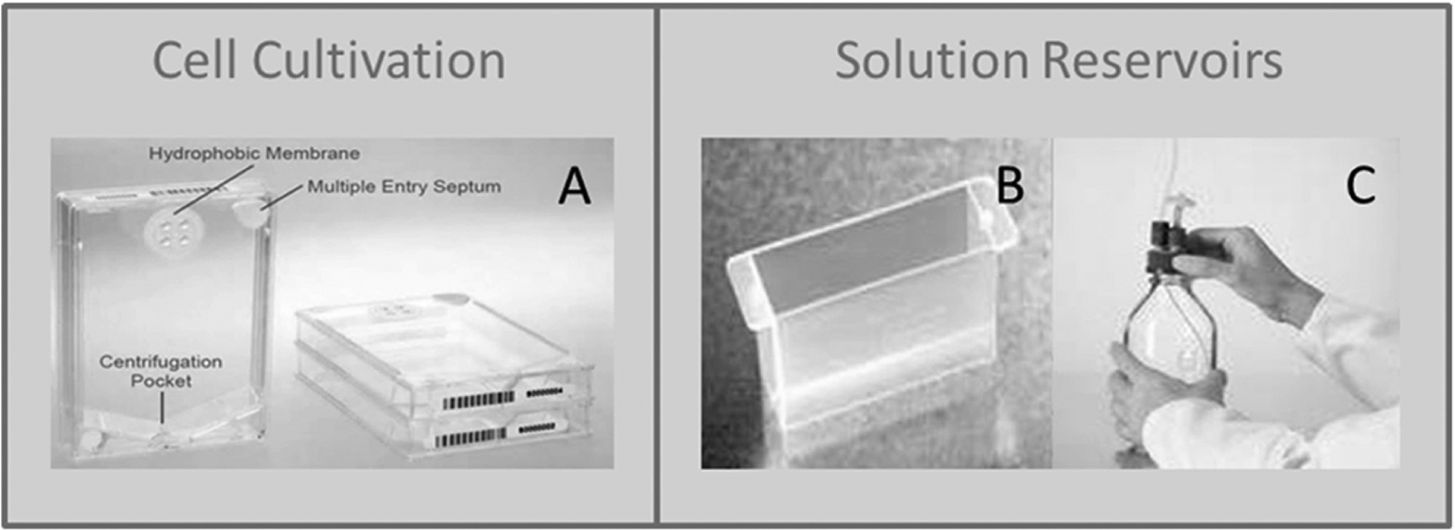
The specific flasks and reservoirs for automated cell cultivation by the Biomek Cell Workstation. The cell culture AutoFlask is for the automated cell cultivation of adherent cells or suspension cells through the 96-well format, the hydrophobic membrane, and the entry septum for liquid transfer (
The Biomek Cell Workstation requires to work with CELLSTAR AutoFlask (Greiner). These sterile flasks have an SBS format 19 (85.48 mm/127.76 mm/19.50 mm) with a growth area of 83.60 cm2, and they are suitable for automated cell handling. Currently, only the Biomek Cell Workstation enables cell handling within the AutoFlask. A membrane and a septum are located upsides of the flask. The hydrophobic membrane allows gas exchange during the cultivation periods. The multiple entry septa enable the transfer of the solution under sterile conditions (see Fig. 2A ). These flasks permit the direct horizontal handling of the cell cultures due to the location of the septum in the top-right corner. This is unique for this format and labware. The cell handling and growth area of cell culture vessels (75 cm2) are very close to the manual process. The footprint of SBS plates is an essential aspect for the standardization of different automation processes and enables automated stacking. 20 The solutions, cell suspensions, and buffers are presented in sterile modular reservoirs (see Fig. 2B ) or bottles with air filters (see Fig. 2C ). The reusable reservoirs for the automated handling are made of polypropylene. 21 The reservoirs placed on the High Temp Peltier Device enable tempering of solutions. The reservoirs are located on specific positions of the liquid handler deck and provide media (up to 40 mL) for the automated liquid transfer of the span-8 pipetting head. The glass bottles are equipped with the port selection valve to transfer higher volumes of media.
Disseminated cells are seeded in 96-well plates (Greiner) for consecutive bioscreening. The well plate format allows completely automated processes with the screening system.
System Design of the Biomek Cell Workstation
The Biomek Cell Workstation is a compatible and flexible system for automated cultivation of various cell types. This complex system is vertically arranged and moveable with a small footprint of 1.49 m/1.64 m/2.63 m. The workstation allows the cultivation of two-dimensional (2D) 22 cell lines as well as the manufacturing of different three-dimensional (3D) cell cultures (alginate beads, pellet cultures, hanging drops) (see Fig. 3 ). Originally, the Biomek Cell Workstation was designed and used for the cultivation of adherent cell cultures. Currently, the enhancement of the system in combination with appropriate programs also enables the handling of suspension cells.
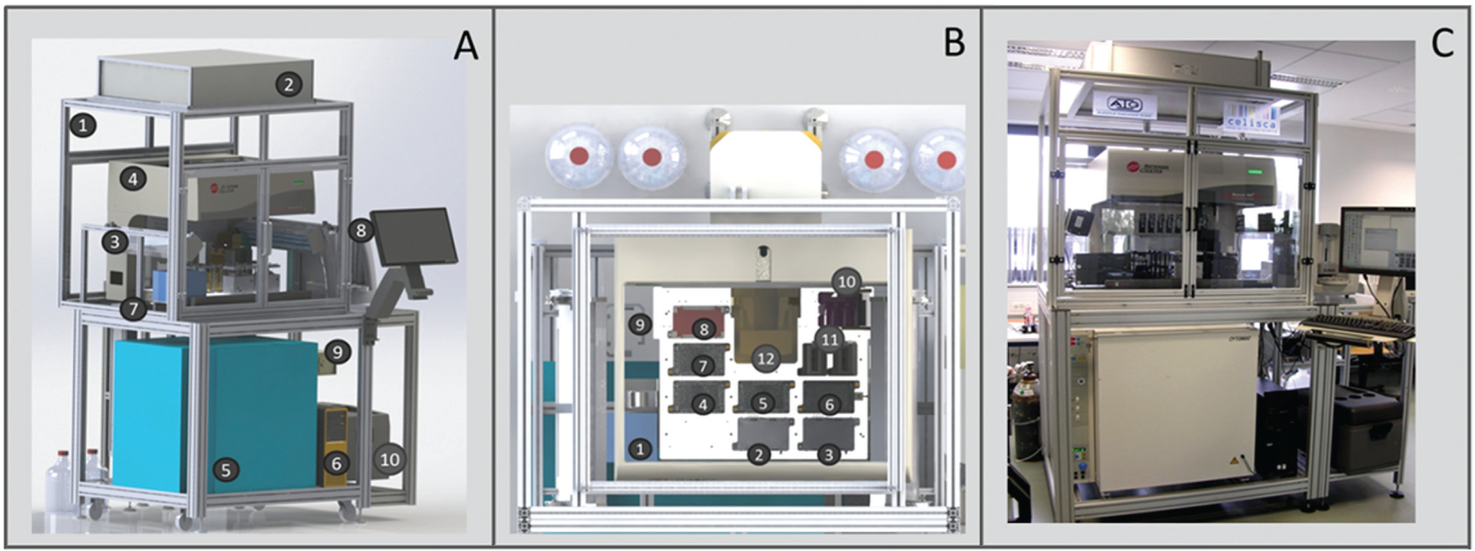
Biomek Cell Workstation and liquid handler deck. Frontal view: The central components of the Biomek Cell Workstation (
According to Figure 3 , the system is set up with a Biomek NX for all liquid handling steps, an incubator (Cytomat; Thermo Fisher Scientific) for the incubation of the different cell lines, a centrifuge (Vspin; Velocity 11), and a cell counter (Vi-CELL XR; Beckman Coulter). The system is designed in a vertical way to realize a small footprint, in contrast to existing systems. Therefore, the devices are arranged one above the other.
The central element of the system is a Biomek NX (Beckman Coulter) for all liquid handling processes. These processes are aspiration, dispensing, and transport of solutions. The Biomek NX is equipped with a span-8 pipetting head and has an integrated gripper at one rail. The one to eight pipetting channels are independent. The cell culture flasks and well plates are positioned by the gripper. The Biomek NX enables the liquid transfer by disposables or steel cannulas. These are associated with syringe pumps. The pumps of channels 1 to 4 associated with steel cannulas have maximum capacities of 1 mL. The pumps from channels 5 to 8 enable the transfer of a maximum of 5 mL in one process step. Channel 7 is associated with a peristaltic pump and the port selection valve. This combination allows the transfer of larger volumes and a change between different media for parallel cell cultivation of different cell lines. System fluid (sterile water) is always in the tubes for the transfer of exact volumes. 23
The intended volumes are controlled by quantifying the volumes by pipettes. The accuracy of the used channels is around 93% for the volume used. Volumes below 10 µL should be avoided due to a decrease in accuracy of the volumes.
The liquid handler deck is equipped according to the requirements of automated cell cultivation (see Figs. 3 , 4 ). Automated laboratory positioners (ALP) and devices are available for specific handling of cell culture flasks or as placeholders.
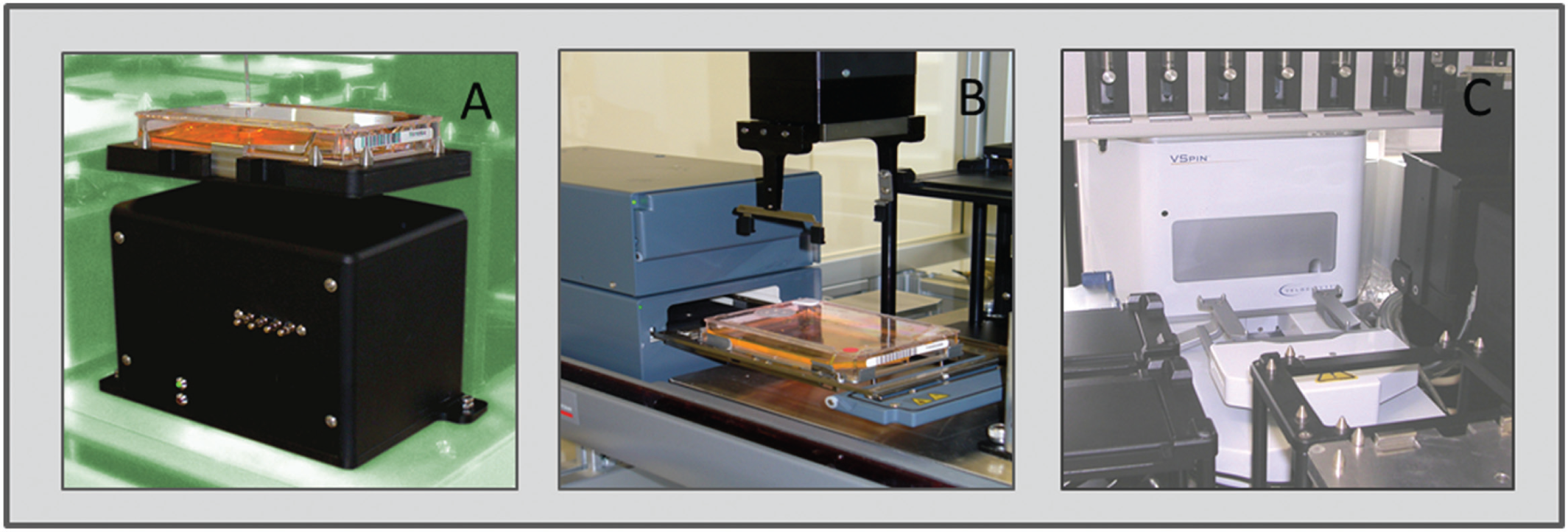
Devices and automated laboratory positioners (ALPs), which are necessary for flexible automated cell cultivation. The important components for cultivation as well as dissemination of adherent cells and suspension cells are the three-dimensional (3D) tilt racks for the automated handling of cell culture flasks (
Furthermore, a microplate shaking ALP is integrated on the liquid handler deck (VARIOMAG Teleshake; Thermo Scientific). This is an active ALP for mixing solutions and cells in microplates or flasks with an adjustable shaking motion. The ALP is a magnetic shaking system for well plates or cell culture flasks in a 96-well format with 100- to 2000-rpm movements and an amplitude of 2 mm. This device has a small footprint (high: <40 mm), minor transference of vibrations, and self-centering to the zero position. The microplates (96- and 384-well) or cell culture flasks (Greiner) can be added to the Orbital Shaker ALP by a lab technician or the gripper.
For short incubation periods of the cell culture flasks at 37 °C, two on-deck incubators (inheco, Martinsried, Germany) are available. These can be accessed directly from the liquid handler deck. The temperature and shaking periods are adjustable.
Two washing stations (Beckman Coulter) have been included on the liquid handler deck for cleaning and disinfection of the steel cannulas. One wash station is associated with autoclaved isopropanol and the other with water for active and passive washing steps. The wash station with water enables the active wash steps to rinse the steel cannulas inside and outside. This step prevents contamination. Two bottles with the specific fluids are connected with the wash stations, and the waste is collected in one bottle. The steel cannulas are cleaned in two ways. The pumps of the Biomek NX deliver water or isopropanol to the cannulas, and system fluid will be dispensed into the wash station. On the other hand, steel cannulas can dispense systems fluid into the wash station. Thus, the solution washes inside the cannulas and the level of the wash fluid rise with increased speed into the cleaning wells to clean the outside of the cannulas positioned in the wash station. 24
For simulating the manual handling of the cell flasks, two 3D tilt racks (AIG, Rostock, Germany) have been integrated. The 3D tilt rack (AIG) is a proprietary component for the automated handling of cell culture flasks. The rack enables the tilting of the flasks in the x, y or x/y axis as well as pivoting and knocking the flask. The tilt angle and speed are adjustable. The flask is fixed on the rack by deadlocks for pipetting steps through the septum (see Fig. 4A ). 10
For the culturing and incubation of the cells, an incubator has been integrated into the system. The Cytomat Automated Incubator (Thermo Scientific, Schwerte, Germany) allows the cultivation of cells at 37 °C and 5% CO2. The temperature and oxygen concentration are controllable. Inside, nine storage modules with 21 places each are integrated for flasks or sheets on a turntable. The plate transfer of the Cytomat to the lift is realized by a plate shuttle system and an X/Y/Z moveable handler through a heated automated access door. 25
Since the system is organized in a vertical way in order to realize a small footprint, a transfer system between the incubator and the liquid handler deck is realized. The proprietary lift enables the transport of one well plate as well as flask per time to bridge a height of 480 mm. The transport of the labware starts with the movement of the labware by the plate shuttle system from the back through the automated access door of the Cytomat to the lift traveling stage and a rotation of 90° by a pneumatic rotation mechanism with adjustable speed. The vertical movement to the liquid handler deck is realized by a linear spindle drive with a time of 10 s. Then, the gripper of the liquid handler is able to locate the labware at the specific positions. The whole process, to move the labware from the Cytomat to the positon of the liquid handler deck, requires 35 s.
A Vspin centrifuge (Velocity 11) is connected to the liquid handler deck with direct access by the Access2 plate transport with integrated grippers. The liquid handler locates the well plates on the Access2 plate transport station, and the gripper transports the plates into the centrifuge. The Vspin device enables the centrifugation of two plates or flasks at the same time. The spindle speed is 0 to 3000 rpm. In addition, the time and the spindle speed are adjustable (see Fig. 3 ).
Cell viability and cell count are evaluated using a ViCell analyzer (Beckman Coulter, Brea, CA). This device is associated with the liquid handler deck over a conduit and has an analysis time of 2.5 min. The cells are analyzed using the Trypan blue method. The viable cells are marked green because they actively exclude the dye instead of dead cells. These cells are marked red.
The viability range of 0% to 100% is detected in a sample volume of 0.5 mL with image technologies of the auto-focus CCD array and firewire camera by the video image through a quartz flow cell. The size range of the cells is 2 to 70 µm. 24
All subsystems, including the Biomek NX liquid handler (Beckman Coulter), Vspin centrifuge (Velocity 11), barcode reader, lift, and a conduit to the ViCell (Beckman Coulter), 10 are covered by a housing. The housing associated with a HEPA filter (Camfil, Stockholm, Sweden) and UV lights (Vilber, Eberhardzell, Germany) guarantees sterile conditions. The HEPA filter system works in continuous operation, whereas the UV lights (Vilber) are used for a time period before and after the cultivation processes.
The integration modules enable the automated control of the different devices of the Biomek Cell Workstation with the automation software (SAMI software; Beckman Coulter). The devices (e.g., ViCell) supported by Beckman Coulter have integration modules. However, external devices (e.g., shaker and incubator) require new proprietary integration modules.
Software
The automated processes for the cell cultivation methods are controlled by specific software. Basically, the devices are controlled using the specific firmware and device control software. The Biomek NX software enables the detailed configuration of all liquid handler steps associated with the specific ALPs and devices on the deck. To edit and manage the complete processes with all necessary devices, we used SAMI EX software (Beckman Coulter). The integrated scheduler is used for prevalidation of the schedules and optimizes the processes. Furthermore, the graphic runtime software of SAMI EX visualizes the whole process. In detail, the graphical interface shows the actual status, live method, labware report, system view, plate/flask location in the system, and the system actions. This is an important instrument in automation to translate manual work steps into automated methods.
Processes for Prevention of Contamination
The automated processes on the Biomek Cell Workstation require different steps to guarantee sterile cell cultivation without cross-contamination. The syringes and the whole tube system of the Biomek NX liquid handler have to be purged with system fluid daily and before every cultivation process. Every week, the system is rinsed with 5% Korsolexplus (Bode Chemie, Hamburg, Germany) to prevent contamination with fungi and bacteria. The port selection valves, the used channels, and the tubes are rinsed with water and isopropanol (Carl Roth, Karlsruhe, Germany) before and after every automated process. The UV lights are used 15 min before every procedure to decontaminate the air and the surface of the liquid handler deck.
Further studies showed that the combination of UV lights and rinsing steps with water, isopropanol, and Korsolexplus prevents contamination with microorganisms and cross-contamination in the parallel cultivation with different cell lines. The membranes of cervix carcinoma cells were labeled with different fluorescence markers (PKH-26, PKH-67) and in parallel cultivated with the Biomek Cell workstation. No contamination was detected in the microscopic evaluation and fluorescence-activated cell sorting (FACS). 26 To exclude cross-contamination, cell samples can be evaluated by FACS analysis routinely. In the case of suspension cells, the different cell lines have specific cell sizes.
Cell Culture Process
The Biomek Cell Workstation allows the cultivation of adherent cells and suspension cells. In this project, we investigated the cultivation and dissemination of four different suspensions cells (Jurkat, SEM, RS4, and Molt4) to visualize the comparability of automated processes with the manual methods.
The cell culture process includes two different automated main processes (see
Fig. 5
). First, the regular automated cell cultivation was done. The AutoFlasks (Greiner) and the SBS plates (Greiner) were stored in the Cytomat (Thermo Fisher). The cell culture flask with the suspension cells and the SBS plates were transferred to the liquid handler deck. The 3D tilt rack in combination with the liquid handler allowed the transfer of a 2-mL/well cell suspension into a deep-well plate (DWP) (Greiner) by aspirating and dispensing steps of the liquid handler with steel cannulas at the AutoFlasks (Greiner). The detailed programmed dimensions of the Biomek liquid handler for the aspirating steps are 0.3 cm and 205° from the center as well as 40 mm from bottom. The dispensing by the liquid handler was performed by the following dimensions: 0.01 cm and 330° from the center as well as 40 mm from bottom (see
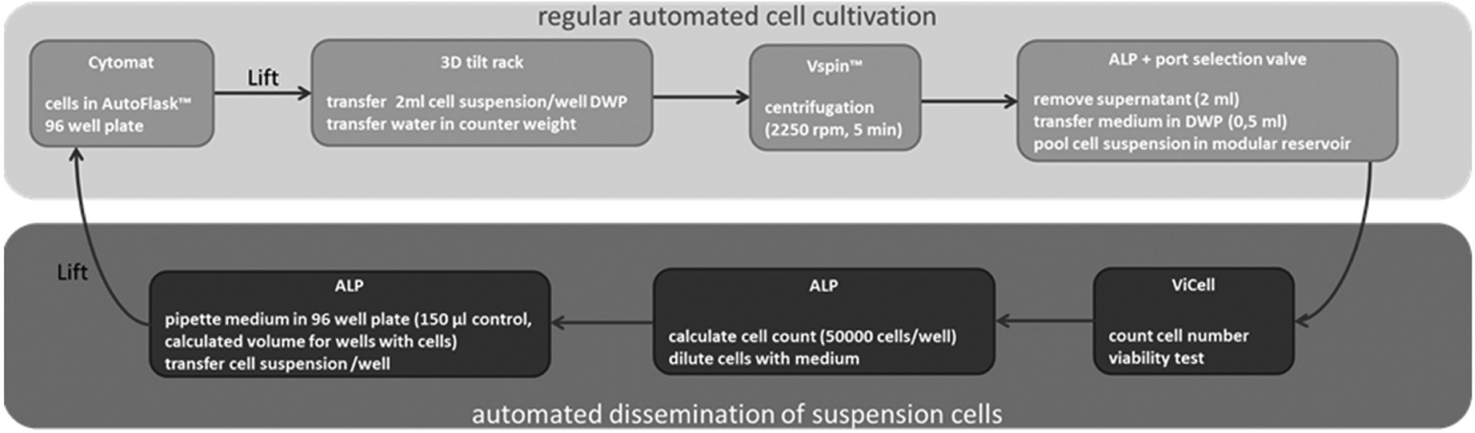
The automated process for cell cultivation and subsequent dissemination of suspension cells. The cell culture process for the investigation of suspension cells is separated into regular automated cell cultivation and automated dissemination of suspension cells.
Then followed the automated dissemination of the suspension cells. The ViCell (Beckman Coulter) detected the viability and counted the cell number. After calculation of the exact cell number (50,000 cells/well), medium and subsequent cell suspension were transferred into the SBS plate with a volume of 150 µL. Incubation of the well plates with cells was realized using the Cytomat (37 °C, 5% CO2). For cell expansion, new AutoFlasks (Greiner) with the specific cell lines were cultivated. The cell suspension and solutions were transferred at a speed of 5%, 5 mm from the bottom of the 3D tilt rack, at an angle of −4° at the pivot (see
Results and Discussion
Evaluation of the Cells
The cells were investigated with the ViCell XR (Beckman Coulter) device to detect the cell count and the cell viability. These are important measurements to evaluate the correct cell amount and to seed the exact cell number automatically. The suspension cells were cultivated in T75-cm2 flasks (Sarstedt, Nümbrecht, Germany) for manual handling and in AutoFlasks (Greiner) for automated management.
Generally, the viability of suspension cells was higher than 74%. The viability of automated handled cells was decreased compared with manually treated cells (Jurkat: 3.6%; SEM: 5.7%; RS4: 5.8%). Instead, automatically seeded Molt4 cells showed an increased viability of the cells (1.8%) (see Fig. 6A ). This cell line seems to be less damageable with the automated procedure. Regularly, automated cell cultivation is related to more stress for cells. The rough cell handling during the automated process reduces the cell viability of automatically treated RS4. This cell line seems to be more sensitive to cell stress by automated pipetting steps with increased shear stress. The cell stress can be reduced by optimization of the automated method by changing the steel cannulas and the pipetting steps. The cell transfer could be performed by disposables with a wide opening, and the speed of pipetting cell solution may be reduced.
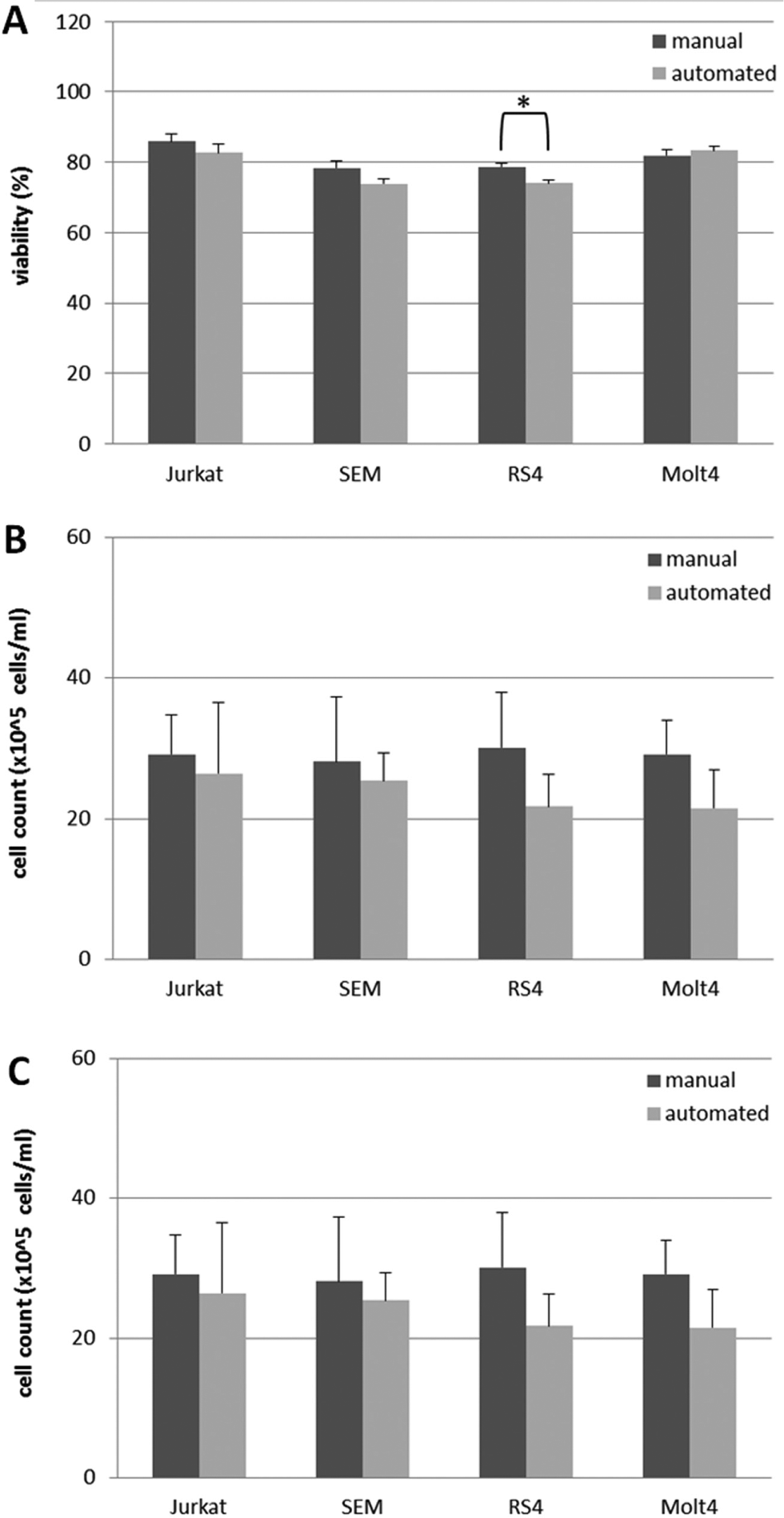
The viability (
The cell count (×105 cells/mL) of the suspension cells cultivated in T75-cm2 flasks was nonsignificantly increased (Jurkat: 2.7 × 10⁵ cells/mL; SEM: 2.8 × 10⁵ cells/mL; RS4: 8.3 × 10⁵ cells/mL; Molt4: 7.6 × 10⁵ cells/mL) compared with the suspension cells in AutoFlasks (see Fig. 6B ). This is caused by the flask geometry and a better gas exchange in the T75-cm2 flask. The flasks for manual handling have a greater height and a lower surface, which seem to support cell proliferation, and a higher media level can increase the cell count. The transfer of cell suspension was performed by tilting the flask and pipetting the solution with steel cannulas in the automated process. The cell loss seemed to be higher with the automated method. Consequently, the number of suspension cells in AutoFlasks should be increased for automated cultivation and dissemination processes.
The cultivation of suspension cells in AutoFlask is associated with a minor reduced cell count and viability compared with cultivation in regular T75-cm2 flasks with manual handling. Schultz et al. 14 published a stable cell count and viability of cells over the time. Furthermore, the automatically evaluated viability and cell count were marked by a constant number of cells without variations. In this project, the low standard error for the viability and mainly cell count of suspension cells support these results.
Proliferation Assay
The proliferation was evaluated using the WST-1 assay (Roche; see Fig. 6C ). The method is based on the conversation of tetrazolium salt to colored formazan and detection of the absorbance.
The results showed the highest proliferation of Jurkat cells, followed by SEM cells, Molt4 cells, and RS4 cells. The proliferation of automatically processed cells was nonsignificantly decreased (Jurkat, 13%; SEM, 12%; RS4, 17%; and Molt4, 16%) compared with the manually treated cells. A nonsignificant decrease in proliferation might be associated with stress on the cells caused by sheer forces and consequently increased cell death and cell loss during the automated transfer steps. Furthermore, Kino-Oka et al. 2 evaluated the performance of myoblasts. The automatically performed myoblasts also showed nonsignificant deficiency of growth activity.
Furthermore, the proliferation standard error of the automated method was mainly lower compared with the manual process (Jurkat, 20%; RS4, 12%; and Molt4, 14%). The SEM cells showed a similar standard error (0.3 units).
Often, the reduced viability of automatically handled cells is connected to the results of the proliferation. The increased stressful conditions during the automated methods were reflected by the results. A fine-tuning of the pipetting steps could reduce the shear stress, but very slow aspirating steps are associated with a long duration of these process steps. However, a benefit of automated methods should be a reduced process time.
The results of manual cell handling can significantly vary between different operators. Additionally, the results of the cell-handling processes also depend on the physical and mental state of the operator. The automation of these processes reduces the risk of an operator influence and supports the same cell handling.
The regular available systems for automated cell culture processes support the cultivation of adherent cell lines.1,2,11,14,17,18 The Biomek Cell Workstation supports the cell cultivation of adherent cells and new adapted suspension cell lines. Nevertheless, the BioCel System 17 and Cellerity 13 in addition enable cell manipulations and analysis (e.g., transfection and flow cytometry), 13 PCR preparations, and ADME tests. 17 But the bioscreening had to be realized using a separate high-throughput screening system (celisca) after the automatic cell cultivation.
The footprint of the moveable Biomek Cell Workstation is 1.49/1.64/2.63 m. Kato et al. 18 reported a smaller footprint of 70/60/86 cm for their cell culture system, but the reduced size is linked to a loss of functionality and integrated devices. The cells have to be inserted manually in the system. Furthermore, the cell counter and incubator are not integrated into the system of Kato et al. Comparable published systems with increased size and similar (passaging, seeding, incubation) functionality are the Cellerity (2.75 × 1.1 m) 13 and the CompacTSelecT (5 × 3 m). 1 A small footprint is a big advantage due to limited space in many laboratories. A footprint of 4 m2 should be tolerable as laboratory equipment.
The movement of the labware between different integrated devices is realized by an integrated gripper and a plate lift at the Biomek Cell Workstation. Robotic arms generally realize the movement of the labware.1,11,13,17 However, robotic arms are associated with high acquisition cost and need specific precautions and space requirements.
The Biomek Cell Workstation enables the detachment, passaging, and dissemination of cells. The CompacTSelecT 1 and systems by Kato et al. 18 or Kino-Oka et al. 2 include manual detachment processes and sample transfer into the system. The Cellerity covers detachment processes, but the cell handling is stressful for the cells due to vertical cell handling with the RoboFlask. 13 Furthermore, the sterile cell handling of the Biomek Cell Workstation is supported by the liquid transfer of the Biomek NX. The CompacTSelecT realizes the liquid transfer by decantation processes. 1 Other systems use eight reusable tips11,13 or serological pipettes 1 for liquid handling to prevent contamination. The strongholds 8-steel cannulas enable the whole-cell culture processes (detachment, seeding, passaging) using the Cellstar AutoFlask but require specific cleaning steps to exclude contamination. Furthermore, the Biomek Cell Workstation enables horizontal cell handling during the whole process.
The new systems used specific cell culture dishes 18 and containers. 2 The Biomek Cell Workstation uses AutoFlasks since cell cultivation processes with HTS require a microplate format for uniformity. Moreover, the CompacTSelecT even allows automated cell cultivation using T75-cm2 flasks 1 as well as the system by Kino-Oka et al. 2
The Biomek Cell Workstation and automation of cell culture processes are required for laboratories with a large number of cell cultivations. The return on investment is about 2 years in various countries (Europe, North America, and Japan). Furthermore, automation increases the independence of the skill level of laboratory staff.
In conclusion, in this work, we presented a flexible system for automated cell cultivation. This flexible system allows the cultivation of different cell types (adherent cells, suspension cells). This system enables the passaging of cells, expansion, media change, and dissemination of exact cell numbers in well plates for further bioscreenings. In particular, the automated cultivation and dissemination of suspension cells have not been published and were realized with the Biomek Cell Workstation. This complex, moveable, and flexible system is vertically arranged with a small footprint. The used AutoFlasks have an almost similar growth area to the commonly used T75-cm2 cell culture vessels. The central component is the liquid handler, which is connected to the integrated devices. The sterile cell handling is guaranteed by the housing with the HEPA filter, the UV light, and the flushing steps for cleaning and decontamination of the tube system for contamination-free cell cultivation. The benefits of the software are the simple optimization of liquid handler steps (Biomek software), the intuitive programming and scheduling of the methods, and the graphical overview and monitoring of the running processes (SAMI Process Definition Editor).
The ViCell XR (Beckman) allows the counting and viability testing of the cells for the calculation of the cell number. The cell counts of the suspension cells in AutoFlasks for automated cell handling were nonsignificantly decreased and associated with a decreased viability compared with T75-cm2 flasks for the manual processes. The proliferation, detected by the WST-1 assay, showed nonsignificant differences between manual and automated processes combined with a lower standard error of the automated method. Thus, the manual method can be replaced by the automated technique.
In the future, we will treat the cells with compounds for screening of the proliferation by using an HTS system. Furthermore, the Biomek Cell Workstation will be configured to cultivate different 3D cell cultures for a better mimicking of in vivo conditions. These cultures are pellet cultures, alginate beads, and hanging drops.
Footnotes
Acknowledgements
We thank Mrs. Grit Koch (University of Rostock) and Mrs. Carolin Gallert for the excellent technical assistance.
Declaration of Conflicting Interests
The authors declared no potential conflicts of interest with respect to the research, authorship, and/or publication of this article.
Funding
The authors disclosed receipt of the following financial support for the research, authorship, and/or publication of this article: We thank the Federal Ministry of Education and Research (BMBF Germany) for the financial support (FKZ: 03ZIK021, 03ZIK022, 03Z1KN11).
References
Supplementary Material
Please find the following supplemental material available below.
For Open Access articles published under a Creative Commons License, all supplemental material carries the same license as the article it is associated with.
For non-Open Access articles published, all supplemental material carries a non-exclusive license, and permission requests for re-use of supplemental material or any part of supplemental material shall be sent directly to the copyright owner as specified in the copyright notice associated with the article.