
Abstract
Lens-free (or lensless) imaging is emerging as a cost-effective, compact, and lightweight detection method that can serve numerous biological applications. Lens-free imaging can generate high-resolution images within a field-portable platform, which is ideal for affordable point-of-care devices aiming at resource-limited settings. In this mini-review, we first describe different modes of operation for lens-free imaging and then highlight several recent biological applications of this emerging platform technology.
Recent advances in miniaturization of laboratory instruments have led to the development of simple, cost-effective, and versatile analytical devices.1–7 Even though a significant amount of attention is being paid for the scaling of laboratory instruments, there have been relatively fewer efforts in miniaturization of the observation tools such as microscopes, which still remain large and bulky.
The progress in semiconductor technology has enabled the introduction of low-cost, compact, and high-performance (better resolution and signal-to-noise ratio, faster frame rate, etc.) image sensors such as charge coupled devices (CCDs) and complementary metal oxide semiconductor (CMOS) detectors. These image sensors combined with conventional magnifying lenses can acquire microscopic images of an object, but without an imaging system in front, they can only obtain indirect spatial information of the specimen, such as its diffraction pattern. Quite interestingly, the limited performance of imaging components in x-ray optics has led to the development of sophisticated signal-processing algorithms that can be used to reconstruct microscopic images from such diffraction patterns. 8 Recently, similar computational techniques have also been applied to the visible part of the electromagnetic spectrum and were used to process diffraction information of a specimen for, for example, microscopy and cytometry applications.9–18 Benefiting from such advanced image-processing methods together with digital image sensors can be used to create powerful microscopic analysis systems that can operate without the use of any lenses or other bulky optical components while still matching the performance of conventional bench-top lens-based microscopes. Therefore, replacing bulky microscopes with lensless (or lens-free) computational imaging systems can enable numerous opportunities, especially in biomedical sciences. Toward this end, lens-free imaging has several key advantages over conventional lens-based optical microscopes, which include the following:
Simplicity, light weight, and compactness: A lens-free imaging system can be easily constructed using a light source, an aperture (pinhole), and an image sensor, making the system compact (a few cubic centimeters) and lightweight and hence portable, which is suitable for point-of-care (POC) diagnostic systems.
Cost-effectiveness: The main components of a lens-free imaging system include off-the-shelf items such as a commercial light-emitting diode (LED) and a CMOS image sensor, making it an affordable imaging technology.
Three-dimensional (3D) imaging: Lens-free imaging can provide a rapid way of screening of large sample volumes in 3D, providing a high-throughput microscopy platform.
In summary, because of its simplicity and cost-effectiveness, lens-free imaging is gaining widespread use in various applications, including interferometry, microfluidics, POC devices, and biology.9–48 In this mini-review, we will mainly focus on biological applications of lens-free imaging systems, especially toward use in POC and field settings.
Overview of Lens-Free Imaging Systems and Their Applications
Lens-free computational imaging systems can be divided into several categories based on the temporal and spatial coherence properties of the illumination light as well as based on their detection/imaging geometries and reconstruction or image-processing algorithms. If spatial and temporal coherence of the source are in place (e.g., by using a laser light that is spatially filtered using a small aperture of ≤1 µm diameter), then digital in-line holography literature would provide a rich source of reconstruction techniques for conducting lens-free computational imaging. On the other extreme, if spatial and temporal coherence of the illumination source is not present (e.g., by using broadband illumination from an extended source), holographic reconstruction techniques would not be of use, and the lens-free imaging platform would then be limited to “spatial signatures,” which, under repeatable experimental conditions, could still be very useful for various applications including, for example, high-throughput cytometry and sensing. It is quite interesting to note that in between these two extreme modes of operation, there exists a sweet spot of spatial and temporal coherence, which still permits the use of various holographic reconstruction techniques to push the limits of the imaging performance (especially in terms of space bandwidth product and throughput) while also mitigating various noise terms such as speckle and multiple-reflection interference.15,32,36,38–40,45 These partially coherent approaches also form an important subcategory of lens-free imaging, which will be discussed briefly in this short review. Starting with the next section, we will review each one of these major modes of operation of lens-free computational micro-analysis techniques.
Digital In-line Holography (DIH) Using Coherent Light
In conventional holography, the light interference between the unscattered waves (acting as the reference) and the scattered object waves is first recorded on a photosensitive film. When this recording film is illuminated by the same coherent reference wave used in recording, an image of the object can then be formed. 9 In DIH-based lens-free computational microscopy platforms, the photosensitive film used in conventional holography is replaced with modern digital image sensors such as CCD and CMOS chips to directly record the lens-free hologram. After the hologram is acquired by an image sensor, it is digitized and then processed to reconstruct the object image. Note that while reconstructing the lens-free image of an object, DIH uses numerical methods instead of the reference wave illumination.21,24,25
In general, coherent point sources that are required for DIH microscopy can be readily implemented using, for example, a laser and a small pinhole (e.g., ~1 µm diameter), making the imaging system relatively easy to construct, ignoring light-coupling optics between the source and the pinhole. The scheme for DIH is shown in Figure 1A , where an object is placed in between the light source and the sensor planes and is usually closer to the source than it is to the sensor. Typical distances between the pinhole and the object (z1) are a few millimeters (~1000 × λ), whereas the distance between the object and the image sensor (z2) is about a few centimeters. Therefore, the fringe magnification (M = (z1 + z2)/z1) that is typically employed in DIH microscopy is on the order of, for example, ~10.21,36
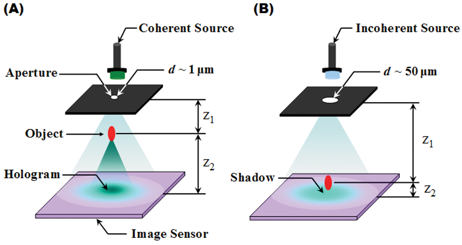
Schematics of digital in-line holography (DIH) (A) and incoherent or partially coherent lens-free on-chip imaging (B) methods. z1 is the distance between the aperture and the object planes, z2 is the distance between the object and the image sensor planes, and d is the diameter of the aperture. In DIH, a small aperture and a coherent light source (e.g., a laser) are used where the object is placed closer to the light source. In incoherent or partially coherent lens-free on-chip imaging platforms, a relatively larger aperture and an incoherent or partially coherent light source is used, and the object is placed much closer to the image sensor plane.
Using DIH under spatially and temporally coherent illumination, one can reconstruct lens-free images of specimens with a resolution of less than a micrometer. For example, Xu et al. reconstructed images of Ditylum brightwellii by stacking series of sectional images with 460 nm intervals. 21 They observed the hollow shell of a cytoplasm and the outline of a frustule. Micó and Zalevsky further modified the original DIH concept using a synthetic aperture scheme. For this purpose, they used a combination of angular multiplexing provided by tilted-beam illumination with time multiplexing of different objects’ spectral regions. 35 Using this approach, they were also able to achieve a resolution of less than 1 µm and observed the thin tail (<1 µm) of a swine sperm. In addition, because of its large depth of field compared with a lens-based microscope, DIH has also been used to record 3D trajectories of moving objects. Accordingly, the swimming behavior of bacteria and marine species such as Ciliate, Didinium, and Rotifer has been observed 25 ( Fig. 2 ). Furthermore, DIH has also been applied to other research fields, such as microfluidics for flow field visualization22,29,41 and interferometry for measurement of deformation of soft materials.13,19,20,26
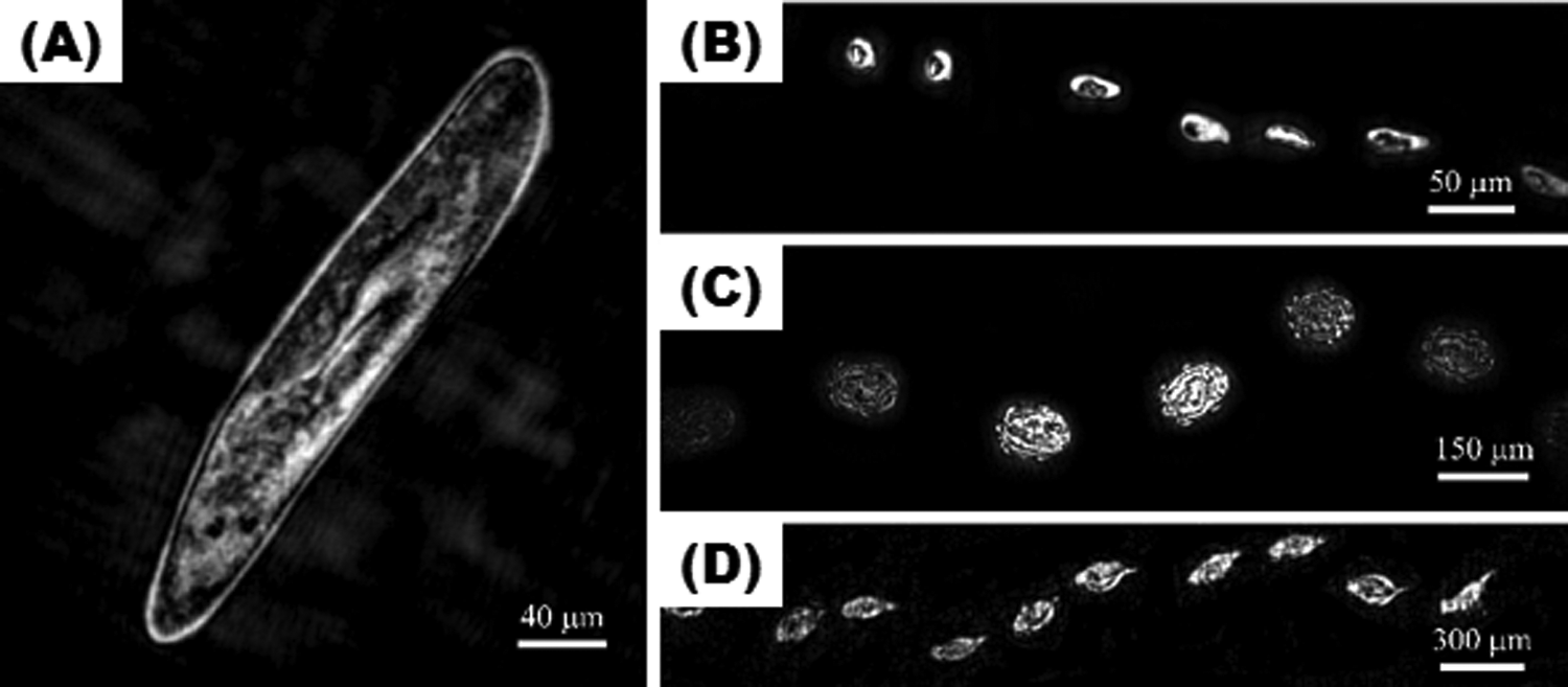
Images of underwater species obtained using digital in-line holography (DIH). 25 (A) Paramecium (length: 320 µm, width: 46.8 µm). Trajectories of various underwater species. (B) Ciliate (length: 25 µm, width: 13 µm), (C) Didinium (length: 133 µm, width: 77 µm), and (D) Rotifer (length: 200 µm, width: 100 µm). Reproduced with permission of Applied Optics.
Incoherent Lens-Free On-Chip Imaging
Lens-free incoherent on-chip imaging forms a very simple method because it does not require a coherent light source, such as a laser or any other bulky/advanced optical components. In lens-free incoherent on-chip imaging, the specimen is placed directly on or close to a digital sensor array (i.e., z1 » z2), and an incoherent light source is used for illumination ( Fig. 1B ). Because this approach uses an incoherent light source (e.g., a broadband extended source), the image sensor records self-interfering classical diffraction patterns or the shadows of the specimen of interest. Because z1 » z2, this lens-free imaging geometry can achieve a wide field of view (FOV, e.g., ~10–20 cm2), which can be used for, for example, high-throughput detection. 16 Accordingly, despite the lack of sufficient spatial resolution, this incoherent platform can still be rather useful as a cytometry tool rather than a microscopic imager. For instance, Lange et al. monitored the temperature dependence of Caenorhabditis elegans. 23 They measured the stroke frequency of C. elegans by monitoring the variations in the shadow images of these animals. In addition, incoherent illumination has also been used to develop a lensless, ultrawide field cell-monitoring array platform based on shadow imaging (LUCAS) that can detect and count cells with a FOV that is two orders of magnitude wider than that of a conventional microscope. 30 Using LUCAS, different cell types were distinguished from each other based on their specific shadow patterns/signatures. After its initial demonstration, Su et al. developed a novel pattern recognition algorithm and further improved LUCAS to handle heterogeneous cell solutions. 16 Later, by incorporating microfluidics onto a LUCAS platform, Moon et al. developed an HIV-monitoring device on a bench-top platform. 31 A similar lens-free incoherent on-chip imaging platform has also been applied to real-time monitoring of squirming objects such as beating cardiomyocytes. For this purpose, Kim et al. extracted a CMOS image sensor from a commercial Web cam and used it to record changes in the beating rates and the beat-to-beat variations of embryonic stem cell–derived cardiomyocytes under treatment of doxorubicin and isoprenaline ( Fig. 3 ). 42
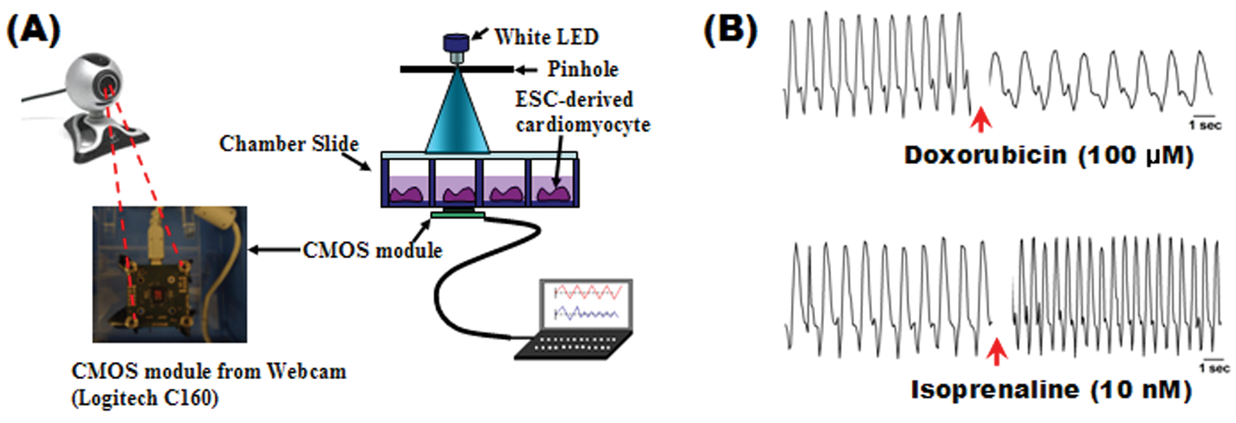
Lens-free imaging for cardiotoxicity testing. 42 (A) Schematic of a cardiotoxicity sensor that uses direct projection. Embryonic stem cell–derived cardiomyocytes are placed on a complementary metal oxide semiconductor image sensor extracted from a Web cam. (B) The effects of two drugs (isoprenaline and doxorubicin) on beating rate variations of cardiomyocytes were investigated. Isoprenaline (doxorubicin) is found to increase (decrease) the beating rates of cardiomyocytes. Reproduced with permission of the Lab on a Chip.
Partially Coherent Digital In-line Holography
Lens-free incoherent on-chip imaging that is discussed in the previous section has a large FOV, but its resolution is significantly limited because of lack of temporal and spatial coherence. However, it turns out that by selecting a quasi-monochromatic light source (e.g., a simple LED) together with a relatively large pinhole (e.g., 0.5–1 mm), one can fine-tune the spatial and temporal coherence properties of the illumination beam at the sensor plane such that the useful lens-free holographic information of the specimens can still be recorded and digitally reconstructed. Further details of this approach and some related reconstruction schemes can be found in, for example, work by Mudanyali et al. 36 and Isikman et al. 45
In this partially coherent approach, the object is also placed closer to the image sensor plane rather than the pinhole. 15 Because of a demagnification factor in its imaging geometry 36 (z1/z2 ~ 40 – 100), spatially incoherent illumination through a large aperture is approximately equivalent to coherent illumination of a micro-object as long as the object’s diffraction pattern is smaller than the coherence diameter at the sensor plane (Dcoh ~ (λ0z1)/D), where D is the width of the aperture (pinhole) and λ0 is the central wavelength of the incoherent light source. 36 The bandwidth of the light source is also critical as it determines the effective numerical aperture of the reconstructed images; however, because of short z2 distances involved in this approach, a simple LED with a bandwidth of ~10 to 20 nm would be sufficient to reach, for example, 0.2 numerical aperture. 36
Using partially coherent DIH, on-chip cytometry has been developed to analyze a heterogeneous solution of red blood cells, yeast, and microspheres using pattern recognition algorithms. 15 Su et al. also applied this approach for automated semen analysis and measured the number, speed, and dynamic trajectories of motile sperms within a field-portable telemedicine microscope that weighs ~45 g. 39 In another study, a pixel super-resolution scheme was developed to improve the resolution of partially coherent DIH to submicrometer. The pixel super-resolution scheme was realized by shifting the light source, which causes subpixel hologram shifting at the sensor plane. Using this approach, Bishara et al. were able to achieve a spatial resolution of <1 µm over an FOV of 24 mm 2 .32,44 They reconstructed images of red blood cells and C. elegans. 32 Stybayeva et al. combined lens-free holographic imaging with micro-arrayed antibodies and performed rapid and multiparametric analysis of leukocytes in human blood. 38 They also measured the CD4/CD8 ratio (HIV/AIDS diagnostic marker) in uninfected and HIV-infected blood samples.
To further improve the axial resolution in partially coherent in-line holography, a multiangle lens-free imaging platform and a lens-free optical tomographic microscope have also been developed.40,45,47 Using this lens-free optical tomographic microscope, Isikman et al. were able to achieve a spatial resolution of <1 µm and <3 µm in the lateral and axial direction, respectively, within a sample volume of ~15 mm3 ( Fig. 4 ). 45
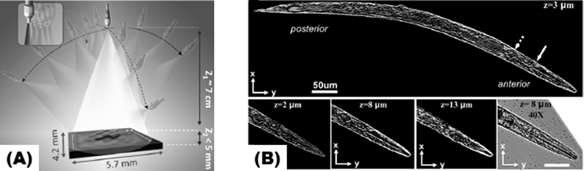
Lens-free optical tomography on a chip using partially coherent digital in-line holography. 45 (A) The sample is illuminated from multiple angles using a partially coherent light source to record lens-free in-line holograms of the specimen on the chip. (B) Images of C. elegans obtained using lens-free optical tomography. Tomogram of an entire worm corresponding to a plane that is 3 µm above the center of the worm (top). Tomograms for the anterior of the worm at different layers (bottom) and a microscope comparison image (40×, NA = 0.65, bottom right). Reproduced with permission of the PNAS.
Other Related Lens-Free Imaging Methods
An optofluidic microscope realized by placing an array of submicron apertures on a CMOS imager has recently been developed. In this approach, a micro-fluidic channel confined C. elegans on the imaging section of CMOS sensors. By confining the motion of C. elegans, a projection image of the specimen was created using the temporal signal emerging from each submicron aperture.18,28,37 Relatively recently, holographic approaches were also applied to opto-fluidics to create lens-free holographic opto-fluidic microscopy and tomography techniques.33,48 Fluorescent microscopy was also recently applied to lens-free imaging to probe transgenic C. elegans, 46 to count fluorescent-labeled white blood cells, 34 to detect C. botulinum toxin activity, 43 and for fluorescent microarray analysis. 27
Summary
In this mini-review, we have discussed some of the emerging lens-free technologies and their biomedical applications. Lens-free imaging, in general, provides field-portable, cost-effective, and a fast imaging toolset, and its applications are on the rise. Recent advances in lens-free imaging include the demonstration of a wider FOV and higher resolution, providing immediate applications in high-throughput biosensing. Therefore, lens-free imaging can reduce the dimensional gap between lab on a chip components and microscopy to provide a powerful approach to a portable POC diagnostic system.
Footnotes
Acknowledgements
This article was supported by the National Institutes of Health (HL092836, EB008392). S.B.K. was partially supported by the National Research Foundation of Korea Grant funded by the Korean government (NRF-2009-352-D00032).