
Abstract
Our present study aimed to reveal the clinical significance of miR-605-5p/STAT2 axis in patients with gastric cancer (GC). The association of STAT2 or miR-605-5p expression with the clinicopathological characteristics and prognosis in patients with GC was analyzed using the tissue microarray and TCGA RNA-seq data. Pearson correlation analysis was used to evaluate the correlation of STAT2 with miRNAs expression in GC tissues. Cox proportional hazard regression model was conducted to assess whether STAT2 or miR-605-5p expressions was an independent prognostic factor in patients with GC. Consequently, we found that STAT2 expression levels were dramatically elevated in GC tissues and acted as an independent prognostic factor of poor survival in patients with GC. The upregulation of STAT2 was attributable to the dysregulation of miR-605-5p rather than its genetic and epigenetic modulation. MiR-605-5p indicated a negative correlation with STAT2 expression and was an independent prognostic factor of poor survival in patients with GC. In conclusion, dysregulation of miR-605-5p/STAT2 axis predicted a poor survival in patients with GC.
Introduction
Gastric cancer (GC) is the third common cancer and the third leading cause of cancer-related death in China, 1 and the prognosis of GC patients in an advanced stage is unfavorable due to its distant metastasis. Numerous studies have shown that the activation of oncogenes or inactivation of anti-oncogenes participates in the multiple stages of GC. Therefore, identification of the potential markers is indispensable for the early detection of GC.
Activation of signal transducers and activators of transcription (STATs) is associated with the tumor growth and progression. Increasing data show that STAT2 expression is increased in cholangiocarcinoma. 2 High expression of STAT2 is associated with the high-risk of lung adenocarcinoma (LAC), 3 and the aggressive phenotype in breast cancer. 4 STAT2 also regulates the tumor growth 5 and IFN-alpha-induced cell apoptosis. 6
MiRNAs/target mRNA axis participates in the progression of multiple malignancies including GC. 7 Some studies have shown that miR-221/222 suppresses glioblastoma tumorigenicity by inhibition of INF-alpha signaling. 8 In the present study, we found that STAT2 expression was dramatically elevated in GC tissues and acted as an independent prognostic factor of poor survival in patients with GC. The upregulation of STAT2 was attributable to the dysregulation of miR-605-5p, which indicated a negative correlation with STAT2 expression, and was an independent prognostic factor of poor survival in patients with GC.
Materials and methods
Clinical samples
The tissue microarray (TMA) including 89 pair-matched GC tissue samples was purchased from Shanghai Outdo Biotech Co. LTD (HStm-Ade180Sur-01, Shanghai, China). The clinicopathological and prognostic data in another cohort for 415 cases of GC samples and 32 adjacent normal tissues as well as the relative expression levels of STAT2 and 10 miRNAs (has-323b-3p, has-3127-5p, has-24-3p, has-502-3p, has-501-3p, has-370-3p, has-3150b-3p, has-3928-3p, has-605-5p has-3074-5p) were downloaded from The Cancer Genome Atlas (TCGA) 2015 RNA-seq database (https://genome-cancer.ucsc.edu). The protocols used in our study were approved by the Ethics Committee of Jingzhou Central Hospital.
Immunohistochemistry analysis
The protein expression of STAT2 was examined using immunohistochemistry (IHC) analysis in 89 pair-matched GC tissues from the TMA. These tissues were stained using anti-STAT2 (ab53132; Abcam, Cambridge, MA, USA). The detailed process of IHC as well as the quantification of STAT2 protein expression was conducted as previously described. 7
Identification of the miRNAs that target STAT2 3′UTR
Ten miRNAs that target 3′UTR of STAT2 gene were identified using the TargetScanhuman7.1 (http://www.targetscan.org/vert_71/) according to the weighted context score.
Statistical analysis
The statistical analyses were conducted using SPSS 20.0 (IBM, SPSS, Chicago, IL, USA) and GraphPad Prism. Student’s t test and chi-square test were used to analyze the statistical significance for the comparisons of two groups. Pearson’s correlation coefficient analysis was used to analyze the correlation of STAT2 with the miRNAs expression in GC tissues. Survival and recurrence curves were plotted using the Kaplan–Meier method and the statistical significance was evaluated using a log-rank test. Statistical significance was set at P < 0.05.
Results
Upregulation of STAT2 expression was associated with a poor survival in GC patients
The protein expression levels of STAT were examined in 89 pair-matched GC samples by IHC analysis, which indicated that the positive expression of STAT2 was increased in GC tissues as compared with the adjacent normal tissues (P < 0.001, Supplementary Figure S1). TCGA cohort further validated that STAT2 expression levels were tremendously increased in paired and unpaired GC tissues (P = 0.0039, P < 0.0001; Figure 1(a)). According to the survival time, survival status, and STAT2 expression levels, a cutoff value of STAT2 was obtained using the Cutoff Finder tool (http://molpath.charite.de/cutoff/load.jsp) in these 301 GC samples (Figure 1(b)) and divided the patients into high and low expression groups (Figure 1(c)).

High expression of STAT2 was associated with a poor survival in GC patients. (a) TCGA cohort analysis of the expression levels of STAT2 in paired and unpaired GC tissues. (b) ROC curve analysis of the cutoff value of STAT2 in GC samples (n = 301). (c) The cutoff value of STAT2 divided the patients into high and low expression groups. (d) The Kaplan–Meier analysis of the association of high or low STAT2 expression with the poor survival and tumor recurrence in GC patients.
Moreover, we analyzed the association of STAT2 expression with the clinicopathological characteristics and prognosis in GC patients and found that high expression of STAT2 was associated with pathological stage (P = 0.002) and lymph node metastasis (P = 0.040), but had no association with other factors (each P > 0.05) in GC patients (Supplementary Table S1). TCGA cohort further evidenced that STAT2 expression was only associated with the age (P = 0.034) rather than other factors in GC patients (Supplementary Table S2). The Kaplan–Meier analysis demonstrated that the patients with high STAT2 expression had a poorer survival, but displayed no difference in tumor recurrence, as compared with those with low STAT2 expression (Figure 1(d)). Univariate and multivariate analysis revealed that high STAT2 expression as well as the pathological stage was an independent prognostic factor of poor survival in GC patients (Table 1).
Cox regression analysis of STAT2 expression as a survival predictor in GC patients.

GC: gastric cancer; CI: confidence interval; NA: not analyzed.
STAT2 was identified to have a negative correlation with miR-605-5p expression in GC tissues
To uncover the cause of STAT2 upregulation in GC samples, we estimated the genetic and epigenetic alterations of STAT2 gene in GC (n = 287), indicating that STAT2 had no significant alterations at the genetic (Supplementary Figure S2A) and methylation levels (Supplementary Figure S2B), and DNA mutation, copy number, and methylation level could not account for the STAT2 upregulation in GC.
We hypothesized that STAT2 upregulation in GC might be modified by miRNAs at post-transcriptional levels. To confirm our hypothesis, we identified 10 miRNAs that may bind to STAT2 3′UTR using TargetScanHuman7.1 and evaluated the expression levels of these miRNAs in GC samples, of which only miR-605-5p had a reduced expression in paired and unpaired GC tissue samples, as compared with the adjacent normal tissues (Figure 2(a)). The downregulation of miR-605-5p was validated in another TCGA cohort in unpaired GC tissues (Figure 2(b)). Pearson correlation analysis showed that STAT2 had a negative correlation with miR-605-5p expression in GC tissues (Figure 2(c)).
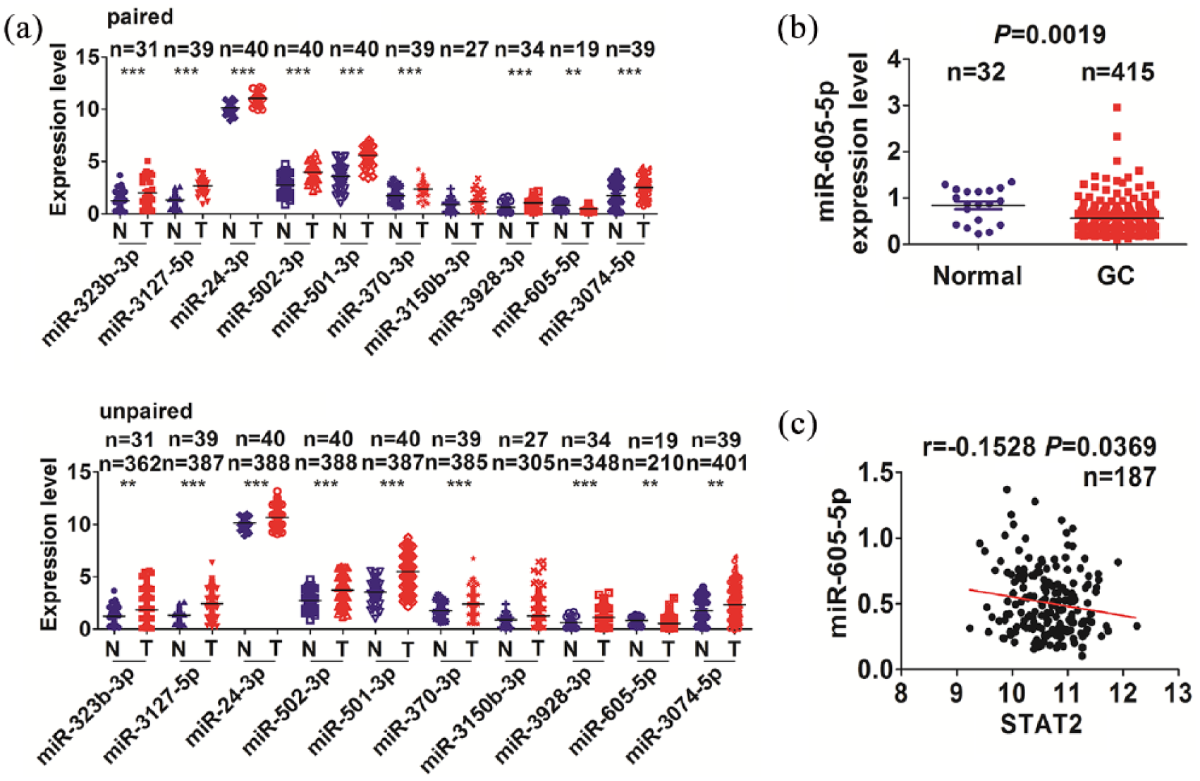
STAT2 was identified to display a negative correlation with miR-605-5p expression in GC tissues. (a) TCGA analysis of the expression levels of 10 miRNAs in paired and unpaired GC tissues. (b) TCGA validation of the expression levels of miR-605-5p in unpaired GC tissues. (c) Pearson correlation analysis of the correlation of STAT2 with miR-605-5p expression in GC tissues.
Low expression of miR-605-5p was associated with a poor survival in GC patients
Having verified the decreased expression of miR-605-5p in GC tissues (Figure 2(a) and (b)), we further analyzed the association between miR-605-5p expression and clinicopathological characteristics and prognosis in GC patients. A cutoff value of miR-605-5p was obtained in GC tissues (n = 160) using the Cutoff Finder tool (Figure 3(a)) and divided the patients into high and low miR-605-5p expression groups (Figure 3(b)). The AUC value, sensitivity, and specificity of miR-605-5p expression in GC patients were 0.51, 95.0%, and 10.0%, respectively (Figure 3(a)).

Low expression of miR-605-5p was associated with a poor survival in GC patients. (a) ROC curve analysis of the cutoff value of miR-605-5p in GC samples (n = 160). (b) The cutoff value of miR-605-5p divided the patients into high and low expression groups. (c) The Kaplan–Meier analysis of the association of high or low miR-605-5p expression with the poor survival and tumor recurrence in GC patients.
Furthermore, miR-605-5p expression had no association with the clinicopathological factors in GC patients (each P > 0.05, Supplementary Table S3). The Kaplan–Meier analysis demonstrated that the patients with low miR-605-5p expression harbored a poorer survival, but had no difference in tumor recurrence (Figure 3(c)), as compared with those with high miR-605-5p expression. Univariate and multivariate analyses revealed that miR-605-5p high expression as well as the distant metastasis was an independent prognostic factor of overall survival in GC patients (Supplementary Table S4).
Discussion
STAT2 is aberrantly expressed in cancers, acting as an oncogenic factor. 2 However, the expression levels of STAT2 in GC remain unclear. In the present study, STAT2 expression was confirmed elevated in GC by TMA and TCGA cohorts. TMA data demonstrated that high expression of STAT2 was associated with pathological stage and distant metastasis, but another cohort from TCGA data validated that STAT2 had no association with the clinicopathological factors except for the age in GC. The upregulation of STAT2 has been reported associated with a poor prognosis in cancers.3,4 We found that high expression of STAT2 was associated with a poor survival and acted as an independent prognostic factor of poor survival in GC patients.
Furthermore, the upregulation of STAT2 was attributable to the post-transcriptional modulation rather than its genetic and epigenetic alterations in GC. MiR-605-5p was identified downregulated in GC tissues, displayed a negative correlation with STAT2 expression, and acted as an independent prognostic factor of poor survival in GC patients. Some studies indicated that the serum levels of miR-605 are associated with the risk of prostate cancer, 9 and miR-605 acts as a tumor suppressor by targeting engrailed homeobox 2 in prostate cancer, 10 and Gankyrin in cholangiocarcinoma. 11 These results suggested that STAT2 expression might be regulated by miR-605-5p in GC.
In conclusion, our findings demonstrate that high expression of STAT2 or low expression of miR-605-5p is associated with poor survival and acted as an independent prognostic factor of poor survival in GC patients. miR-605-5p/STAT2 axis may represent potential markers for poor survival in GC patients.
Supplemental Material
Figure_S1_pdf – Supplemental material for Dysregulation of miR-605-5p/STAT2 axis predicts an unfavorable survival in patients with gastric cancer
Supplemental material, Figure_S1_pdf for Dysregulation of miR-605-5p/STAT2 axis predicts an unfavorable survival in patients with gastric cancer by Yang Teng, Rong Tang and Shao-Jie Jiang in European Journal of Inflammation
Supplemental Material
Figure_S2_pdf – Supplemental material for Dysregulation of miR-605-5p/STAT2 axis predicts an unfavorable survival in patients with gastric cancer
Supplemental material, Figure_S2_pdf for Dysregulation of miR-605-5p/STAT2 axis predicts an unfavorable survival in patients with gastric cancer by Yang Teng, Rong Tang and Shao-Jie Jiang in European Journal of Inflammation
Supplemental Material
Supplementary_data – Supplemental material for Dysregulation of miR-605-5p/STAT2 axis predicts an unfavorable survival in patients with gastric cancer
Supplemental material, Supplementary_data for Dysregulation of miR-605-5p/STAT2 axis predicts an unfavorable survival in patients with gastric cancer by Yang Teng, Rong Tang and Shao-Jie Jiang in European Journal of Inflammation
Footnotes
Acknowledgements
Y.T. and R.T. contributed equally to this article.
Declaration of conflicting interests
The author(s) declared no potential conflicts of interest with respect to the research, authorship, and/or publication of this article.
Funding
The author(s) disclosed receipt of the following financial support for the research, authorship, and/or publication ofthis article: This work was supported by grants from the Jingzhou Municipal Science and Technology Bureau (no. 2016086).
Supplemental material
Supplemental material for this article is available online.
References
Supplementary Material
Please find the following supplemental material available below.
For Open Access articles published under a Creative Commons License, all supplemental material carries the same license as the article it is associated with.
For non-Open Access articles published, all supplemental material carries a non-exclusive license, and permission requests for re-use of supplemental material or any part of supplemental material shall be sent directly to the copyright owner as specified in the copyright notice associated with the article.