
Abstract
Background:
Survivin is a protein that inhibits apoptosis and regulates cell division. Studies examining the expression of survivin in oral cancer are limited. Correlation between the expression of survivin in dysplasia and different grades of oral squamous cell carcinoma (OSCC) could help us to assess its use as a biomarker.
Objectives:
The objectives of this research were to evaluate the expression of survivin in dysplasia and in different grades of OSCC keeping in mind that when comparison is made between these two groups, it may highlight the use of this biomarker.
Method:
Forty samples divided into four groups; 10 each of dysplasia, well-differentiated OSCC, moderately differentiated OSCC and poorly differentiated OSCC were selected for the study. Immunohistochemical staining for the expression of survivin protein was performed.
Results:
Survivin showed increased expression in OSCC as compared to dysplasia, although the result was not statistically significant. The distribution of survivin showed significant differences between dysplasia and moderately differentiated OSCC.
Conclusion:
A higher expression of survivin was seen in OSCC as compared to dysplasia, hence it could be used as a marker for proliferation and invasion, but the immunohistochemical expression of survivin overall shows variable expression. To correlate the expression of survivin with the grade of the tumour, a bigger sample size is desirable.
Introduction
Oral squamous cell carcinoma (OSCC) constitutes the most frequent malignancy of the oral cavity. Carcinogenesis is a multistage process involving the activation of oncogenes and inactivation of tumour suppressor genes. 1 It is a multifactorial disease and one of the main factors that contribute to tumour development is an imbalance of regulatory mechanisms controlling cell cycle progression, cell death, viability balance and apoptosis. 2 An important reason for poor prognosis of OSCC is the lack of molecular prognostic markers available to assess the aggressiveness of this tumour, probability of recurrence and disease progression. 3
Apoptosis has become one of the basic tools in cancer research and establishing cancer pathways. 4 Survivin is a protein that inhibits apoptosis and regulates cell division. It is categorized as a member of the inhibitor of apoptosis protein (IAP) family. Survivin inhibits apoptosis, by either directly or indirectly interfering with the function of caspases. Survivin is expressed in embryonic tissues as well as in majority of human cancers but is not expressed in most normal adult tissues, except in thymus, placenta and basal colonic epithelium and CD34+ stem cell also express survivin. 4
The cancer-specific expression of survivin coupled with its importance in inhibiting cell death and in regulating cell division makes it a useful prognostic marker for cancer and a potential target for cancer therapy.
Recent research has shifted focus towards survivin as a target for cancer therapy. This is because there is evidence suggesting its role limited not only to apoptosis inhibition but to its association with angiogenesis and tumour invasiveness. 5,6 Survivin is known to be expressed in the G2/M phase of the cell cycle to support rapidly dividing cell machinery and helps in proper segregation of chromosomes during cell division. 7,8 Several protocols have been developed to examine the therapeutic potential of survivin in cancer treatment. These include suppressing survivin expression by antisense, ribozyme, siRNA or shRNA approaches or antagonizing survivin function by dominant-negative survivin or by Cdk inhibitors. Anti-survivin therapy has been evaluated in several preclinical models using mice harbouring pre-established tumours. 9
However, there are relatively few studies correlating the expression of survivin in dysplasia and different grades of OSCC. The present study aims at assessing the expression of survivin in potentially malignant lesions and different grades of OSCC and thereby, evaluating its potential role as a prognostic marker.
Material and methods
Case selection
The study group consisted of 40 samples. Paraffin embedded archival tissue blocks of histopathologically confirmed dysplasia and OSCC were selected for this study. The sample was divided into four groups consisting of 10 cases each of histopathologically confirmed dysplasia, well-differentiated OSCC, moderately differentiated OSCC and poorly differentiated OSCC. The hematoxylin and eosin stained slides were reviewed to confirm the diagnosis.
Immunohistochemistry
Immunohistochemical study using an antibody to survivin protein was performed on 4 µm sections on poly-lysine coated slides. Antigen retrieval was performed by heat-induced epitope retrieval technique using a pressure cooker. Anti-survivin rabbit monoclonal antibody (Path N Situ) was used for immunostaining.
Scoring and grading
In this study, we recorded the sections as positive when either/both nuclear or cytoplasmic expression of survivin was visible. Expression of survivin positive cells in dysplasia and different grades of OSCC were detected (Figure 1). To quantitate the expression of survivin, 300 cells were examined in at least five areas at 40× magnification and a mean percentage of positive tumour cells was determined, allowing the tumours to be graded into the following categories.

Immunohistochemical patterns observed with survivin staining. (a) Dysplasia with focal nuclear and cytoplasmic staining scored as 1; (b) well-differentiated oral squamous cell carcinoma with cytoplasmic staining scored as 3; (c) moderately differentiated oral squamous cell carcinoma with nuclear and cytoplasmic staining scored as 4; (d) poorly differentiated oral squamous cell carcinoma with no staining scored as 0; (e) negative control of normal oral mucosa with the absence of staining.
Grading of survivin positive tumour cells
Grading of survivin positive tumour cells is as follows: (a) 0 = <5%, (b) 1 = 5–25%, (c) 2 = 26–50%, (d) 3 = 51–75% and (e) 4 = >75%. Cases with a score zero were considered as negative and cases with scores of 1–4 as positive.
The percentage of survivin positive cells was calculated according to the method of Lo Muzio et al. 1 All grading was done independently by three experienced observers to eliminate bias and a mean of three scores was assigned to each case.
Statistical analysis
The data were subjected to statistical analysis. The differences noted were recorded. The statistical method used was χ 2 test and p value. P value of less than 0.05 is considered as statistically significant.
Results
Immunohistochemical staining patterns for survivin observed in tissue sections of normal mucosa, dysplasia and OSCC are shown in Figure 1. Normal oral mucosa which was taken as negative control did not show positivity for survivin. This was in accordance with previous studies. 1
Figure 1(a) shows dysplasia with focal nuclear and cytoplasmic staining scored as 1 and Figure 1(b) shows well-differentiated OSCC with cytoplasmic staining scored as 3. Figure 1(c) shows moderately differentiated OSCC with nuclear and cytoplasmic staining scored as 4, Figure 1(d) shows poorly differentiated OSCC with no staining scored as 0, and Figure 1(e) shows negative control of normal oral mucosa with absence of staining.
Expression of survivin in dysplasia
All 10 cases of dysplasia showed variable survivin expression. Staining observed was both focal nuclear and cytoplasmic. Of 10 cases of dysplasia, 7 cases showed less than 5% of survivin expression (score 0), 2 cases showed 5–25% of survivin expression and 1 case showed 51–75% of survivin expression (Table 1).
The frequency distribution of survivin expression in the two study groups.a

OSCC: oral squamous cell carcinoma.
a p Value = 0.765 (Not significant).
Expression of survivin in OSCC
Out of the 30 cases of OSCCs, 16 cases showed less than 5% of survivin expression (score 0), 5 cases showed 5–25% of survivin expression (score 1), 2 cases showed 26–50% of survivin expression (score 2), 6 cases showed 51–75% of survivin expression (score 3) and 1 case showed over 75% of survivin expression (score 4). Well-differentiated OSCC showed cytoplasmic staining for survivin. Moderately differentiated OSCC showed both nuclear and cytoplasmic staining for survivin. Poorly differentiated OSCC showed nuclear and cytoplasmic staining with survivin (Table 1).
Group comparisons showed a statistically significan difference in survivin expression only between dysplasia and moderately differentiated OSCC (Table 2).
The comparison of expression of survivin between three groups of OSCC and group of dysplasia cases.a
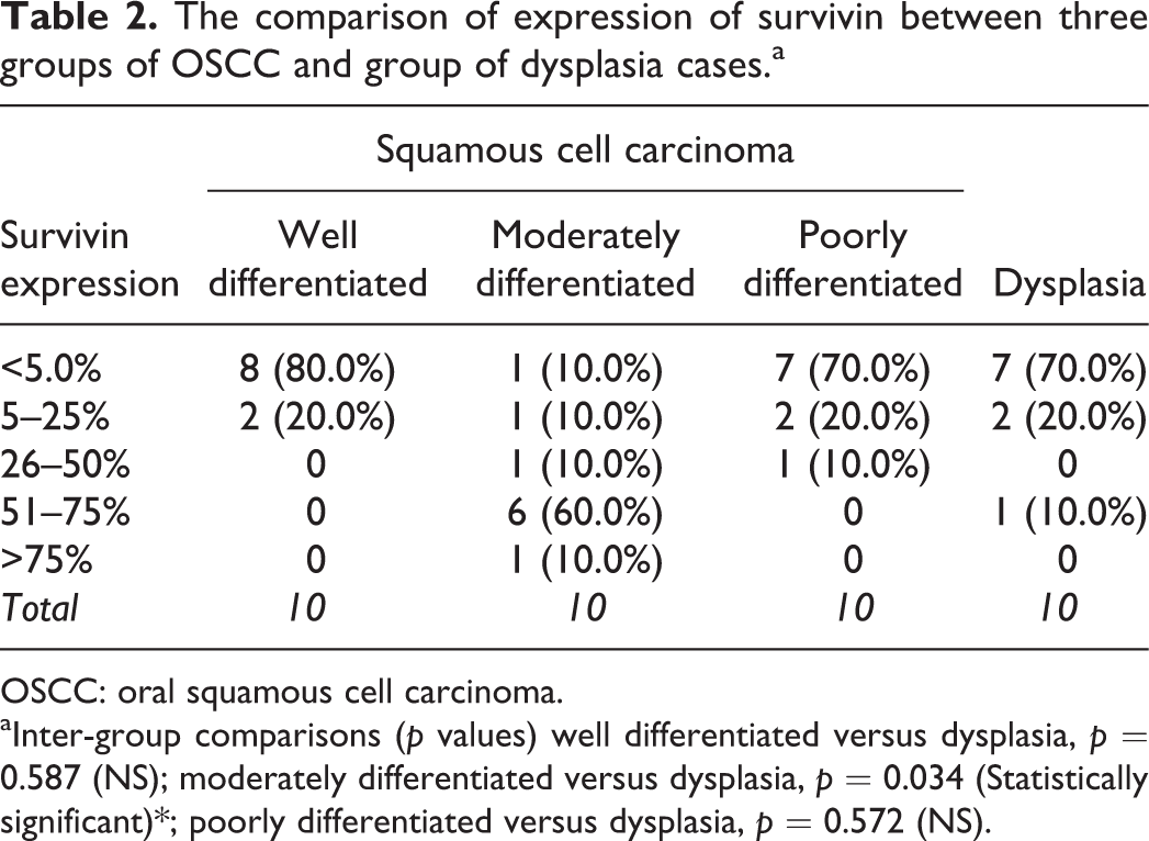
OSCC: oral squamous cell carcinoma.
aInter-group comparisons (p values) well differentiated versus dysplasia, p = 0.587 (NS); moderately differentiated versus dysplasia, p = 0.034 (Statistically significant)*; poorly differentiated versus dysplasia, p = 0.572 (NS).
Discussion
OSCC is a malignant neoplasm exhibiting aggressive phenotypes with unpredictable biological behaviour. 10 The accumulation of neoplastic cells can occur through enhanced proliferation, diminished cell turnover or combination of both processes. 11
Biomarkers for OSCC may exist as genetic or molecular indicators identified as mutated, silenced, overexpressed, amplified or altered genes or gene products. These biomarkers may represent the loss of functional tumour suppressors, cell cycle regulators or apoptosis regulators resulting in dysregulation of cell growth and/or cell death. 12
Apoptosis or programmed cell death results from the activation of molecules belonging to a family of 14 cysteine proteases called caspases, enzymes that cleave cellular proteins. 13
The structure of survivin protein is intimately linked with its function as an inhibitor of apoptosis. 4 Several mechanisms are explained by which survivin suppresses apoptosis. Direct suppression of caspase-3 by survivin has been speculated by some investigators; yet survivin lacks structural components present in other IAPs that allow their direct binding to caspase-3. Survivin may indirectly inhibit caspases via intermediate proteins. 14 Survivin has a versatile role in modulating cell division and apoptosis in cancer. Low survivin expression is statistically correlated significantly with better survival rates but not with age, sex, tumour size, presence of lymph nodes and distant metastasis. 11
Survivin was found to be preferentially expressed in non-advanced, non-metastatic and chemotherapy sensitive OSCC. On the contrary, increased expression of survivin was found in high-grade tumours suggesting its contribution to apoptosis resistance in response to therapy. Also significant correlation between survivin expression, lymph node metastasis and proliferation was revealed but not with differentiation, micro-vessel density or Tumour/Node/Metastasis (TNM) staging. 11
According to the results of this study, survivin showed increased expression in OSCC as compared to dysplasia. Of the three groups of OSCCs, moderately differentiated OSCC showed increased expression as compared to dysplasia which was statistically significant.
Previous studies by Jane et al. who examined 38 cases of OSCCs and 17 cases of leukoplakia found increased expression of survivin in high-grade OSCC and it was statistically significant. 3
Lo Muzio et al., who studied 110 cases of OSCCs, found that survivin expression was increased in poorly differentiated tumours, but the result was not statistically significant. Their data suggested that survivin expression may identify cases of OSCC with more aggressive and invasive phenotype only. 1
The results of our study differed from the results of Jane et al. and Lo Muzio et al. 1,3 The cases studied included by us may not have had an aggressive phenotype. Standardization of the immunohistochemistry technique and external quality assurance on immunohistochemistry data may help to reduce the variations in the reported study.
Conclusion
According to the results of our study, survivin showed increased expression in OSCC than dysplasia. This expression may not necessarily increase with the increase in the grade of the tumour but may show variations with aggressive phenotypes. Survivin may be used as a prognostic marker for proliferation and invasion. This differential expression of survivin in squamous malignancies may make it an attractive target for selection of cases for molecular-based cancer therapies.
Footnotes
Acknowledgements
We are extremely thankful to Dr. Vinay Hazare, Senior Oral Pathologist, India for encouraging us to publish this research work in the esteemed journal Translational Research in Oral Oncology. This endeavour would not have been possible without the blessings of Professor Saman Warnakulasuriya and Professor Vinay Hazare.
Declaration of Conflicting Interests
The author(s) declared no potential conflicts of interest with respect to the research, authorship, and/or publication of this article.
Funding
The author(s) received no financial support for the research, authorship, and/or publication of this article.