
Abstract
Objective
This study builds on brief focused mindfulness meditation (BFMM) to examine its associations with physiological indices and electroencephalographic (EEG) neural features related to stress in young adults. In addition, deep learning models are employed to identify complex, nonlinear patterns in EEG signals during BFMM, aiming to determine the most effective classification model.
Methods
Twenty-nine participants (n=29) were enrolled in a before-and-after study of the same cohort. Participants underwent a 10-min resting state, then were instructed to perform BFMM for 10 min. Physiological indices were recorded pre- and post-BFMM, while EEG signals were captured during both the resting and BFMM states. Deep learning techniques including multi-layer perceptron (MLP), long short-term memory (LSTM), convolutional neural network (CNN), and ensemble models were subsequently employed to classify EEG signals.
Results
Final Twenty-four participants (n=24) were included in the analysis. The differences in both heart rate (t = 4.22, p < 0.001) and respiratory rate (t = 5.05, p < 0.001) were significant between pre- and post-BFMM levels. Results showed significant power spectral density differences between the resting and BFMM states in the theta (Z = 3.17, q = 0.039, beta (Z = 3.17, q = 0.049) bands of the right frontotemporal region (T8) and the theta (t = 3.41, q = 0.039) band of the right frontocentral region (FC6). In addition, the ensemble model (MLP+LSTM+CNN) outperformed other methods, achieving an accuracy of 79.0% in classifying EEG signals.
Conclusion
These finding suggested that a single session of BFMM may regulate the autonomic nervous system and modulate neural activity. The proposed ensemble model shows promise in distinguishing BFMM from resting-state EEG, providing a foundation for future EEG-based assessment of mindfulness meditation.
Introduction
In contemporary psychological research, mindfulness carries multiple layers of meaning. As a transient mental state, mindfulness is defined as the act of paying attention to present-moment internal and external experiences with an attitude of curiosity, non-judgment, and acceptance. 1 As a stable personality trait, mindfulness reflects the frequency and intensity of individuals’ experiences of present-moment awareness in daily life. 2 State and trait mindfulness can influence each other: sustained engagement in state mindfulness practice enhances individuals’ trait mindfulness, while higher trait mindfulness facilitates easier access to mindful states. 2
Meditation refers to a class of practices aimed at promoting mind-body integration, calming the mind, and enhancing well-being. These practices often involve focusing attention on specific objects such as the breath, sounds, or visual images. 3 Mindfulness meditation, a subcategory of meditation, typically involves attentional training in mindfulness. It is one of the most widely used methods for cultivating state mindfulness and, over time, facilitates the development of trait mindfulness.3,4
Mindfulness meditation typically comprises two fundamental forms: focused attention (FA) and open monitoring (OM). 5 FA mindfulness meditation requires practitioners to sustain attention on a specific focus (e.g. the breath), thereby training voluntary attentional control and reducing mind-wandering. 6 In contrast, OM mindfulness meditation encourages practitioners to maintain open, accepting, and non-reactive awareness of any arising experience. 5 These two forms of mindfulness meditation exert differential effects on attention and emotional regulation. Neuroimaging studies have shown that FA meditation primarily enhances activity in the ventral attention network and reduces mind-wandering with the default mode network, while OM meditation involves broader monitoring of internal and external stimuli and fosters a non-judgmental, non-reactive attitude. 5 Moreover, empirical research indicates that 8 weeks of FA mindfulness meditation significantly improves self-reported attentional control, whereas OM training is more effective in increasing emotional non-reactivity. 5 Notably, FA meditation strengthens FA and executive functioning, providing beginners with a clear attentional anchor and making it easier for them to enter a mindful state.7,8
In recent years, with the accelerating pace of life and growing attention to mental health among young people, brief focused mindfulness meditation (BFMM) has gained increasing recognition among youth. BFMM typically denotes to FA mindfulness meditation practices lasting from a few minutes to approximately 10 minutes. 9 This format allows individuals to experience the benefits of mindfulness meditation even within a busy daily schedule, such as physical and mental relaxation, stress reduction, enhanced attentional focus, and improvements in emotional regulation and cognitive functioning. 10 Given BFMM's real-world feasibility and potential stress-reducing effects, the present study investigates associations between the BFMM and physiological, electroencephalographic (EEG) neural correlates in young adults.
A growing body of research has investigated the effects of FA mindfulness meditation on stress in young individuals. Studies have shown that FA mindfulness meditation reduces heart rate, heart rate variability, respiratory rate, blood pressure, and skin conductance in youth.11,12 These physiological indicators—particularly heart rate, heart rate variability, and respiratory rate—are commonly regarded as markers of autonomic nervous system (ANS) functioning. 13 The ANS is critical for modulating emotional experiences, while cognition-driven neural states exert significant influence on its activity. 14 These findings suggest that FA mindfulness meditation may reduce physiological stress by eliciting a “resting state” response at the bodily level. 15 However, it is worth noting that research on the associations of BFMM on both physiological stress in young people remains limited.
At the neural level, EEG is commonly used to identify brain activity associated with FA mindfulness meditation. 16 Existing studies have shown that, compared to the resting state, FA mindfulness meditation is associated with increased alpha and theta power in the brain.17,18 The simultaneous enhancement of alpha and theta waves reflects a state of “relaxed alertness,” which is believed to contribute to stress reduction. 16 In addition, FA mindfulness meditation has been linked to increased activation in the premotor cortex, right dorsolateral prefrontal cortex, dorsal anterior cingulate cortex, and insula—regions associated with cognitive control. Such activations are thought to improve attentional regulation, facilitate psychophysical relaxation, and lower heart rate, anxiety, and overall stress levels. 19 Research has also found that reduced cerebral blood flow in the superior temporal gyrus, cingulate cortex, and prefrontal cortex during FA meditation is significantly associated with increased alpha power. This neural pattern may reflect decreased energy expenditure in the brain, leading to a deeper sense of relaxation and enhanced positive affect and calmness.20,21 In sum, previous EEG studies have not only identified characteristic changes in alpha and theta power during FA mindfulness meditation but also found that increases in meditation depth are typically accompanied by sustained elevations in these oscillatory activities.22,23 However, to the best of our knowledge, no study to date has examined brain activity during BFMM.
Previous studies on FA mindfulness meditation have primarily employed traditional power spectral density (PSD) analysis to examine EEG changes during meditative states.24–26 However, PSD analysis relies on the assumption of linearity, ignores phase information, and cannot capture cross-frequency interactions—an approach that contradicts the inherently nonlinear and dynamic nature of brain activity. 27 Recent findings suggest that linear PSD indices fail to systematically track meditative depth. For example, heartbeat-evoked potentials have been shown to correlate with self-reported meditation depth, whereas commonly used PSD markers, such as midline theta power, do not exhibit covariation with depth. 28 Similarly, Reggente et al. 29 analyzed EEG data from Vipassana meditation experts and found that conventional channel-level spectral features were insufficient to predict depth, while multivariate machine learning models successfully decoded variations in meditative depth. Building on these findings, we conceptualize BFMM as a continuously deepening process and propose the use of deep learning models to detect complex, nonlinear patterns in EEG signals associated with BFMM states.
Recent studies have conceptualized mindfulness meditation as a continuously deepening process (continuum) and have employed deep learning techniques to model and identify meditative states based on nonlinear patterns in EEG activity. De Los Angeles et al. 30 reported that self-report measures such as the MEDEQ can quantify meditation depth through multi-level scoring, with deeper meditative states typically associated with increased theta power and decreased beta/gamma power. In related work, Sharma et al. 31 used an artificial neural network to distinguish between meditators and non-meditators. Reggente et al. 29 further applied EEG and deep learning to predict self-reported, time-varying meditation depth in Vipassana meditation experts. Their results showed that traditional channel-level spectral analyses and pre-defined default mode network regions failed to capture the complex neural dynamics associated with depth fluctuations. In contrast, machine learning models that integrated spatial, spectral, and connectivity features achieved high predictive accuracy for expert-reported depth. 32 This study demonstrated the feasibility of decoding individualized meditative depth from EEG signals. Nevertheless, to date, no research has examined the use of EEG and deep learning to identify BFMM states, leaving a gap in our understanding of how brief meditative experiences are reflected in brain activity.
In summary, previous research has primarily focused on the effects of FA mindfulness meditation on physiological stress and EEG activity in young adults, while the impact of BFMM in these domains remains underexplored. Moreover, although some studies have demonstrated that deep learning outperforms traditional methods in EEG feature classification, it is still unclear which model is most effective for distinguishing the BFMM state from resting-state EEG signals.33,34 To address these gaps, the present study focuses on BFMM and investigates its associations on physiological stress responses and EEG activity in young adults following a single-session intervention. Furthermore, we employed multiple deep learning models to classify EEG features from BFMM and resting states, aiming to identify the most effective model. This approach may provide a methodological foundation for future EEG-based tracking and assessment of FA mindfulness meditation states.
Subjects and methods
Subjects
The G*Power 3.1.9.7 program was used to determine the required sample size. Refer to analogous studies,35,36 in the G*power test, when “Paired samples t test, two-tailed, effect size 0.60, alpha error probability 0.05, power 0.80” was set, the total number of samples was 22. Ultimately, 29 participants were recruited accounting for a 20% dropout rate and unforeseen factors.
This was a before-and-after cohort study. Participants were recruited through flyers, with 29 young adults from Shanghai University of Traditional Chinese Medicine enrolled from 1 November to 31 December 2022. Five participants were excluded: three due to poor data quality caused by distraction from physical movements and two for falling asleep during practice. The final analytic sample comprised 24 participants (14 females, 58.33%; 10 males, 41.67%), with a mean age of 20.13±1.85 years. None had prior experience with mindfulness or other forms of meditation. The demographic characteristics of participants are shown in Table 1.
The demographic characteristics of participants.

The inclusion criteria were (1) full-time undergraduate students aged 18–22 years; (2) no prior experience with meditation practices (including self-study); (3) no language or communication barriers; (4) provided of informed consent and adherence to ethical guidelines. Exclusion criteria were (1) history of major cardiovascular, hepatic, renal, pulmonary, hematologic, gastrointestinal, endocrine, immunologic, dermatologic, or neurological diseases prior to enrollment; (2) consumption of alcoholic or caffeinated foods/beverages within 48 h before the mindfulness meditation session; (3) history of severe psychiatric comorbidities other than anxiety and stress (e.g. bipolar disorders, psychotic disorders) or substance use disorder; (4) noncompliance with the experimental protocols.
All participants were fully informed of the experimental protocols and provided voluntary written informed consent. Upon completing the experiment, they received monetary compensation. The study complied with the Declaration of Helsinki and was approved by Yueyang Hospital of Integrated Traditional Chinese and Western Medicine affiliated to Shanghai University of Traditional Chinese Medicine (No. 2020-178).
Experiment paradigm
Upon entering the test room, participants were first briefed on the experimental procedure and then instructed to sit quietly for 5 min to achieve full relaxation. Firstly, resting-state EEG signals were recorded for 10 min while participants maintained a resting posture. Following the resting state, physiological indices were recorded, including blood pressure, heart rate, and respiratory rate. Next, BFMM state EEG signals were recorded during a 10-min focused mindfulness meditation session guided by study-specific prompts. EEG signals were continuously recorded throughout the session. Post-BFMM, the same physiological indices were remeasured. The study's experimental protocol is illustrated in Figure 1.

The experiment protocol.
Brief focused mindfulness meditation intervention
The 10-min BFMM exercise for this study was an audio recording of focused mindfulness meditation by a qualified mindfulness teacher. The content of the guides was derived from the course exercises of mindfulness-based cognitive therapy for life in the Oxford Center for mindfulness.
Focused mindfulness meditation was performed in a quiet room with moderate temperature and humidity. Participants adopted a standardized seated posture: feet flat on the floor, hips firmly positioned on the chair, and hands resting lightly on the abdomen. The room was lit by natural light, and participants kept their eyes closed throughout the 10-min session.
The intervention protocol consisted of three sequential steps: First, participants were instructed to relax muscle tension, attune to the environment, soften their gaze, and notice the weight of the entire body, remaining present with the sensation of quiet sitting (2 min); next, participants focused their attention on their breath, selecting a comfortable sensory anchor (e.g. nostrils, abdomen) to track inhalations and exhalations. They were instructed to notice variations in airflow, speed, temperature, and humidity with each breath, attending to the temperature and speed of air entering the nostrils during inhalations (7 min); finally, participants gradually redirected their attention from the breath to their bodies, noticing bodily stability. Upon readiness, they opened their eyes (1 min). The specific intervention instruction is provided in the Supplementary material 1.
To mitigate assessor bias, interventions were documented by a certified mindfulness instructor. Audio interventions were delivered by a single experimenter, while intervention evaluations were conducted by two independent authors (not involved in intervention). Consensus ratings from the two assessors served as the final assessment; in cases of disagreement, a third assessor was consulted to resolve discrepancies. Training adherence was evaluated via self-report, in which participants were asked whether they had fallen asleep during the meditation or experienced persistent distractions.
Physiology indices recording
YE660D arm-type electronic sphygmomanometer produced by Yuwell Company was used to measure the participants’ blood pressure and heart rate (pulse rate) after resting state and mindfulness meditation. Studies have shown that pulse rate in young people can be an effective substitute for heart rate.37,38 The operator observed the number of thoracic movements (ups and downs) for 30 s and then calculated the respiratory rate by multiplying the count by two. 39
Electroencephalographic recording
We used a 32-channel non-invasive saline electrode cap manufactured by Greentek Pty Co., Ltd (Wuhan, China). To ensure accurate electrode cap placement, the subject aligned the Cz electrode with the front midline of the head and the line connecting the tips of the ears, with electrode placement following the International 10–20 system. Additionally, throughout the EEG recording session, strict measures were taken that saline-soaked (0.9% NaCI) electrode cotton pads were carefully attached to the electrode to optimize contact with the scalp. EEG signals were recorded with the Smarting Pro amplifier (mBrainTrain, Serbia) at 500 Hz sampling rate and 24-bits resolution. 40 The reference electrode represents the average potential of all recording electrodes. Impedances were set no more than 10kΩ during EEG recording.
Statistical Analysis
Physiology indices analysis
The SPSS 25.00 was utilized to analyze physiological differences, including blood pressure, heart rate, and respiratory rate, before and after focused mindfulness meditation. The normality of the data was ensured prior to the analysis, then we performed paired t-tests. A significance level of α = 0.05 was established for the test.
Electroencephalographic preprocessing and spectral analysis
The EEGLAB v2023.0 toolbox was employed to determine the channel locations using the provided experimental channel location file. Subsequently, the signals were filtered into 0.1–100 HZ (notch at 48–52 HZ). Based on the pre-annotated time markers, continuous time segments of the rest and BFMM states were, respectively, extracted. Then, each segment was divided into consecutive non-overlapping 2s time windows. Visualize the EEG time series using the pop_eegplot tool in EEGLAB, removing manually the bad segments. Moreover, the pop_runica tool was used to perform Independent Component Analysis on EEG signals for artifact detection and removal. Finally, a whole-brain average re-referencing procedure was executed on the EEG signals.
In this study, a total of 32 channels were effectively utilized for analyzing. The Fast Fourier Transform was applied to the EEG signals, with the PSD (uV2/Hz) subsequently computed channel by channel. PSD analysis is a fundamental technique in EEG quantification that involves decomposing the intricate EEG signal into its constituent frequencies by applying the Fourier transform.41,42 EEG signal was segmented into four distinct frequency bands: delta (0.1–4 Hz), theta (4.5–8 Hz), alpha (8.5–12 Hz), beta (12.5–36 Hz), and gamma (36.5–100 Hz), enabling the assessment of PSD differences across varying frequency ranges. 43
To compare differences in PSD between rest and BFMM states, the Lilliefors test was employed to assess data normality, after which paired t-tests were used for normally distributed data while Wilcoxon signed-rank tests were applied to non-normally distributed data.44,45 Additionally, the Benjamini–Hochberg correction was subsequently implemented to correct errors between multiple tests (q<0.05).
Deep learning analysis
The Python 3.8 programming environment within PyCharm Community Edition 2024.3.1 software was used in the deep learning analysis. The study involved a comparative analysis of three deep learning models, specifically the multi-layer perceptron (MLP), long short-term memory (LSTM), and convolutional neural network (CNN). The proposed integrated deep learning frameworks in this study productively combine complementary sub-model advantages.
The cohort of 24 participants was analysis, and each participant's brain activity was recorded using 32 EEG channels, covering a span of 2050 discrete time points, during both the resting and BFMM states. For dataset preparation, time-series data from the 32 EEG channels of 22 randomly selected participants were concatenated. The corresponding labels, indicating whether the brain activity corresponded to a resting or BFMM states, were also appended to each time point in the concatenated dataset. Subsequently, this combined dataset was then randomly split into training and validation sets in an 8:2 ratio. Furthermore, the EEG data from the remaining two participants were withheld and used as unseen test data to evaluate model performance. Notably, normalization was omitted during preprocessing, as empirical experiments showed no improvement in model accuracy with normalized data.
For the deep learning analysis, a single data sample (one trial of the subject) is initially represented as a 2D array with a shape of (m,n), where m represents the number of time steps in the sequence, and n denotes the number of physiological channels. To meet the specific input requirements of each model architecture, this base data structure is transformed as follows:
The MLP model: The MLP processes data in the form of a flat feature vector. Therefore, the input 2D array of shape (m,n) is flattened into a 1D vector of size m×n. This transformation allows the model to learn from the entire set of features concurrently, without considering their temporal sequence. The LSTM model: The LSTM network is designed to process sequential data. It requires a 3D input tensor with the standard format of (batch size, time steps, channels). Our raw data shape (m,n) directly corresponds to the (time steps, channels) part of this requirement. During training and inference, the data loader automatically groups samples into batches, forming the complete 3D tensor for the model. The CNN model: We employed a 1D CNN architecture, which is well-suited for feature extraction from sequential data. A 1D CNN expects an input tensor in the format (batch size, time steps, channels), where channels refers to the number of features at each time step. Similar to the LSTM, our data with shape (m,n) naturally fits this structure, with m as time steps and n as channels. Thus, the data is fed directly into the 1D convolutional layers without needing a dimensional expansion. This allows the network to apply filters along the time axis to learn temporal patterns from the multivariate signals. The ensemble model: This model functions as a meta-classifier that aggregates the outputs from the MLP, LSTM, and CNN base models. During inference, the input data is simultaneously processed by all three models, each independently computing a prediction probability vector. The ensemble then combines these vectors by averaging them to yield a single, more robust final prediction. This approach synthesizes the distinct perspectives of each model by combining the MLP's non-temporal feature mapping, the LSTM's sequential dependency modeling, and the CNN's local pattern detection to improve overall classification accuracy and generalization.
Results
The results of physiological indices between pre- and post-brief focused mindfulness meditation
The result found that at the end of BFMM, there was a significant decrease in both heart rate (t = 4.22, p < 0.001) and respiratory rate (t = 5.05, p < 0.001) compared to pre-BFMM levels. However, there was no significant changes observed in systolic and diastolic blood pressure levels pre- and post-BFMM. These results are presented in Table 2.
The results of physiological indices between pre- and post-BFMM.

***p<0.001.
BFMM: brief focused mindfulness meditation.
The results of power spectral density between resting and brief focused mindfulness meditation states
The comparison results of the PSD in each band at each channel between the rest and BFMM state are shown in Tables 3 and 4 and Supplementary Tables 1 to 3. The results of multiple comparison correction revealed significant differences between the resting and BFMM states in the theta band at T8 (Z = 3.17, q = 0.039, median 95% CI: Rest = 0.045–0.061, BFMM = 0.039–0.049) and FC6 (t = 3.41, q = 0.039, mean 95% CI: Rest = 0.038–0.051, BFMM = 0.039–0.050), as well as in the beta band at T8 (Z = 3.17, q = 0.049, median 95% CI: Rest = 0.024–0.035, BFMM = 0.022–0.025). Otherwise, there were no other significant between rest and BFMM state in EEG.
The results of PSD between resting and brief focused mindfulness meditation states in theta band.

*q<0.05.
BFMM: brief focused mindfulness meditation; PSD: power spectral density.
The results of PSD between resting and brief focused mindfulness meditation states in beta band.

*q<0.05.
BFMM: brief focused mindfulness meditation; PSD: power spectral density.
In the theta and beta bands of right temporal pole of the prefrontal region (T8), the PSD during the BFMM state was inferior to resting state. In addition, within the right frontocentral region (FC6), the PSD in the beta band during the BFMM state was lower than that during the resting state. The topographic distribution during the resting and BFMM states is shown in Figure 2.

The distribution of PSD during resting and BFMM states.
The accuracy of prediction by deep learning for brief focused mindfulness meditation
The performance of deep learning models was rigorously assessed through comprehensive testing; the progression of predictive accuracy over training iterations for three deep learning models is shown in Figure 3.
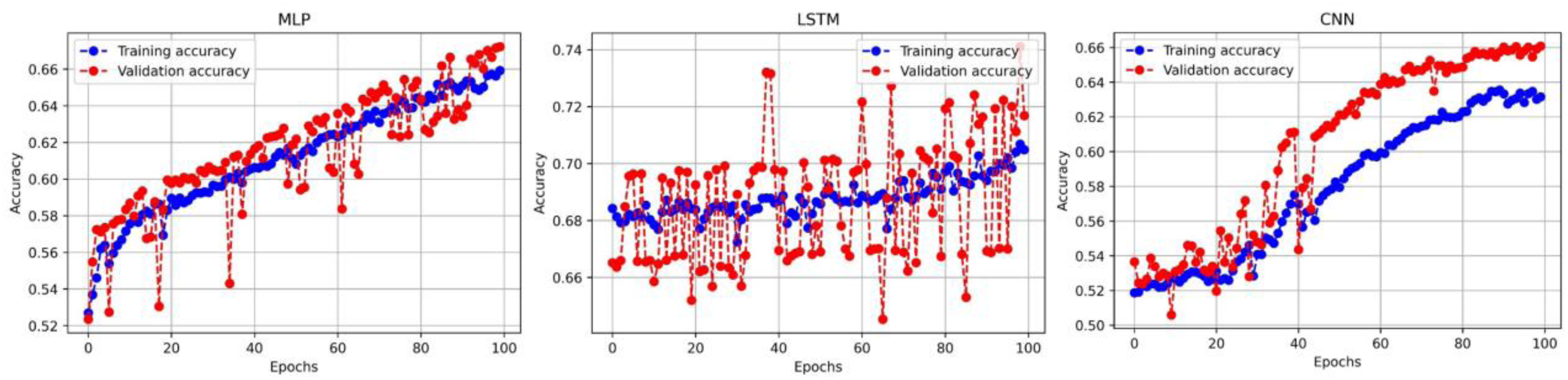
The progression of predictive accuracy over training iterations for three deep learning models.
The comprehensive results of the cross-validation are detailed in Table 5. These findings demonstrate that the ensemble model achieved the best overall performance, with an average training accuracy of 79.0%±8.0%, a test accuracy of 77.0%±9.0%, a mean training AUROC of 0.84±0.06, and a test AUROC of 0.92±0.05. For comparison, the LSTM, CNN, and MLP models yielded lower mean test accuracies (73.0%±10.0%, 64.0%±12.0%, and 62.0%±13.0%) and corresponding lower mean test AUROCs (0.91±0.06, 0.88±0.08, and 0.84±0.09). These results underscore the superior performance and generalization of the ensemble approach for BFMM.
Performance of each model evaluated using leave-one-subject-out cross-validation.

MLP: multi-layer perceptron; LSTM: long short-term memory; CNN: convolutional neural network.
Discussion
BFMM is a commonly used intervention for alleviating psychological stress among contemporary youth. This study found that, at the physiological level, a single session of BFMM significantly reduced respiratory and heart rate, suggesting a regulatory association with the ANS. At the neuroelectrophysiological level, the PSD in the theta and beta bands of the right frontotemporal region, as well as in the theta band of the right frontocentral region, was lower during the BFMM state compared to the resting state. This suggests that BFMM may relieve stress by suppressing the excitability of relevant brain regions. In addition, the deep learning results showed that the ensemble model achieved a prediction accuracy of 79.0% for the BFMM state, indicating that EEG features extracted through deep learning can effectively distinguish between the BFMM and resting states. These findings collectively reveal the potential mechanisms of BFMM in autonomic regulation and neural activity modulation, providing neurophysiological evidence supporting its broader application among youth populations.
Firstly, consistent with previous studies,3,12,15 the present study found that a single session of BFMM can reduce respiratory rate and heart rate among young adults. BFMM emphasizes deliberately slowing down the breath during practice. Physiological research has shown that when respiratory rate decreases from 16 to 8 breaths per minute, heart rate significantly decreases. 46 This suggests that slow breathing during BFMM may enhance vagal tone and increase baroreceptor sensitivity, thereby reducing heart rate. Moreover, BFMM emphasizes lengthened inhalation and exhalation, which promotes active diaphragmatic movement and greater lung expansion, facilitating increased venous return and cardiac diastole. This, in turn, activates baroreceptor-mediated vagal reflexes. The vagus nerve then inhibits sinoatrial node pacemaker activity, ultimately leading to a decrease in heart rate. However, our findings differ from those of Walsh et al., 47 who reported no significant difference in heart rate between the mindfulness training group and the control group. Moreover, existing research suggests that in real-world settings (e.g. outside of laboratory conditions), focused meditation practice does not necessarily lead to a significant reduction in heart rate and may, in some cases, even result in an increase in heart rate.47,48 The discrepancy between our findings and previous studies may be attributed to differences in experimental conditions, such as participants’ baseline heart rates, prior meditation experience, and variations in mindfulness meditation protocols. Further research is needed to clarify the effects of BFMM on heart rate in young individuals.
Secondly, the present study found that a single session of BFMM did not significantly reduce blood pressure in young adults, which is consistent with previous findings.49,50 The intervention used in this study was a one-time, brief mindfulness meditation practice. Such a short-term intervention is unlikely to produce rapid reductions in blood pressure. Blood pressure is a relatively stable physiological indicator influenced by a wide range of factors, and significant changes typically require longer duration and higher frequency practice to accumulate sufficient effects. Therefore, this brief intervention may not have been sufficient to elicit marked autonomic regulation within a limited timeframe. Future research should consider implementing multi-session, systematic intervention protocols to more effectively assess the regulatory impact of mindfulness meditation on blood pressure and related physiological parameters.
Thirdly, we found that during the BFMM state, the PSD in the theta and beta bands of the right frontotemporal region, as well as the beta band in the right frontocentral region, was lower compared to the resting state. This finding is consistent with previous studies,30,51 suggesting that the BFMM state may suppress neural activity in brain regions associated with attentional and emotional processing. Theta and beta oscillations are two reliable neurophysiological markers during meditation. 16 Theta activity in the frontotemporal region is typically associated with emotional processing, self-referential cognition, and the shift of attention inward. 52 In deep meditative states, external distractions are generally well suppressed, the demand for emotional regulation diminishes, and attentional processes become more automatic—an effect that may manifest as a reduction in theta power. 51 Furthermore, research has shown that theta activity increases when individuals are more distracted and is positively associated with shallower levels of meditation. 51 This suggests that even shallow BFMM practice may help reduce distraction among young individuals. In contrast, beta waves are the most common high-frequency brain oscillations during wakefulness and are broadly involved in attentional monitoring, cognitive control, and selection processes.53,54 Research has shown that beta activity is negatively correlated with meditation depth. A reduction in beta power in the right frontocentral region may reflect decreased cognitive control and preparatory processing of external stimuli during meditation, thereby facilitating a more relaxed mental state. However, other studies have reported increased theta power during mindfulness meditation.16,30,55 One possible explanation for this discrepancy is that the participants in our study were meditation novices, engaging in a session of BFMM. They may not yet have developed a stable pattern of internally directed attention, which could influence the neural dynamics typically observed in more experienced practitioners.
Finally, exploratory analysis showed that the ensemble model achieved a prediction accuracy of 79.0% for the BFMM state, representing improvements of 4.6% and 4.4% compared to the LSTM model on the validation and test sets, respectively. This indicates that EEG features extracted through ensemble modeling can effectively distinguish between the BFMM and resting states. Previous studies have reported classification accuracies ranging from 78% to 90% in identifying meditative states.27,56,57 The present study demonstrates that each of the three deep learning models—MLP, LSTM, and CNN—has its own strengths and limitations. In practical applications, an integrated strategy that uses CNN to extract spatial features and combines LSTM or MLP to process time-series information is expected to enhance the representation learning ability of EEG signals.32,58 Our research result shows that the ensemble model constructed by fusing the different base models can effectively leverage their respective synergistic advantages, and its overall performance is superior to that of a single model structure, which is consistent with recent research findings59–61. Future research should focus on constructing the optimal fusion framework to more effectively utilize the synergistic potential of these models.
Limitation and future work
This study has several noteworthy limitations that warrant further investigation and resolution in future research. Firstly, the sample size in the present study was relatively limited, which may have resulted in insufficient statistical power and a risk of overlooking potentially significant effects. Moreover, employing deep learning methods under small-sample conditions increases the likelihood of overfitting, thereby reducing the generalizability of the model on new data. Future research should aim to expand the sample size, which would not only improve statistical power but also facilitate a more appropriate division of data into training, validation, and test sets, ultimately enhancing the model's applicability in real-world settings.
Secondly, all participants in the current study were mindfulness meditation-naïve beginners. As novices are typically in an adaptive phase when first engaging in BFMM, their EEG activity may reflect responses to novelty rather than stable patterns of mindfulness meditation. 62 This participant characteristic may affect the stability of EEG signals and limit the interpretability of the findings. Therefore, future research should compare experienced and inexperienced mindfulness meditators to examine how prior mindfulness meditation experience may modulate EEG responses. 54
Third, this study adopted a single-session BFMM design, which limits the external validity and generalizability of the findings. A one-time intervention may only capture short-term physiological and neural responses, making it difficult to reveal the sustained effects of FA mindfulness meditation on brain function. Future research should employ longitudinal designs involving repeated interventions to investigate how long-term FA mindfulness practice influences neural and physiological mechanisms.
Fourth, although appropriate multiple comparisons correction was applied to control for Type I error, the significance of some findings was reduced after correction. This suggests that certain effects may be relatively weak or sensitive to sample size and statistical power. Future studies should aim to replicate these results with larger samples to evaluate their robustness.
Fifth, regarding the analytical approach, the current cross-validation strategy has certain limitations. Given that deep learning models are prone to overfitting on small datasets, the present validation method may not adequately assess the model's performance on unseen data. Future research is advised to adopt more robust cross-validation techniques, such as nested cross-validation, to enhance the accuracy and reliability of model evaluation.
Sixth, this study primarily used frequency-domain analysis to compare EEG activity between the resting state and the BFMM state. Prior research has suggested that low-frequency oscillations are more prominent during light meditation, whereas high-frequency activity tends to emerge in deeper meditative states. However, the present study did not further examine the relationship between specific frequency bands and BFMM depth. Future studies could employ time–frequency analysis to explore the dynamic EEG characteristics associated with light, moderate, and deep levels of BFMM.
Seventh, our study focused exclusively on EEG activity during BFMM, which may limit a comprehensive understanding of the meditative state. Future research should consider adopting multimodal approaches, integrating EEG with physiological signals such as heartbeat-evoked potentials, to enhance the accuracy of BFMM state identification. 28 In addition, future work should broaden the scope to examine other dimensions of the BFMM experience, such as interoception and emotional processes, to achieve a more holistic understanding of BFMM.3,63
Last but not least, future research should also explore the differential neural effects of various mindfulness meditation forms (e.g. FA vs. OM) and practice durations (e.g. short-term vs. long-term). 64 Combining deep learning techniques with multimodal data to systematically investigate the mechanisms through which different mindfulness meditation approaches influence mental health in young adults represents a promising direction for further inquiry.
Conclusions
Collectively, our study demonstrates that a single BFMM is instrumental in the ANS and potential modulatory on neural activity, specifically manifested as decreases in respiratory rate and heart rate, alongside reduced PSD in the theta and beta bands of the right frontotemporal region and the theta band of the right frontocentral region. Furthermore, we show that the ensemble model exhibits robust performance in detecting distinct EEG signal features associated with BFMM. These findings provide neurophysiological evidence supporting the use of BFMM in young adults and validate the feasibility of the ensemble model for distinguishing between BFMM and resting states.
Supplemental Material
sj-docx-1-dhj-10.1177_20552076251388138 - Supplemental material for Electroencephalographic neural correlates and deep learning analysis of a single brief focused mindfulness meditation in young adults: A pilot study
Supplemental material, sj-docx-1-dhj-10.1177_20552076251388138 for Electroencephalographic neural correlates and deep learning analysis of a single brief focused mindfulness meditation in young adults: A pilot study by Yongxin Luo, Yaoyao Zhang, Yuqing Zhai, Zhixiang Lu, Lujia Shou, Li Hu, Wei He, Yue Qin, Nafisa Anwar, Zhongfu Zhang, Shutian Xu, Pingping Sun and Jianwei Lu in DIGITAL HEALTH
Supplemental Material
sj-docx-2-dhj-10.1177_20552076251388138 - Supplemental material for Electroencephalographic neural correlates and deep learning analysis of a single brief focused mindfulness meditation in young adults: A pilot study
Supplemental material, sj-docx-2-dhj-10.1177_20552076251388138 for Electroencephalographic neural correlates and deep learning analysis of a single brief focused mindfulness meditation in young adults: A pilot study by Yongxin Luo, Yaoyao Zhang, Yuqing Zhai, Zhixiang Lu, Lujia Shou, Li Hu, Wei He, Yue Qin, Nafisa Anwar, Zhongfu Zhang, Shutian Xu, Pingping Sun and Jianwei Lu in DIGITAL HEALTH
Supplemental Material
sj-m-3-dhj-10.1177_20552076251388138 - Supplemental material for Electroencephalographic neural correlates and deep learning analysis of a single brief focused mindfulness meditation in young adults: A pilot study
Supplemental material, sj-m-3-dhj-10.1177_20552076251388138 for Electroencephalographic neural correlates and deep learning analysis of a single brief focused mindfulness meditation in young adults: A pilot study by Yongxin Luo, Yaoyao Zhang, Yuqing Zhai, Zhixiang Lu, Lujia Shou, Li Hu, Wei He, Yue Qin, Nafisa Anwar, Zhongfu Zhang, Shutian Xu, Pingping Sun and Jianwei Lu in DIGITAL HEALTH
Footnotes
Acknowledgements
We would like to thank the direct support professionals who participated in this study. Additionally, we thank students for her help on this project.
Ethical approval
This study was approved by Yueyang Hospital of Integrated Traditional Chinese and Western Medicine affiliated to Shanghai University of Traditional Chinese Medicine (No. 2020-178). The involvement of human participants was reviewed and approved by ChiCTR2000040609.
Informed consent statement
Informed consent was obtained from the participants before the study, and the participants received some payment after the experiment.
Author contributions
Conceptualization did by JWL, PPS, STX, and YYZ; data curation did by WJL; investigation did by ZFZ and LJS; methodology did by WH, YQ, and YQZ; software did by YXL and XZL; validation did by NA, LH and XZL; formal analysis did by YYZ and YXL; supervision did by PPS and JWL; writing—original draft preparation did by YYZ and XYL; writing—review and editing did by XYL, YQZ, and YYZ. All authors have read and agreed to the published version of the manuscript.
Funding
The authors disclosed receipt of the following financial support for the research, authorship, and/or publication of this article: This work was funded by the National Key R&D Program of China (grant number: 2024YFC3507100); the National Natural Science Foundation of China (grant number: 82472623); Postdoctoral Fellowship Program of CPSF (grant number: GZB20230600); the Shanghai Oriental Scholar Top Talent Program (grant number N/A); the High-Quality Development Project of Shanghai Economic and Information Commission (grant number: 2023-GZL-RGZN-01012); and the Ministry of Education of China (grant number: 2023ZY028).
Declaration of conflicts interest
The authors declare no conflict of interest.
Data and code availability statement
The data and code presented in this study are available upon request from the corresponding author.
Guarantor
JWL.
Supplemental material
Supplemental material for this article is available online.
References
Supplementary Material
Please find the following supplemental material available below.
For Open Access articles published under a Creative Commons License, all supplemental material carries the same license as the article it is associated with.
For non-Open Access articles published, all supplemental material carries a non-exclusive license, and permission requests for re-use of supplemental material or any part of supplemental material shall be sent directly to the copyright owner as specified in the copyright notice associated with the article.