
Abstract
Diagnosis and treatment of endometrial diseases are crucial for women's health. Over the past decade, ultrasound has emerged as a non-invasive, safe, and cost-effective imaging tool, significantly contributing to endometrial disease diagnosis and generating extensive datasets. The introduction of artificial intelligence has enabled the application of machine learning and deep learning to extract valuable information from these datasets, enhancing ultrasound diagnostic capabilities. This paper reviews the progress of artificial intelligence in ultrasound image analysis for endometrial diseases, focusing on applications in diagnosis, decision support, and prognosis analysis. We also summarize current research challenges and propose potential solutions and future directions to advance ultrasound artificial intelligence technology in endometrial disease diagnosis, ultimately improving women's health through digital tools.
Introduction
Endometrial disease represents a significant cause of female infertility, and has garnered considerable attention due to its potential impact on fertility. Epidemiological studies of infertility have demonstrated that approximately 10–15% of couples in developed countries encounter fertility challenges. Among these couples, female infertility accounts for 37% of cases. 1 Endometrial disease represents a significant contributor to female infertility. 2 The epidemiological profile of typical endometrial diseases indicates that endometriosis is a prevalent gynecological disorder globally, 3 affecting 10–15% of women of childbearing age and 35–50% of women with pelvic pain and infertility. The peak incidence of endometriosis occurs between the ages of 25 and 45 years, although both postmenopausal and adolescent women are at risk of developing this condition. 4 Endometrial polyps are also a common endometrial disease, affecting up to 34.9% of women throughout their lives. More importantly, endometrial polyps are common in women with infertility, up to 32% in patients undergoing in vitro fertilization, and malignant transformation occurs in about 3.1% of patients. 5 Endometrial cancer represents the sixth most common cancer in women globally, accounting for 2% of new cancer cases and 1% of deaths in 2020. 6 Research has indicated that fertility factors, including the number of births, age at menarche, and timing of menopause, may be linked to inter-individual variations in unopposed estrogen exposure and may contribute to differing endometrial cancer risks. 7 Other pathologies such as fibroids and endometrial inflammation should also not be ignored. The aforementioned endometrial diseases impact a range of pathological conditions pertaining to the structure and function of the endometrium. For example, endometriosis may result in the formation of pelvic adhesions, elicit an inflammatory response, and impair ovarian function. These changes may, in turn, affect the quality of eggs and the transport of sperm. Similarly, endometrial hyperplasia, endometrial polyps, and endometrial adhesions may result in alterations to the morphology of the uterine cavity, thereby affecting the environment in which the fertilized egg attaches. Furthermore, it is crucial to evaluate the influence of malignant tumors, such as endometrial cancer, on fertility, particularly in young women. The early detection and treatment of endometrial cancer is of the utmost importance in order to maintain fertility, despite the fact that it is a relatively uncommon cause of infertility. In light of the significant risks posed by endometrial disease to women's reproductive and physical wellbeing, the medical community has been engaged in efforts to identify more effective diagnostic and therapeutic approaches for conditions associated with the endometrium.
Diagnostic ultrasound technology is a non-invasive means of medical imaging that offers reproducible and safe characteristics. 8 Patients can undergo multiple examinations without worrying about surgical risks or ionizing radiation. Additionally, ultrasound equipment is highly portable, relatively low cost, and simple to maintain, making it widely used in daily medical diagnosis, telemedicine, and emergency medical assistance. Ultrasound technology provides a range of imaging modalities that cover organ structure and hemodynamics. 9 It is particularly effective in high-resolution soft tissue imaging. 10 Ultrasound imaging has demonstrated unique cost-effectiveness and safety advantages in diagnosing specific areas, such as endometrial disease, compared to other imaging techniques like CT and MRI. Therefore, it is an essential tool for screening and diagnosis.
Although ultrasound technology has advantages in diagnosing endometrial disease due to its non-invasive, safe, and cost-effective nature, there are still several challenges and limitations. Expertise and clinical experience are required for interpreting ultrasound images. Furthermore, ultrasound images are rich in information and can contain intricate details, which may lead to the oversight of subtle lesions, particularly when the image quality is poor or the operator lacks experience. AI technologies, particularly deep learning, present novel solutions to the diagnostic challenges of ultrasound by enhancing the precision and efficiency of ultrasound image interpretation. AI-assisted diagnostic ultrasound not only reduces the physician's workload but also provides a more efficient approach to diagnosing endometrial diseases in resource-limited environments. As AI technology continues to advance, it is expected to further improve the process of endometrial disease detection and treatment, as well as the quality of medical services in resource-limited environments.
Despite the paucity of publicly available ultrasound datasets for analysis, AI algorithms have demonstrated a notable growth trend in the field of ultrasound endometrial disease diagnosis, as illustrated in Figure 1. The main areas of application include:
Disease classification: The utilization of AI algorithms to extract and analyze features from ultrasound images, with the objective of differentiating between various types of endometrial diseases, including the distinction between benign and malignant tumors. Prognosis prediction: AI technology is employed to extract and analyze alterations in pivotal features derived from the images, thereby anticipating the trajectory or outcome of the disease. This facilitates more precise treatment recommendations for patients by enabling physicians to better assess the disease's progression. Feature extraction: Artificial intelligence (AI) is employed to automatically identify salient features in an image, such as shape, texture, and size, for subsequent analysis. Image segmentation is a process whereby the image is divided into distinct regions based on visual characteristics. AI algorithms are capable of automatically segmenting specific structures in ultrasound images, such as the boundaries of the endometrium, thereby enhancing diagnostic accuracy. Endometrial tolerance assessment: To assess the ability of the endometrium to accept embryo implantation during a given menstrual cycle, AI technology can assess the thickness, morphology, and dynamic characteristics of the endometrium.

Number of AI papers published on ultrasound endometrium per year.
In the field of gynecological health, our team has been engaged in research aimed at improving the diagnosis and management of endometrial diseases through the application of advanced technologies. For instance, our team examined the efficacy of four-dimensional hysterosalpingography (4D HyCoSy) in evaluating the patency of the fimbria of the fallopian tubes in women experiencing infertility. 11 Our findings revealed an overall accuracy of 92.9% and sensitivity and specificity exceeding 90%. Subsequently, our team conducted a pioneering pilot cohort study to develop a deep learning-based ultrasound radiomics model that integrated clinical data to predict clinical pregnancy following frozen embryo transfer. 12 The study's key innovation was the integration of ultrasound radiomics features with clinical parameters. The findings demonstrated that the proposed multi-modal fusion model exhibited superior efficacy in predicting the incidence of clinical pregnancy following FET compared to models utilizing images or quantitative variables alone.
Building upon the foundations of our previous work, we will examine the application of AI in ultrasound imaging analysis of endometrial diseases. A review of the paper on the subject was conducted and the findings were collated and summarized in the databases PubMed and Google Scholar. Subsequently, the collected literature was subjected to screening, the process of which is illustrated in Figure 2. The specific method is as follows: Firstly, a Python script was employed to identify and remove duplicate entries based on document titles. Specifically, the SequenceMatcher method from the difflib module was employed for the calculation of the similarity between file names. A similarity threshold of 90% was set, enabling the automatic identification and categorization of duplicates. Within each group of duplicates, only one document was retained, and the similarity between the retained file and the duplicates was recorded in order to ensure transparency and reproducibility of the screening process. Secondly, we read the article's title and abstract and delete any literature not related to the theme “Ultrasound endometrium combined with AI digital health research.” Thirdly, the remaining papers were read in full, and those with overly simplistic artificial intelligence model structures or research methods were excluded. Finally, the 46 English papers were read carefully. The screened paper was classified according to the type of disease studied and subsequently subdivided and summarized according to the application field of AI. The results were presented in tabular form. To the best of our knowledge, there is currently no extensive and comprehensive survey that provides a summary of the application and future trends of AI technology in the diagnosis of endometrial diseases. The objective of this article is to address this gap in the existing literature. This article makes the following contributions:
This paper provides a comprehensive review of the application and technological advancements of AI in ultrasound imaging, specifically focusing on traditional machine learning and deep learning methods for diagnosing endometrial diseases. It compares the effectiveness of traditional machine learning techniques with deep learning approaches in key areas such as ultrasound image feature extraction, disease classification, and prognosis assessment, offering a scientific basis for selecting the most suitable technological path for these applications. Additionally, the paper addresses unresolved challenges and existing methodologies in this field. It proposes potential solutions and outlines future development trends in response to the identified issues, emphasizing the ongoing progress of integrating AI with ultrasound technology for endometrial disease diagnosis.

Paper screening process.
Ultrasound images for diagnosing endometrial diseases
Ultrasound image characteristics
Endometrial diseases are a group of conditions that can significantly impact women's reproductive and physical health. This group includes endometrial polyps, endometrial tuberculosis, endometrial cancer, and endometriosis. These conditions can cause menstrual disorders, dysmenorrhea, infertility, and even life-threatening complications. Therefore, it is crucial to diagnose endometrial diseases accurately and promptly to ensure effective treatment. In the field of ultrasound medicine, techniques such as B-mode ultrasound, vaginal ultrasound, 3D ultrasound, and color Doppler flow imaging provide crucial information for diagnosing endometrial diseases.
Although ultrasound techniques are commonly used in clinical practice, the development of deep learning algorithms is limited by the availability of sufficient image datasets. As demonstrated in Figure 3, color Doppler flow imaging is essential for diagnosis, but its application in deep learning is restricted due to the scarcity of datasets. In this paper, we will focus on introducing and analyzing medical images that are commonly used in ultrasound AI in order to advance practical technology development and clinical applications.

Distribution of types of datasets used in research papers.
Abdominal B-mode ultrasound is a fundamental imaging technique that provides two-dimensional gray-scale images for assessing endometrial thickness, morphology, and potential growths and lesions. It is a crucial initial screening tool. The endometrium appears moderately bright on ultrasound images and is affected by various factors, including the menstrual cycle, thickness, and underlying lesions such as polyps, hyperplasia, and leiomyomas. If a lesion is present, the echo characteristics will vary depending on the type of lesion. It is important to note that subjective evaluations have been excluded from this description. However, image quality may be reduced and artefactual interference increased due to limitations in image resolution and abdominal wall fat in obese patients.
Vaginal ultrasound is the main data source for machine learning and deep learning analysis, as shown in Figure 4. It provides two-dimensional grey-scale images similar to those of abdominal ultrasound but with reduced interference from abdominal fat and intestinal gases due to the use of intra-vaginal probes. This result in a clearer image of the uterus. Vaginal ultrasound is a valuable diagnostic tool in gynecology and obstetrics as it can reveal abnormal echogenic features of the endometrium more clearly than abdominal ultrasound, especially in cases where abdominal ultrasound is not effective. Vaginal ultrasound is a valuable diagnostic tool in gynecology and obstetrics as it can reveal abnormal echogenic features of the endometrium more clearly than abdominal ultrasound, especially in cases where abdominal ultrasound is not effective. However, it may cause discomfort and is not applicable to certain patient groups.
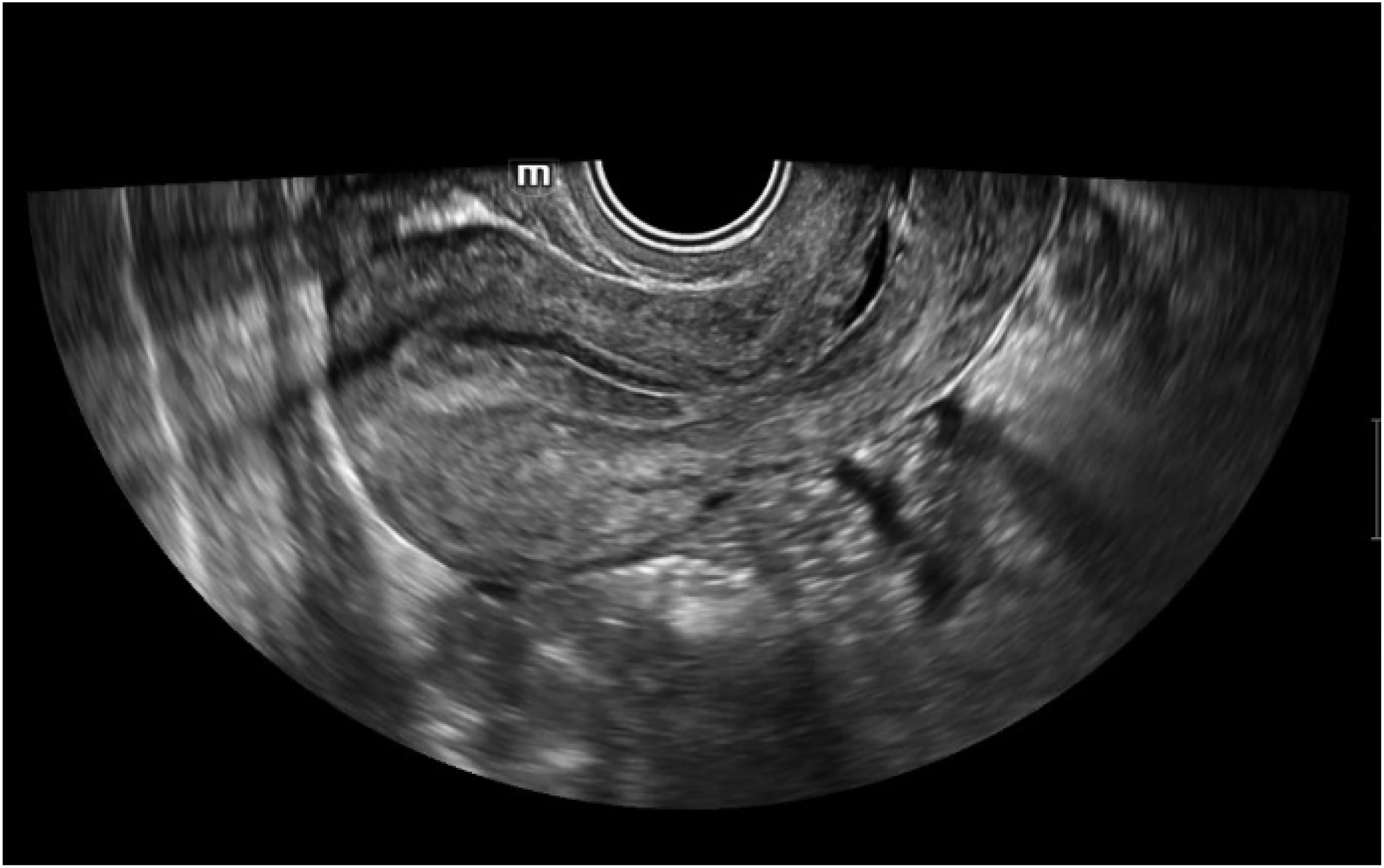
Endometrial vaginal ultrasound image.
Three-dimensional ultrasound technology creates a 3D model by combining multiple 2D images, 13 as demonstrated in Figure 5. This method provides more comprehensive spatial information than conventional 2D ultrasound. This technique accurately reveals the localization, size, and morphology of lesions, enabling physicians to review the endometrial structure from multiple perspectives and depths. This allows for a more accurate assessment of its thickness, morphology, and potential for abnormal proliferation and lesions. Although the high cost and operational complexity of 3D ultrasound currently limit its widespread use, technological advances are expected to gradually reduce these barriers.
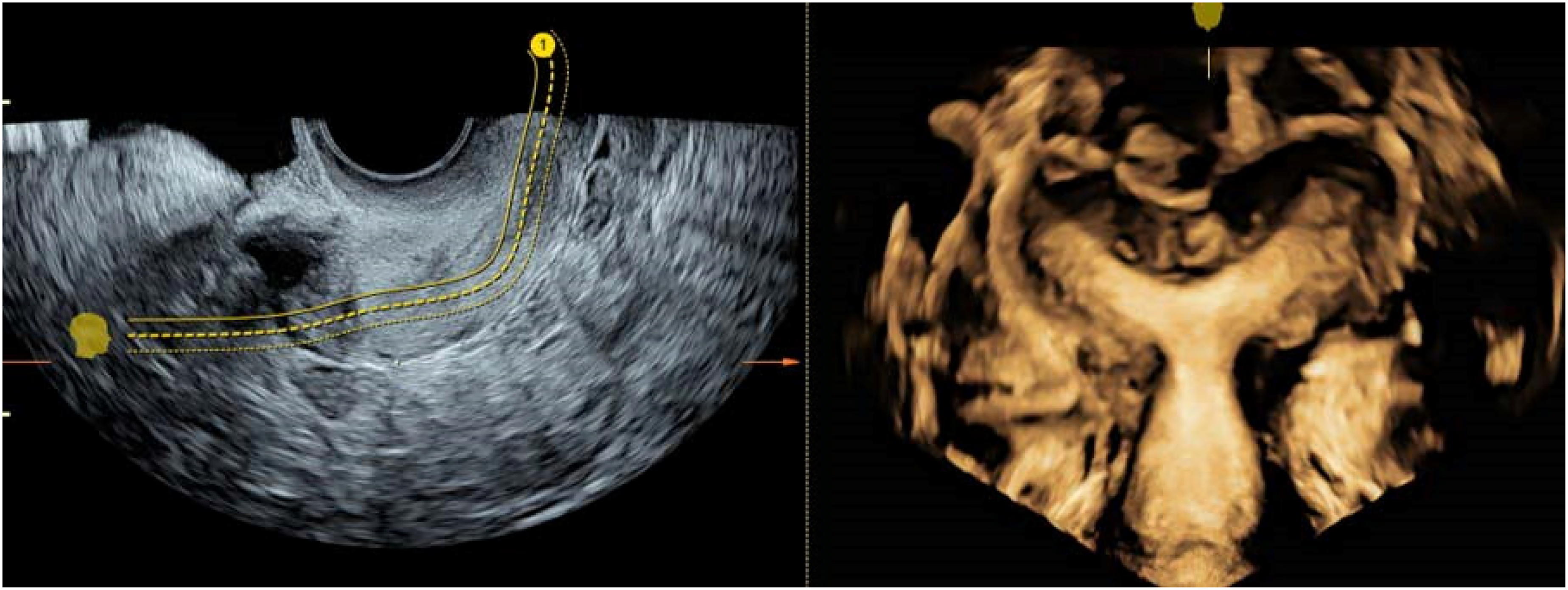
3D multi planar ultrasound sonogram of unicornuate uterus.
The importance of ultrasound AI
Various ultrasound techniques provide a wealth of image information for the detection of endometrial disease. However, these techniques also pose challenges. The traditional method of relying on the physician for visual examination suffers from unavoidable problems:
The challenges in diagnosing various endometrial ultrasound image diseases include a lack of specialists with sufficient domain knowledge, differences in the level and number of specialists across regions, and the inexperience and long growth cycle of young doctors. Prolonged traditional examination and diagnosis can lead to a decrease in doctors’ attention, increasing the risk of misdiagnosis and diagnosis time. The diverse manifestations of endometrial diseases can complicate visual diagnosis.
The emergence of these problems increases the burden on patients. To improve the accuracy and efficiency of diagnosis, reduce the burden on specialists, and provide opportunities for young doctors to learn and improve, the field of medicine has been exploring integration with other fields. AI is undoubtedly one of the most promising applications in assisted diagnosis.14–16 Compared to manual ultrasound image diagnosis, AI can process and analyze large amounts of data quickly, improving work efficiency. Its ability to process high-dimensional data helps to discover details that may be overlooked by human experts. As technology advances, AI will play an increasingly important role in future medical practice.
The use of AI in endometrial disease diagnosis via ultrasound
Figure 6 shows that AI techniques are divided into two main categories: traditional machine learning and deep learning. Traditional machine learning relies on manual feature extraction, which has the advantage of model interpretability and fast inference. 17 In contrast, deep learning automatically learns complex features from data without the need for tedious feature engineering. Ultrasound image analysis presents several challenges, including high-dimensional data, dynamic changes, and noise interference. Efficient analysis methods capable of handling large numbers of pixels and capturing subtle time-varying patterns are required. Furthermore, the subjectivity and inconsistency of ultrasound image interpretation can increase diagnostic difficulty. In response to these challenges, deep learning has shown significant advantages due to its ability to automate feature extraction and process high-dimensional data. This paper will focus on deep learning, while traditional machine learning is preferred in some cases for its interpretability and efficiency. 18 When applying AI techniques to ultrasound imaging, the appropriate method should be selected based on the task requirements and available resources.

Categories of AI algorithms.
Applying conventional machine learning to endometrial disease imaging
Traditional machine learning, as a branch of AI, originated from the checkers program developed by Samuel in 1959. 19 In the field of endometrial imaging, traditional machine learning methods face challenges, particularly when dealing with image data. Figure 7 illustrates that these methods typically require feature engineering to manually extract key features in the image, 20 such as regions, shapes, and textures. Subsequently, a feature selection algorithm filters the features most relevant to disease diagnosis. 21 These features are then combined with the patient's clinical data to form the input for the machine learning model. This section evaluates these methods according to the algorithms’ objectives, which aim to address the problem of insufficient data for a single disease and explore the commonalities between different diseases to find a universal machine learning strategy.
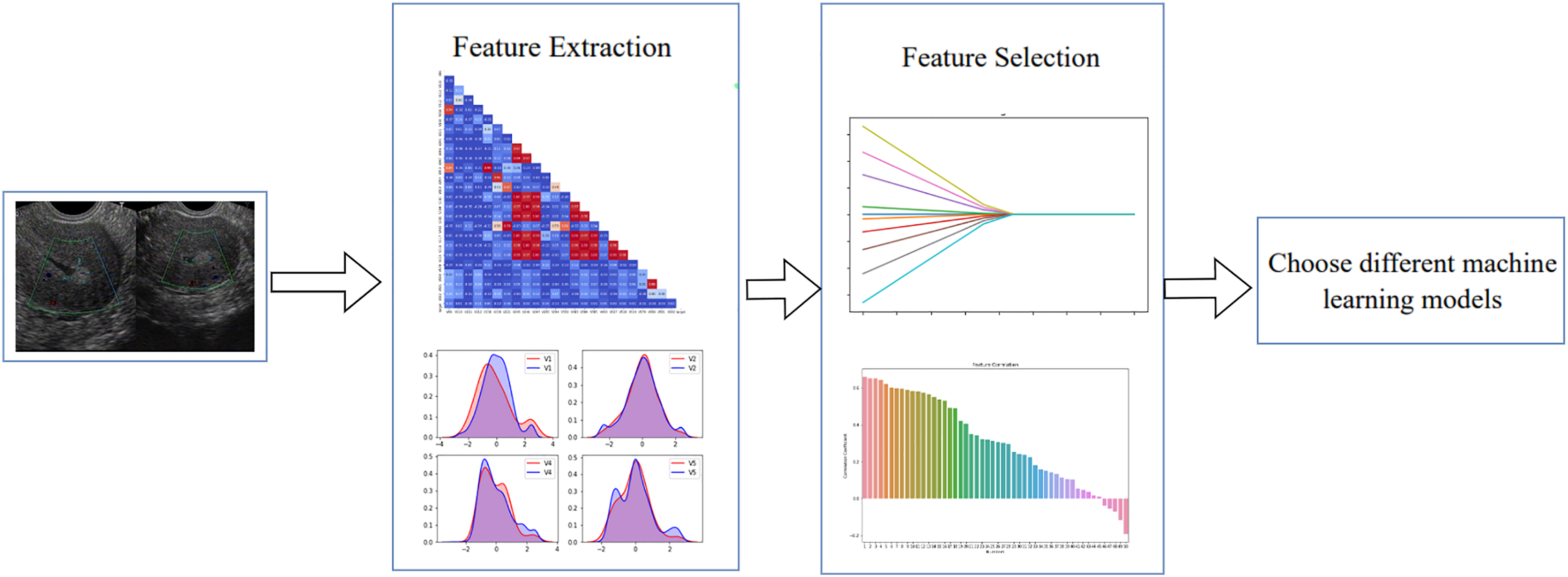
Schematic diagram of machine learning algorithm processing flowchart.
Traditional machine learning for prognostic analysis
Prognostic analysis enables the stratification of patients into different risk groups, aiding physicians in providing more precise treatment recommendations and improving treatment outcomes. 22 Prognostic analyzes aim to assess the likely outcome or course of a disease. Research in this area can be categorized into four types as shown in Figure 8.

Categories of prognostic analysis research.
In prognostic analysis of ultrasound endometrial images using traditional machine learning, it is essential to first perform feature extraction and feature screening on the images. This is because traditional machine learning algorithms typically handle numerical or tabular data, whereas images are complex collections of elements. 23 Upon completion of the feature extraction and screening process, the most suitable machine learning algorithm can be selected based on the specific application scenario. This selection process requires comprehensive consideration of factors such as model performance, complexity, training time, and adaptability to data to ensure that the selected algorithms can effectively solve real-world problems while maintaining high operational efficiency. Table 1 presents the research on machine learning in the field of prognostic analysis.
Machine learning algorithms for ultrasound endometrial prognostic analyses—development, strengths, and weaknesses.

SP: survival prediction; RC: risk classification; PD: predictor discovery.
The field of ultrasound endometrial prognostic analysis is increasingly utilizing machine learning techniques, with a shift towards multi-model integration. Previous studies have primarily employed single models, such as logistic regression, to analyze disease stages by combining ultrasound features with clinical data. As research progressed, researchers explored multi-model integration strategies such as Random Forest, 32 Support Vector Machine (SVM), 33 and Hybrid AdaBoost. 34 These strategies enhance prediction accuracy and robustness due to their ability to effectively deal with data uncertainty and noise.
Furthermore, in prognostic analyzes, feature extraction methods for machine learning have shifted from traditional manual measurements to deep learning automated techniques. Convolutional neural networks (CNNs) and U-Net are examples of deep learning techniques that can automatically extract complex features from ultrasound images, 35 making them particularly effective for small sample datasets. The ability of feature extraction and generalization performance of deep learning models, such as variants of U-Net, deep convolutional networks, and migration learning, has been further improved.
The focus of research has shifted from utilizing a large number of features to identifying key features. Early studies extracted a large number of features through manual measurement or machine learning methods, but the model results were unsatisfactory. With the advancement of technology, researchers have begun to employ feature screening techniques, such as ANOVA, 36 ridge regression, 37 recursive feature elimination (RFE), 38 and Pearson's correlation coefficient, 39 to decrease the number of features and enhance model efficiency and generalization. These methods aid in identifying the features that have the most significant impact on the prediction results, constructing streamlined and efficient models, and enhancing the adaptability of non-linear relationships between features. This provides a more comprehensive perspective on endometrial prognostic analyzes.
Machine learning for endometrial disease recognition and diagnosis
Traditional machine learning methods are crucial in identifying and diagnosing endometrial diseases. These methods construct efficient diagnostic models by extracting features from ultrasound images and combining them with clinical data to determine whether a patient has the disease. Figure 9 shows the varied applications and effectiveness of machine learning techniques in diagnosing a wide range of endometrial diseases across different pathological conditions.

Percentage of machine learning applications for endometrial disease diagnosis.
In the diagnosis of endometrial cancer, machine learning models are used to improve prediction accuracy. Guerriero et al. investigated single models such as k-NN and Naive Bayes. 40 They stated that Naive Bayes works well with low feature correlation, while k-NN is suitable for small data sizes. Wang et al. developed an integrated model, TJHPEC, which fuses logistic regression, gradient-boosted decision trees, and random forests. 41 This model demonstrated high accuracy and AUC values but may increase computational cost. Studies on endometriosis have aimed to improve the sensitivity and specificity of diagnostic models. According to a systematic review by Sivajohan et al., the random forest model has diagnostic potential, particularly when symptoms are not evident. 42 Logistic regression was found to perform well in the diagnosis of the disorder by Bendifallah et al., 43 but selecting the model and feature engineering remain a challenge. He et al. investigated the use of machine learning to enhance the diagnostic efficiency of 3D transvaginal ultrasound imaging. 44 However, this approach may be restricted by equipment and expertise.
This development in technology has allowed for machine learning to be applied from a single model to multiple models. The integration of multiple models, such as Random Forest and Gradient Boosting Decision Tree, 45 has improved the accuracy and robustness of predictions for ultrasound endometrial disease diagnosis. Furthermore, researchers have started to consider factors beyond models, such as enhancing image quality. This implies that, in addition to selecting a model, other techniques like image processing and data preprocessing are equally crucial and deserving of attention in diagnosing endometrial disease.
Machine learning for endometrial disease classification
Machine learning has various applications in classification, including distinguishing between benign and malignant tissues of endometrial cancer (EC), classifying endometrial tuberculosis stages, and predicting pregnancy success. Conventional machine learning methods are crucial in improving the accuracy and efficiency of endometrial disease classification. These methods offer new insights for clinical diagnosis by analyzing ultrasound image features. Although there has been some progress, machine learning studies for other endometrial diseases are still relatively limited. This may be due to sample size, the complexity of disease features, or uneven allocation of research resources. Therefore, this paper will focus on the progress of machine learning classification research for these three diseases.
In studies on the classification of endometrial cancer, Capasso et al. achieved high AUC values by extracting radiomics features from ultrasound images and applying a support vector classifier. 46 Karahaliou et al. successfully distinguished between benign and malignant tissues by employing wavelet transform texture enhancement. 47 Bogani et al. combined radiomics and molecular characteristics to provide personalized treatment decisions for patients. 48 In a study on the classification of endometrial tuberculosis, Garg et al. used the gradient local autocorrelation covariance model (GLAC) and non-subsampled contour wavelet transform (NSCT) to extract texture features and improve classification accuracy. 49 Sahoo et al. compared various models and found that the NSCT-based GLCM model performed the best in terms of classification performance. 50 These studies demonstrate the benefits of using multiscale and multidirectional transforms for image feature extraction, providing new technical tools for image analysis. Li et al. developed a logistic regression model to predict pregnancy success by combining clinical indicators and ultrasound image features. 51 Similarly, Mori et al. used local binary pattern (LBP) analysis of ultrasound image features to predict pregnancy outcomes. 52
These studies improve the accuracy of prediction and provide a precise basis for clinical decision-making. Texture feature extraction, enhancement, and multimodal data fusion have become key methods in the classification of endometrial diseases. 53 The combination of clinical and molecular genetic features can further improve classification accuracy and the development of personalized treatment plans. This provides new perspectives and directions for future research.
Application of deep learning algorithms
Deep learning algorithms are a type of machine learning algorithm that originated from artificial neural networks (ANN). They simulate the way the human brain processes information by building deep neural networks. In the context of diagnosing endometrial disease using ultrasound, deep learning has advantages over machine learning. It can automatically learn features from raw image data, eliminating the need for complex feature engineering, reducing data processing time, and enabling training on large-scale datasets.
Deep learning in endometrial cancer
Endometrial cancer is a prevalent gynecological malignancy. Early detection and treatment are crucial to improve patient survival rates and quality of life. Currently, most studies in this field focus on MRI and hysteroscopic images. Ultrasound images are still a minority of applications, and research on deep learning in endometrial cancer mainly centers on three key aspects: disease classification, development trend prediction, and diagnosis of the degree of myometrial infiltration. 54 Figure 10 shows the proportion of deep learning applications in endometrial cancer research in the existing Literature.
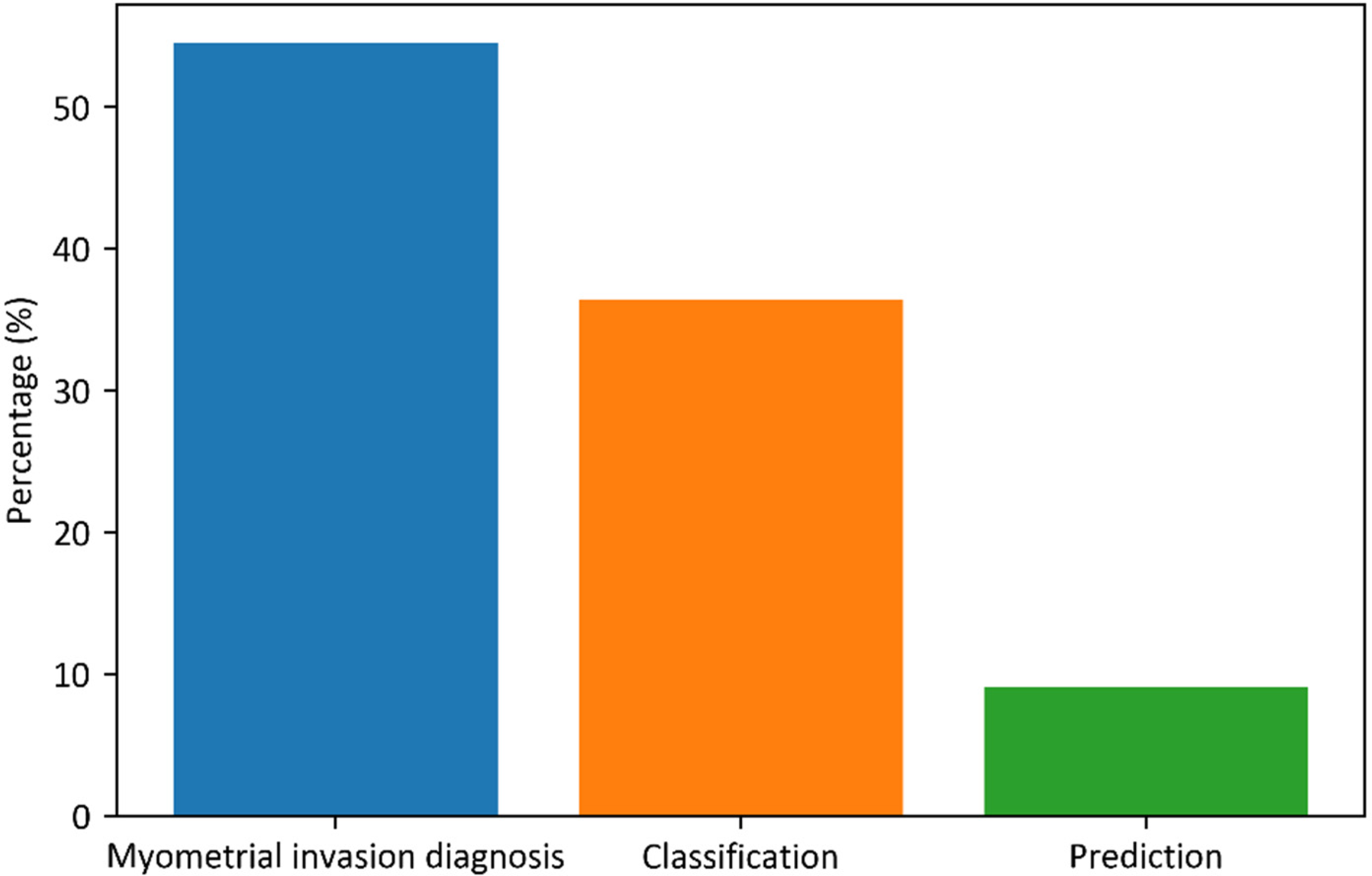
Share of major applications of deep learning in endometrial cancer.
The use of deep learning in endometrial cancer classification involves analyzing data such as ultrasound images to identify the features that distinguish endometrial cancer from other gynecological diseases. This classification encompasses not only the determination of whether endometrial cancer is benign or malignant, but also the classification of a wide range of diseases that include endometrial cancer, depending on the study's objective. This classification aids doctors in making more precise diagnoses and providing more accurate treatment options for patients.
Predictive applications in deep learning are primarily used to forecast the development of endometrial cancer and the prognosis of patients. Deep learning models can predict the likelihood of cancer recurrence, metastatic trends, and the type of lesion by analyzing patients’ clinical data and ultrasound imaging features. Detecting myometrial infiltration is a crucial tool for evaluating the aggressiveness of endometrial cancer. Deep learning models can assist doctors in determining whether a tumor has invaded the myometrium and to what extent. The presence of myometrial infiltration typically indicates a more advanced stage of the disease. This information is crucial in deciding the extent of surgery and the treatment options available. Table 2 presents the utilization of deep learning in the diagnosis of endometrial cancer through ultrasound imaging. This research is categorized into three sections, each pertaining to a distinct diagnostic objective: classification, prediction, and myometrial invasion detection.
Deep learning in ultrasound for endometrial cancer.

CL: classification; PR: prediction; MD: myometrial infiltration detection.
In research on the classification of endometrial disease, deep learning techniques, particularly convolutional neural networks (CNNs), are commonly used for extracting image features and recognizing diseases. The research process typically involves two stages: first, deep learning models automatically extract features from ultrasound images; second, diseases are accurately classified based on these features. This approach enhances diagnosis accuracy and improves the efficiency of handling large data sets. In the field of lesion prediction and analysis, deep learning techniques demonstrate great potential to enhance prediction accuracy and segmentation efficiency. Researchers have explored a variety of model design and optimization strategies by combining various deep learning architectures, such as different types of CNNs, Transformer, 63 LSTM, 64 and Bi-LSTM. 65 Future research will aim to develop efficient algorithms to reduce computational burden, explore data augmentation and regularization techniques to enhance generalization, and utilize multimodal data and interdisciplinary approaches to improve prediction accuracy. In the field of muscle infiltration detection, automated detection research follows an image recognition plus extent assessment strategy. Deep learning techniques are utilized to precisely identify and analyze muscle infiltration features in transvaginal ultrasound images, followed by an assessment of the extent of infiltration. The main objective is to enhance the accuracy of the image recognition algorithm and the reliability of the extent assessment method. Future research will focus on the generalization ability and adaptability of deep learning models to different patient populations. Model training and validation using cross-center, multi-population large-scale datasets can effectively enhance the generalization ability of models.
Deep learning in endometriosis
Endometriosis is a prevalent gynecological condition that occurs when endometrial tissue, which typically grows inside the uterine cavity, appears outside the uterus. This ectopic tissue can grow in various locations, including the ovaries, fallopian tubes, and pelvic peritoneum, causing symptoms such as pain, irregular periods, and infertility. The use of deep learning techniques in this field concentrates on three primary areas: segmentation, classification, and assisted diagnosis of endometriosis. Figure 11 illustrates the percentage of each study in the surveyed literature.

Deep learning research in endometriosis.
Deep learning segmentation methods accurately identify ectopic tissues from complex backgrounds, providing physicians with critical visual information to improve disease examination results. The classification of endometriosis is mainly based on the lesion's morphological characteristics and the precise classification of multiple diseases containing endometriosis. The use of deep learning for assisted diagnosis of endometriosis involves altering data representation methods, integrating additional images, and creating systems for multiple assisted diagnoses. Table 3 provides a detailed summary of the three primary research methods employed in the field of deep learning for endometriosis.
The use of deep learning in ultrasound for endometriosis.

SE: segmentation; CL: classification; AD: assisted diagnosis.
Image segmentation technology is crucial in diagnosing endometriosis. Its purpose is to accurately identify and locate diseased structures in images. Recent studies have shown that combining U-Net and its variants with convolutional neural networks like DenseNet has resulted in remarkable segmentation performance, 77 as evidenced by higher intersection ratios and Dice coefficients. Transfer learning and multi-scale feature extraction are effective in improving the accuracy of pixel-level classification. 78 However, success often depends on high-quality data annotation, which can be a limiting factor in clinical settings where data is scarce.
In medical image analysis, the focus of image classification technology is primarily on identifying and classifying diseases. Research indicates that models such as Inception, 79 ResNet, 80 and EfficientNet are advantageous in extracting multi-scale features and can produce rich classification results. By comparing multiple models and utilizing data augmentation, these models can improve their generalization capabilities and robustness. However, increasing model complexity and the number of parameters can lead to overfitting, particularly when the sample size is limited. Therefore, researchers must pay attention to data diversity and model regularization to address the risks of data bias and overfitting.
The evolution of auxiliary diagnosis technology is demonstrated by the transition from traditional CNN to Transformer architecture, and then to models that combine the two. Initially, CNN models like VGGNet-16 made significant strides in recognizing common lesions due to their strong feature extraction capabilities. Later, the Transformer architecture utilized the self-attention mechanism to capture global dependencies and enhance diagnostic accuracy. Hybrid models of CNN and Transformer are currently receiving attention. These models combine the local feature extraction of CNN with the global information processing capabilities of Transformer. The aim is to improve recognition and classification performance, reduce dependence on a large amount of high-quality annotated data, and reduce computational costs. Technologies such as knowledge distillation and image fusion can also help improve model performance. 81
Application of deep learning in endometrial receptivity assessment
The assessment of endometrial receptivity refers to the ability of the endometrium to accept embryo implantation at specific times in the menstrual cycle. This is a complex biological process that involves the morphology of the endometrium, endometrial movement, and endometrial blood supply. It is important to note that this assessment involves not only ultrasound image data but also the design of clinical text data. Currently, there are few studies that use deep learning models to evaluate endometrial receptivity by ultrasound. Literature research suggests that receptivity assessment can be divided into thickness, morphology, dynamic characteristics, and comprehensive assessments. Figure 12 illustrates the distribution and proportion of tolerance assessment types in the literature.

Distribution of tolerance assessment studies.
Endometrial thickness is a crucial factor in predicting the success of embryo implantation. Assessing endometrial thickness can aid in determining the optimal time for uterine reception and optimizing embryo transfer strategies. Additionally, for patients experiencing postmenopausal bleeding, measuring endometrial thickness is a vital screening method for diagnosing endometrial lesions, including endometrial cancer. Furthermore, the morphology of the endometrium, specifically whether it is leafed before ovulation or uniform after ovulation, has a significant impact on the success rates of pregnancy. Ultrasonography is used to quantify endometrial peristaltic characteristics, such as frequency, direction, intensity, and speed, which are crucial for sperm transport, menstrual excretion, and embryo implantation and development. Table 4 outlines the research models and methods of deep learning used in the personalized comprehensive assessment, which combines ultrasound data with clinical parameters to provide an in-depth understanding of endometrial receptivity.
Research methods for deep learning in endometrial receptivity assessment receptivity.

TE: thickness evaluation; ME: morphology evaluation; DC: dynamic characterization evaluation; CE: comprehensive evaluation.
The measurement of endometrial thickness is automated using the “segmentation-measurement” framework. The accuracy of the image segmentation technology and the suitability of the measurement method are crucial. U-Net and its variants are highly effective in controlling errors, and 3D U-Net further enhances accuracy through multi-scale feature fusion. However, the model still encounters difficulties in dealing with fine thicknesses and boundaries. The assessment of endometrial morphology utilizes the “feature extraction-pattern recognition” strategy. The accuracy of feature extraction and the recognition power of the deep learning model are crucial. The combination of 3D point cloud with U-Net enhances the visualization accuracy of uterine structural abnormalities, 91 but it has high computational requirements and is sensitive to noise. Future research will aim to improve model generalization, reduce reliance on large amounts of annotated data, and explore new data representations and network architectures. With regard to dynamic feature evaluation, deep learning technology is used to extract initial dynamic features, such as peristaltic waves, and analyze them through specific algorithms. The combination of CNN and the optical flow method demonstrated high consistency in assessing intimal peristalsis, highlighting the potential of the model for multi-dimensional assessment. 92 Comprehensive evaluation involves combining clinical and ultrasound image data, which demonstrates the advantages of complementary multimodal information, despite challenges in modal data alignment. To enhance the model's generalization ability and evaluation accuracy, it is expected that a comprehensive analysis will be conducted using additional modal information.
Research on deep learning in the field of endometrial receptivity assessment is still in its early stages but is rapidly developing. Technological advancements are mainly seen in three areas: the transition from 2D to 3D, the evolution of deep learning models, and the exploration of multi-modal data fusion. These advancements offer the potential for more accurate endometrial assessment and can aid in improving the precision of clinical decision-making.
Application of deep learning in other endometrial diseases
The use of deep learning technology in diagnosing endometrial diseases has expanded. It has potential value for various endometrial-related diseases, including endometrial cancer, endometriosis, and endometrial receptivity. Although there have been few studies on conditions such as endometrial tuberculosis, adhesions, hyperplasia, and uterine fibroids, the clinical significance of these diseases cannot be ignored.
Endometrial tuberculosis is an infectious disease that can cause symptoms such as irregular menstruation, infertility, and lower abdominal pain. Endometrial adhesions, which are often caused by surgery, infection, or inflammation, can lead to problems such as light menstruation, dysmenorrhea, and infertility. Endometrial hyperplasia may manifest as irregular menstruation and abnormal uterine bleeding. Uterine fibroids, a common benign tumor, may cause symptoms such as menorrhagia, pelvic pain, and infertility. Through a comprehensive analysis of existing research results, deep learning technology offers new ideas and methods for the early diagnosis and treatment of diseases. Table 5 demonstrates the various aspects of deep learning application in ultrasound diagnosis, including identification and detection of endometrial tuberculosis and uterine fibroids, classification of endometrial hyperplasia, and diagnosis of endometrial adhesions. These advances offer clinicians more accurate diagnostic tools and lay the foundation for future research directions and clinical applications.
The research methods used for deep learning in ultrasound for endometrium and other diseases.

ET: endometrial tuberculosis; EY: endometrial hyperplasia; UF: uterine fibroids; EA: endometrial adhesions.
In the field of endometrial lesion detection, the combination of deep learning technology and image feature enhancement algorithms has proven to be effective. For instance, the Region Proposal Network (RPN) combined with ResNet50 performs exceptionally well in detection performance, 98 highlighting its efficiency in candidate region generation and localization. The Non-subsampled Contourlet Transform combines the ResNet50 method to effectively retain image details through multi-scale analysis. 99 Furthermore, the model that combines geometric features with NSCT achieved an accuracy of 0.91 on ResNet50, highlighting the benefits of multi-scale analysis.
When it comes to the automatic segmentation and classification of endometrial hyperplasia, U-Net and its variants can be combined with different CNN backbone networks to improve segmentation accuracy and reduce the need for manual intervention. The ensemble learning method can enhance the model's generalization ability and robustness by fusing different network architectures. The YOLO series model combined with CNNs such as EfficientNet has excellent accuracy and detection speed for detecting uterine fibroids, 100 making it suitable for real-time clinical applications. Additionally, the Vision Transformer model provides a new perspective for feature extraction through its self-attention mechanism, 101 but further optimization is needed for hyperparameter adjustment and local feature capture. The model demonstrated high accuracy in detecting endometrial adhesions. However, its limitations in model structure suggest the need for future research to explore more efficient network structures and feature extraction technologies. These research results provide a foundation for the application of deep learning in diagnosing endometrial diseases and point toward future technological development.
Research directions and challenges
Main research directions and model analysis of endometrial diseases combined with AI
The previous discussion explored the various applications of ultrasound AI in diagnosing endometrial diseases. This included an in-depth examination of the latest progress and specific practices of traditional machine learning and deep learning technologies for different disease types. In order to facilitate a more intuitive understanding of the research results and technical features, a summary of the principal research directions pertaining to endometrial diseases and the corresponding AI models is presented in Table 6. This summary is designed to assist in the comprehension of the relative advantages and limitations of the various technologies in practical applications.
Research on the application of AI in the diagnosis of endometrial diseases.
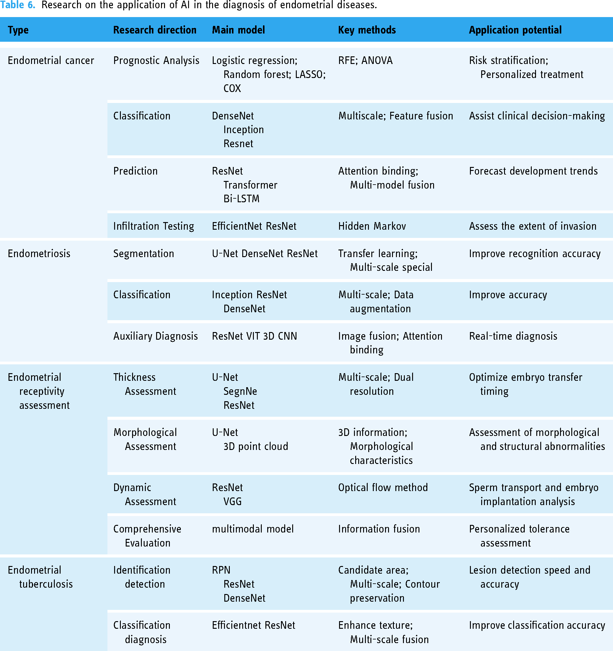
AI technology is becoming increasingly important in the research of ultrasound endometrial diseases. Its application covers key areas such as disease prognosis, classification, prediction, infiltration detection, auxiliary diagnosis, and receptivity assessment, providing a full range of clinical solutions. Deep learning models, including DenseNet, Inception, ResNet, Transformer, and Bi-LSTM, have been used by researchers to achieve outstanding results in image recognition, sequence data processing, and feature extraction. This has enabled accurate identification of disease characteristics and prediction of trends.
Although these models have achieved remarkable results in ultrasound diagnosis, researchers are constantly exploring new technologies and methods to further improve accuracy and practicality. These innovations not only target the optimization of model structure but also aim to enhance the model's ability to process complex data to meet diverse needs in clinical practice. In this process, the key to improving model performance is through the use of technologies such as multi-variable interaction analysis, multi-scale feature fusion, the combination of global and local attention mechanisms, and multi-model fusion strategies. By applying these technologies, the prospects for AI in the field of ultrasound endometrial diseases will be broader.
Existing challenges of endometrial disease combined with AI
Although deep learning models and key technologies have led to significant advancements in the field of ultrasound endometrial diseases, it is important to note that various diseases, such as endometrial cancer, endometriosis, and endometrial receptivity assessment, all use ultrasound endometrium datasets and face similar challenges. Therefore, it is crucial to develop effective solutions that can be applied across different diseases and applications. In order to facilitate a comprehensive examination of the challenges encountered by the diverse range of applications, as illustrated in Table 7, we have undertaken a systematic categorization and synthesis of the applications of AI in endometrial disease. Despite targeting different diseases, the technical challenges and solutions faced are largely interrelated. Next, we will provide an in-depth analysis of the challenges encountered and existing solutions in promoting the translation of technology into clinical practice in these application areas.
Challenges faced by common areas.
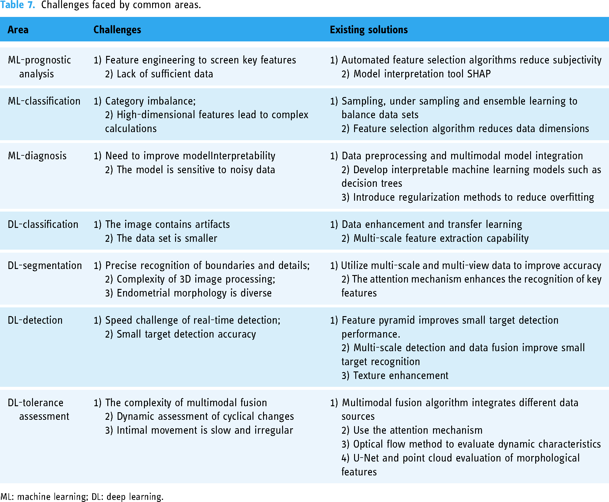
ML: machine learning; DL: deep learning.
After discussing the application of AI in diagnosing endometrial disease using ultrasound, and analyzing the challenges and coping strategies, it has been observed that researchers and developers have successfully developed innovative technologies such as automated feature selection, multi-scale feature extraction, and attention mechanisms. These technologies have effectively improved the diagnostic performance and clinical applicability of the model.
Although these technologies have achieved initial results, it is necessary to continue optimizing and experimentally verifying them to ensure their stability and effectiveness in clinical practice. The rapid development of technology and the growing clinical needs indicate that this field has broad prospects for future development. The new directions and development trends of future research mainly include the following four aspects:
Scarce Dataset: Insufficient medical data limits the performance of the model. 102 In the future, implicit data enhancement techniques (such as BatchFormer and BatchFormer V2103,104) can be used to improve the diversity of ultrasound image data without modifying the original data. In addition, multimodal data fusion can make up for the shortcomings of single-modality information and enhance the accuracy of diagnosis. Feature Extraction Optimization: The dedicated extraction module for endometrial structural features needs to be further optimized. Use attention mechanism and deformable convolution (such as row, column convolution, and snake convolution 105 ) to extract linear features and improve diagnostic accuracy. 106 Model Lightweight: Combining lightweight Transformer models (such as MobileBERT and DistilBERT107,108) with local attention mechanisms can reduce parameters while maintaining the efficiency of feature extraction and improving the application efficiency of the model. Multimodal data fusion and alignment: Strengthen the feature fusion and context alignment of different modal data, especially at the channel and spatial levels, optimize image segmentation and auxiliary diagnosis, and improve the recognition ability of complex lesions.
Conclusion
In conclusion, the implementation of AI in the diagnosis of endometrial diseases has notably enhanced the diagnostic precision of ultrasound imaging, reduced the workload of medical professionals, and facilitated expeditious and accurate decision-making through automated analysis and pattern recognition. In particular, AI technology has been shown to enhance the precision of lesion identification in the context of endometrial cancer and endometriosis. Additionally, it furnishes physicians with invaluable insights into disease progression and prognosis, thus aiding in the creation of bespoke treatment plans. Despite the considerable advances made by AI in the diagnosis of endometrial diseases, there is still a need to enhance the accuracy, relevance, and interpretability of the models in use. It is anticipated that through continued innovation and improvement, AI digital health technology will have a more profound positive impact on clinical practice, improve patient experience, reduce medical costs, improve the efficiency of medical services and ultimately benefit more patients.
Footnotes
Contributorship
Qiao Wei: writing—original draft, investigation, conceptualization, visualization. Zhang Xiao: writing –original draft, writing—review & editing, validation. Xiaowen Liang and Zhili Guo: writing—review & editing, visualization. Yanfen Zhang: visualization. Zhiyi Chen: conceptualization, writing—review & editing, visualization, supervision, funding acquisition.
Declaration of conflicting interests
The authors declared no potential conflicts of interest with respect to the research, authorship, and/or publication of this article.
Funding
The authors disclosed receipt of the following financial support for the research, authorship, and/or publication of this article: This work was supported by the Clinical Research 4310 Program of the Affiliated Changsha Central Hospital of the University of South China, National Natural Science Foundation of China, Health Research Project of Hunan Provincial Health Commission (grant numbers 20214310NHYCG06, 82102054, 82402372, W20241010).
Guarantor
ZC.