
Abstract
Case summary
A female intact domestic shorthair kitten was evaluated at 8 months of age for bilateral mucopurulent nasal discharge, stertor, open-mouth breathing and difficulty eating. Imperforate nasopharynx (INP) was diagnosed on oronasal examination under anesthesia. An extended palatoplasty was performed and resulted in resolution of the clinical signs.
Relevance and novel information
The extended palatoplasty procedure is relatively simple and very similar to the correction of soft palate elongation in brachycephalic dogs. It does not require special equipment and materials; therefore, it represents a viable, more readily available and cost-effective option for the treatment of INP. To the author’s knowledge, this is the first case of INP that has been treated with extended palatoplasty.
Introduction
Nasopharyngeal stenosis (NPS) is a rare condition in cats that is defined as a complete (INP) or partial obstruction of the nasopharynx by a soft tissue membrane. Most cases are acquired, either secondarily to upper respiratory tract inflammatory diseases or previous surgery of the nasopharynx. The cats usually present with stertorous breathing, snoring, open-mouth breathing, reverse sneezing, chronic nasal discharge or lack of nasal airflow. Concurrent abnormalities can be found in cats suffering from nasopharyngeal stenosis, such as choanal atresia. CT can identify and differentiate those abnormalities and evaluate the magnitude of their significance. 1 The stenosis is more commonly seen in the caudal aspect of the nasopharynx, but can also be seen more rostrally. Treatment for this condition is done mostly via stenting and balloon dilatation, but can also be treated with extended palatoplasty. Balloon dilatation is a safe and effective treatment option of relief in clinical signs of NPS in cats, but may require multiple treatments. The dilatation can be followed by temporary stenting with a silicone stent. Stenting is recommended when the lesion is more than 1 cm rostral to the end of the soft palate, to allow closure of the caudal end of the nasopharynx while eating. 2 In the case presented, balloon dilatation was considered inappropriate owing to the nasopharyngeal imperforation. Extended palatoplasty is more available and affordable then stenting and balloon dilatation, and therefore more relevant in multiple cases.
Case description
A female intact domestic shorthair kitten was evaluated at 8 months of age for bilateral mucopurulent nasal discharge, stertor, open-mouth breathing and difficulty eating. The cat was a shelter cat and was found 1 month before presentation with a litter of four kittens. One of the kittens exhibited similar signs and died at the age of 3 weeks. The cat had not responded to an antibiotic treatment followed by a steroid treatment prescribed by the shelter veterinarian.
On presentation, the cat was bright, alert and responsive, with a body condition score of 3/9. Bilateral gelatinous nasal discharge was present with no airflow detected through either nostril. Stertor and open-mouth breathing without apparent distress were noted. On oral examination, the soft palate was bulging (Figure 1). As a result of the bulge, signalment and clinical signs, a nasopharyngeal polyp was suspected.

A bulge in the soft palate was seen in an oral exam
Other differentials for the respiratory condition included a congenital anatomical defect, such as nasopharyngeal stenosis (NPS) or choanal atresia, upper respiratory infection or foreign body. The results of a complete blood count, serum chemistry profile and urinalysis were within the reference intervals, and the cat was negative for feline immunodeficiency virus and feline leukemia virus based on an in-house ELISA. These tests were performed before inducing anesthesia for a more detailed examination, inspection for a nasopharyngeal polyp and possible viral, bacterial and cytological sampling, if indicated. A CT study was not performed owing to financial constraints.
An intravenous catheter was placed, and the cat received bupernorphine 0.015 mg/kg IV and propofol 3 mg/kg IV, and was intubated with a 3.5 endotracheal tube.
The patient was then placed in dorsal recumbency and a spay hook was used to try and retroflex the soft palate. Attempts to retroflex the soft palate were unsuccessful, as no nasopharyngeal opening could be located. A small blind opening was seen at the caudal aspect of the nasopharynx, between the end of the soft palate and the choanae (Figure 2); therefore, imperforate nasopharynx (INP) was suspected. The retroflexion attempts resulted in drainage of a gelatinous, rubber-like material from both nostrils, which led to resolution of the bulge (Figure 3). To differentiate between INP and NPS, the area was probed and found to be imperforate. A further investigation was performed by introducing urinary catheters into both nostrils and gently flushing with NaCl solution, which resulted in nasal drainage only. Flushing while occluding the nostrils resulted in inflation of the soft palate, demonstrating a complete isolation of the nasal and oral cavities with no nasopharyngeal communication.

A small blind opening was seen at the nasopharynx, diagnosed as an imperforate nasopharynx

A gelatinous, rubber-like material was drained through the nostrils
An extended palatoplasty was performed with a monopolar sealing device. The incision was made rostral to the tonsils when crossing the mid-sagittal line and passed medially to the tonsils, with an arch shape (Figure 4). Excision of the entire stenotic region was performed and verified by direct visual inspection after soft palate removal. The oronasal and pharyngeal mucosa were apposed with simple continuous suture pattern with a 4.0 monofilament absorbable poliglecaprone 25 suture material (Monocryl; Ethicon) (Figure 5).

An extended palatoplasty was performed with a monopolar sealing device. The incision was made rostral to the tonsils when crossing the mid-sagittal line and passed medially to the tonsils, with an arch shape
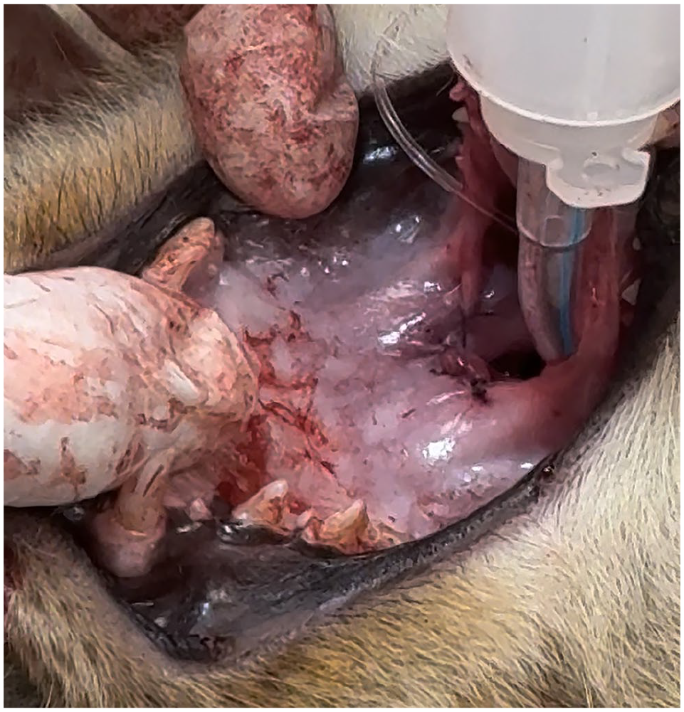
Immediate postoperative view showing the nasopharyngeal opening that was created by extended palatoplasty
The cat recovered from the anesthesia and showed an immediate improvement of its respiratory signs. It was sent back to the shelter with a prescription for buccal buprenorphine 0.015 mg/kg administered 2–3 times/day for analgesia over a few days. One month later, the cat returned for ovariohysterectomy. The caretaker reported a complete resolution of clinical signs and significant weight gain (1.5 kg). The cat’s nasopharynx was examined under anesthesia and was patent and healed (Figure 6).

Postoperative image taken 1 month later, showing a patent nasopharyngeal opening
Discussion
NPS and INP are uncommon disorders in cats that narrow or occlude the nasopharynx, limiting or preventing airflow through the nasal passages. 3 These disorders may be congenital, acquired secondary to an infectious, inflammatory, caustic (eg, aspiration rhinitis) or traumatic etiology, benign or malignant masses, or scarring after nasopharyngeal surgery. 1
Both NPS and INP can result in stertorous nasal breathing, dyspnea, open-mouth breathing, gagging, repeated swallowing, sneezing, and chronic serous or mucopurulent nasal discharge. A definitive diagnosis can be made via retropharyngoscopy, CT of the nasopharynx or both.1,3
Reported treatment options for NPS include surgical resection (extended palatoplasty), laser ablation, mechanical dilatation with a Kelly forceps or balloon, and stenting with a metallic stent or silicone tubing.1,3,4 Balloon dilatation and stenting is less available and more costly than the other treatment options. Extended palatoplasty was found to be a successful and repeatable method to treat caudal NPS, with low risk of complications. 5
In the case presented, the CT option and a referral were discussed with the caretaker; however, since the cat was a shelter cat, there were financial concerns. Extended palatoplasty therefore was chosen as the treatment for this cat. Extended palatoplasty is more affordable and usually does not require multiple procedures. In addition, since the nasopharynx in this cat was imperforated, it seemed more appropriate than the other treatment options. The procedure resulted in a complete resolution of the cat’s clinical signs. Four months postoperatively, the cat was still showing complete resolution.
Conclusions
The extended palatoplasty procedure is relatively simple and very similar to the correction of soft palate elongation in brachycephalic dogs. It does not require special equipment and materials; therefore, it represents a viable, more readily available and cost-effective option for the treatment of INP.
Footnotes
Conflict of interest
The author declared no potential conflicts of interest with respect to the research, authorship, and/or publication of this article.
Funding
The author received no financial support for the research, authorship, and/or publication of this article.
Ethical approval
The work described in this manuscript involved the use of non-experimental (owned or unowned) animals. Established internationally recognized high standards (‘best practice’) of veterinary clinical care for the individual patient were always followed and/or this work involved the use of cadavers. Ethical approval from a committee was therefore not specifically required for publication in JFMS Open Reports. Although not required, where ethical approval was still obtained it is stated in the manuscript.
Informed consent
Informed consent (verbal or written) was obtained from the owner or legal custodian of all animal(s) described in this work (experimental or non-experimental animals, including cadavers, tissues and samples) for all procedure(s) undertaken (prospective or retrospective studies). For any animals or people individually identifiable within this publication, informed consent (verbal or written) for their use in the publication was obtained from the people involved.