
Abstract
A 5-year-old male neutered Siamese cat was referred because of nasal swelling, nasal discharge, and oral respiration. Computed tomography and necropsy following euthanasia revealed a firm tan mass in the nasopharynx, occluding the nasal passage. Histologically, the nasopharyngeal mass was composed of solid nests, anastomosing cords, and closely packed glands separated by a delicate fibrovascular stroma. Individual neoplastic cells were cylindrical to polyhedral in shape, had distinct cell borders, and contained moderate amounts of finely granular eosinophilic cytoplasm and round to oval nuclei. The cytoplasmic granules were positive on phosphotungstic acid–hematoxylin staining. Immunohistochemically, the neoplastic cells were positive for cytokeratin and negative for chromogranin A, neuron-specific enolase, and vimentin. Ultrastructurally, the neoplastic cells contained numerous mitochondria.
Tumors of the nasal cavity and paranasal sinuses are rare in cats, accounting for only 1–8% of all feline tumors. 14,19 Although the majority of tumors are malignant and locally invasive, distant metastases are rare. 14,19 Among nasal and paranasal tumors of epithelial origin, adenocarcinomas are the most common, followed by squamous cell carcinomas 14 ; whereas, among nonepithelial tumors, malignant lymphomas are the most common. 7 No clear pattern with respect to breed or gender has been documented in feline nasal tumors, except for a higher incidence of squamous cell carcinomas in male neutered cats. 14
Oncocytomas, neoplastic proliferations of oncocytes, are rare tumors in both domestic animals and human beings. 5,12 Oncocytes are large polygonal granular cells containing numerous mitochondria appearing as moderate amounts of eosinophilic granular cytoplasm. 5,12 The World Health Organization (WHO) has classified oncocytic tumors into 3 histological categories: oncocytosis, oncocytomas, and oncocytic carcinomas. 3 In human beings, oncocytomas have been reported in a wide variety of tissues, including the parotid gland, 3 nasal cavity, 6 adrenal gland, 11 kidney, 11 and thyroid gland. 11 To date, only a few oncocytomas have been reported in dogs 2,8,13,16 and cats. 1,4,9
A 5-year-old male neutered Siamese cat was referred to the Veterinary Medical Teaching Hospital of Seoul National University (Seoul, Korea) with clinical signs of nasal swelling, nasal discharge, oral respiration, and slight loss of coordination. Hematology and serum chemistry values were unremarkable. Initial nasal radiography revealed a soft tissue opacity just caudal to the tympanic bullae and dorsal to the nasopharyngeal region, with occlusion of the normal airway in the region of the nasopharynx. Differential diagnoses of nasopharyngeal polyp, granuloma, and neoplasia were considered. Computed tomography (CT), a including an initial survey scan and pre- and postcontrast axial scans from the external nares region to the third cervical vertebral level, was performed, yielding axial and reformatted images. Caudal CT images showed a heterogeneous, hyperattenuating mass that filled the nasopharyngeal region (Fig. 1). Moreover, the airway was not visible, except for that portion filled by the inserted endotracheal tube. No invasion of the soft tissue into the nasal cavity or vertebrae was detected. Shortly after the imaging procedure, the cat began to exhibit neurologic signs of entasia and loss of coordination. Because of poor prognosis, the animal was euthanized, and a postmortem examination was performed immediately thereafter.
At postmortem examination, as noted on CT scanning, the nasopharynx was completely occupied by a firm, tan–red mass, with partial destruction of the overlying basisphenoid and presphenoid bones (Fig. 2). However, no gross evidence of tumor infiltration into the adjacent brain tissue was observed, nor was obvious extension or invasion of the main nasopharyngeal mass into the nasal or oral cavities noted on serial sagittal sections. No other significant gross abnormalities were noted in other organs, including regional lymph nodes.
For histopathology, tissue samples except brain were fixed for 3 days in neutral phosphate buffered 10% (v/v) formalin solution, then processed in a routine manner, embedded in paraffin, and stained with hematoxylin and eosin. Replicate sections of selected tumor samples were used for immunohistochemistry and phosphotungstic acid–hematoxylin (PTAH) staining. b To delineate the possible origin of neoplastic cells, a standard avidin–biotin–peroxidase method c and commercially available antibodies against chromogranin A, d cytokeratin (CK), d neuron-specific enolase (NSE), d and vimentin d were used. Feline skin and adrenal gland were used as positive control tissues. For negative control, the slides were treated identically, except omitting primary antibody. Electron microscopy (EM) e was performed on slices prepared from paraffin-embedded tumor blocks as previously described. 18
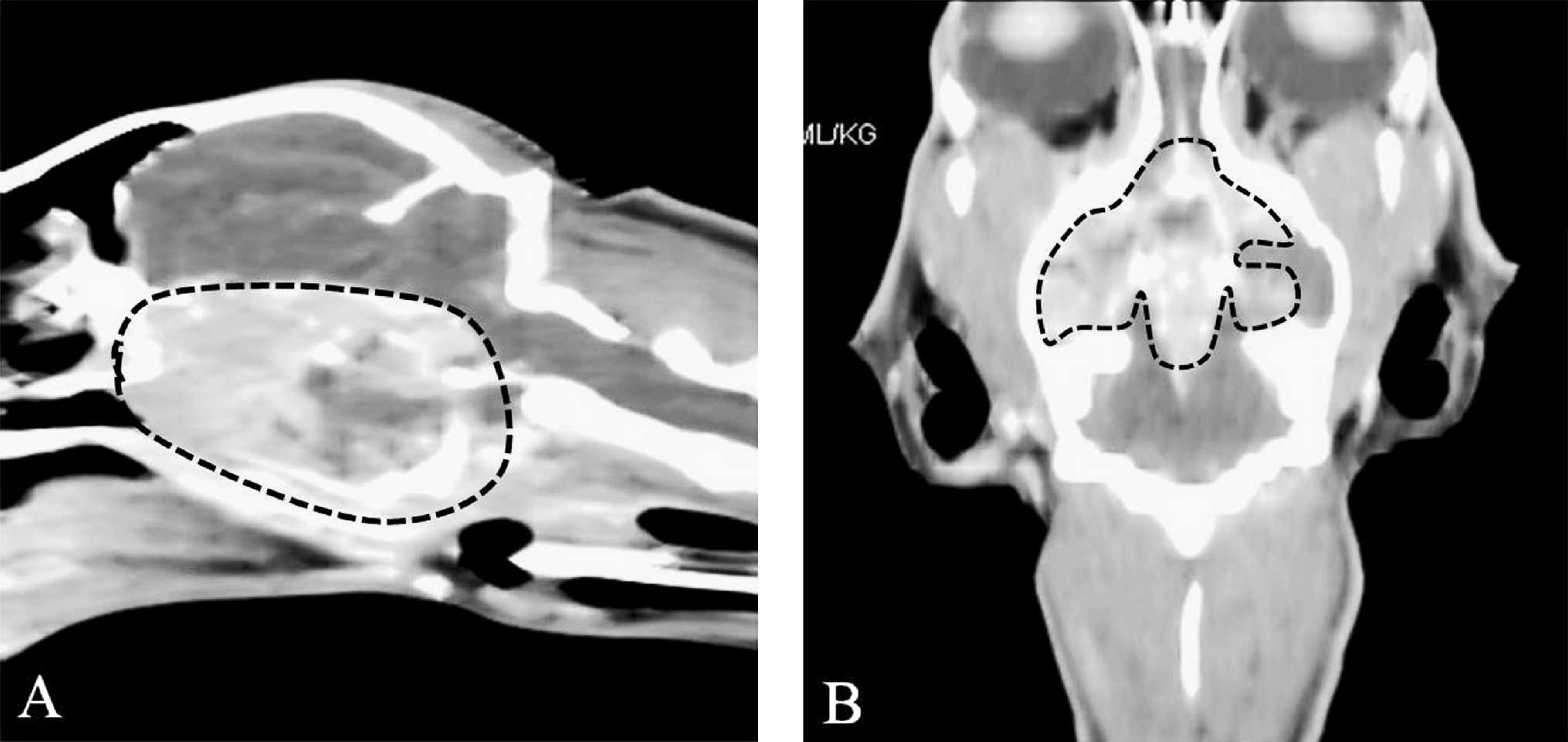
Computed tomographic imaging of head; Siamese cat. Reformatted sagittal (
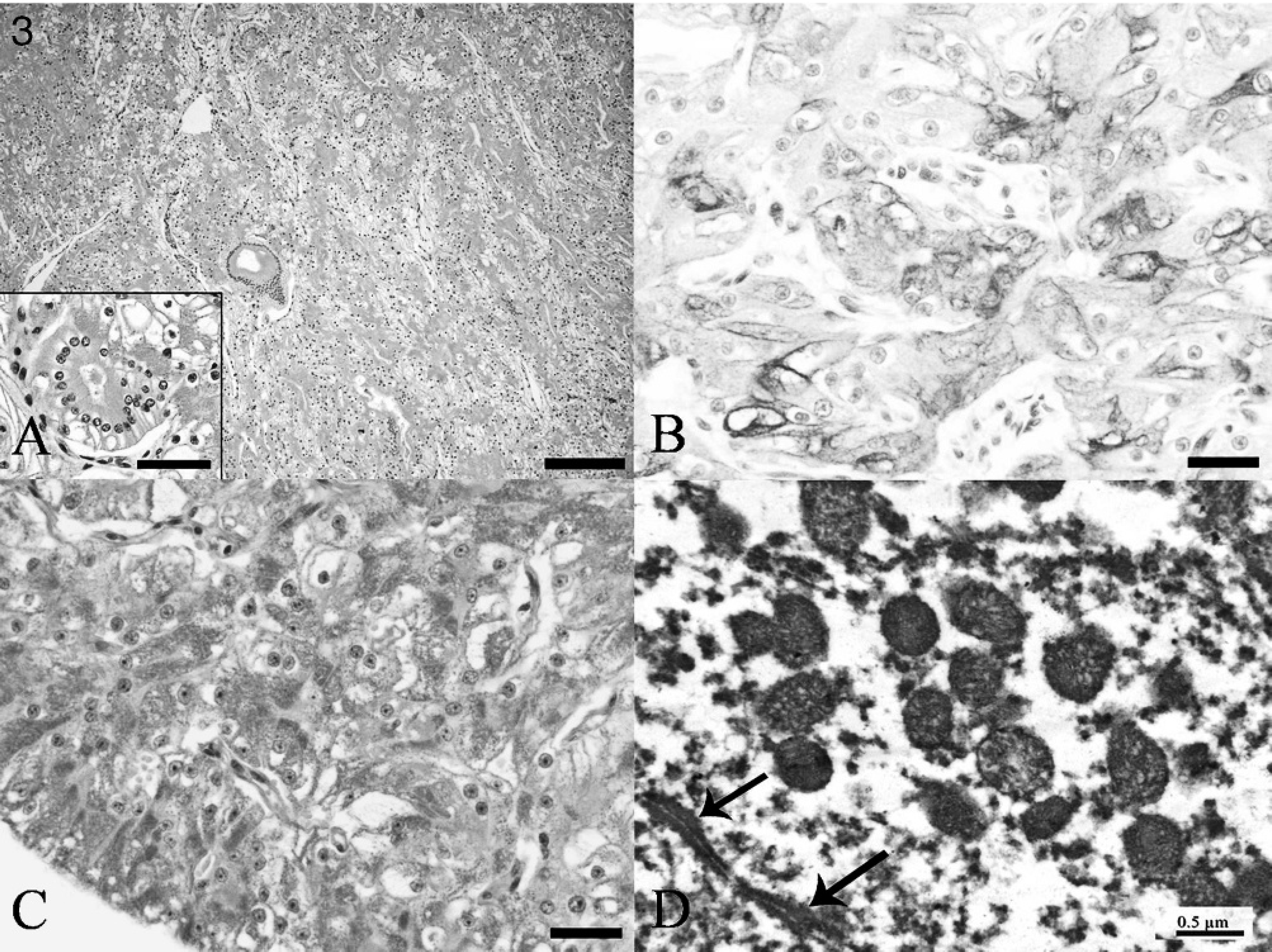
Microscopically, the nasopharyngeal mass consisted of cylindrical to polyhedral neoplastic cells arranged in solid nests, anastomosing cords, and closely packed glands separated by fine fibrovascular septa (Fig. 3A). Individual neoplastic cells had moderate to abundant, finely granular eosinophilic, occasionally vacuolated, cytoplasm with distinct cell boundaries and round to oval, central to basally located nuclei with 1 prominent nucleolus. Zero or 1 mitotic figure was visible per 10 high-power fields, and vascular invasion was not observed. Neoplastic cells extended into and destroyed the adjacent basisphenoid and presphenoid bones but did not involve or extend into the meninges or brain parenchyma. Additionally, the tumor invaded into the distal portion of the nasal cavity. Scattered mild lymphocytic, plasmacytic, and neutrophilic infiltrations were noted in the tumor tissue, mainly around the blood vessels.
Differential diagnoses of nasal neuroendocrine tumor, granular cell tumor, tumor of neuroepithelial origin, and undifferentiated carcinoma were considered. Immunohistochemically, the cytoplasm of neoplastic cells was strongly and diffusely positive for CK (Fig. 3B) but was consistently negative for chromogranin A, NSE, and vimentin. These results helped to rule out neuroendocrine, neuroepithelial origin, and granular cell tumors, suggesting a likely epithelial neoplastic cell origin. To identify the nature of the fine cytoplasmic granules, PTAH staining and EM evaluation were conducted. In the PTAH stain, cytoplasmic granules stained dark blue (Fig. 3C). Ultrastructurally, neoplastic cells were interconnected via junctional complexes and contained numerous round to oval, electron-dense mitochondria (Fig. 3D). On the basis of the immunohistochemical staining profile of the neoplastic cells, together with PTAH staining and EM results, a diagnosis of nasopharyngeal oncocytoma was made.
The precise histogenesis of oncocytes remains unclear. Oncocytes were first identified in the canine thyroid gland and have since been found in the ducts and acini of various endocrine and exocrine glands, presumably as a result of age-related changes. 5 In rats treated with N-nitrosomorpholine, renal oncocytomas developed from the renal cortical collecting duct system. 15 Oncocytogenesis has been considered a compensatory cellular response seeking to overcome mitochondrial dysfunction associated with mitochondrial DNA errors. 3 Such mitochondrial DNA mutations might result from aging or oxidative damage. 17 Additional work is required before more general conclusions can be drawn for pathogenesis of feline nasal oncocytoma.
Histologically, oncocytic tumors are classified into 3 categories: oncocytosis, oncocytoma, and oncocytic carcinoma according to the WHO classification scheme. 3 Oncocytosis is a nonneoplastic nodular hyperplasia of oncocytes similar to nodular hyperplasia of endocrine glands. 3,5 Oncocytic carcinomas have destructive and infiltrative growth, cellular pleomorphism, vascular and lymphatic invasion, and regional and distant metastases. 3 Most human oncocytic tumors are benign, and only a few oncocytic carcinomas have been reported to date in the salivary gland, kidney, thyroid gland, and nasal cavity. 6,10,11 Except for bilateral renal oncocytomas in dogs, 2 all canine and feline oncocytomas found to date have been pathologically and biologically benign.
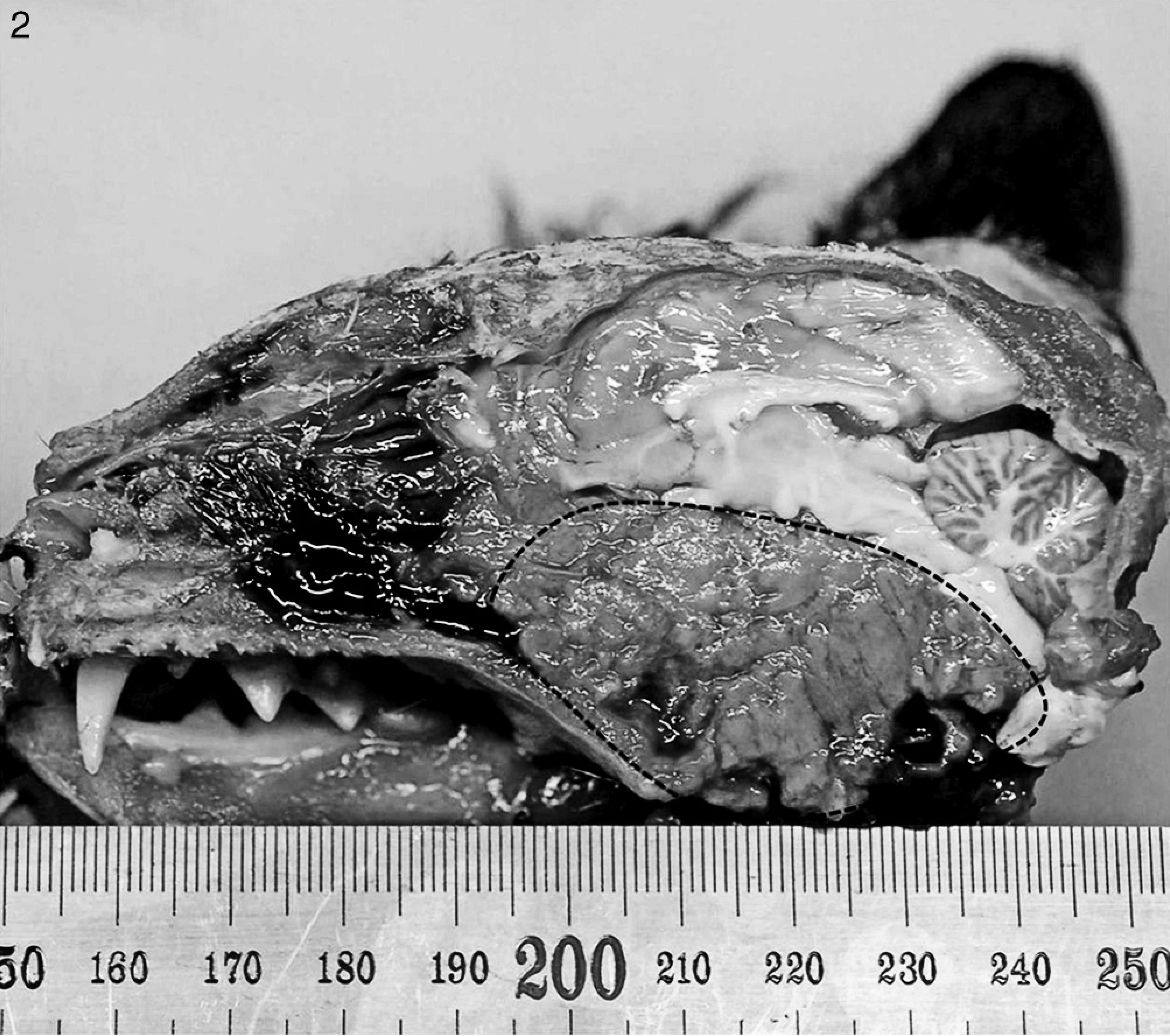
Sagittal section of head; Siamese cat. The nasopharynx is completely obliterated by a firm tan–red mass with partial destruction of the overlying basisphenoid and presphenoid bones. The margins of the tumor are delimited by a dotted line.
Nasopharyngeal mass; Siamese cat.
The size and opacity of the cat tumor described herein, together with local invasiveness to the overlying basisphenoid and presphenoid bones and the distal portion of the nasal cavity, were indicative of malignancy. Nevertheless, the tumor was diagnosed as a benign oncocytoma because it was histologically well differentiated with a very low mitotic index and minimal pleomorphism and did not show any indication of vascular invasion or metastasis. As observed in the present case, extension into the surrounding bony structures can potentially occur in any benign nasal tumor or space-occupying mass that obliterates the nasal passages; such behavior is not necessarily an indication of malignancy. A human patient with a nasal oncocytoma similar in nature to the tumor of the cat described in the current case experienced a good prognosis on follow-up. 10 Additionally, a similar tumor reported in a 12-year-old female spayed Domestic Shorthair cat was benign. 9 Magnetic resonance imaging of the published case showed that the tumor mass occupied the entire nasal cavity and frontal sinus. A full postmortem examination was not performed in that animal, but pathologic findings of the mass were very similar to those of the cat presented in the current report.
Nasal oncocytomas in animals are rare tumors of unknown etiology, similar to the human nasal oncocytoma. 6 Because of the limited number of cases, age, breed, or sex predilection have not been established. To the authors' knowledge, the current case is the second report of a nasal oncocytoma in animals. In the current report, the tumor occurred in the nasopharynx, with no involvement of the nasal cavity, in a relatively young animal, in contrast to the 12-year-old cat from the previously published feline case, 9 and caused severe clinical signs that lead to its death. In contrast to the previous case, 9 a full postmortem examination was performed, and metastases were not observed despite an extensive search. Oncocytomas should be considered as a differential diagnosis of a tumor found in the nasopharynx of domestic animals.
Footnotes
a.
HiSpeed CT/e, GE Medical Systems, Milwaukee, WI.
b.
Sigma-Aldrich, St. Louis, MO.
c.
ABC Elite kit, Vector Laboratories Inc., Burlingame, CA.
d.
Dako North America Inc., Carpinteria, CA.
e.
Philips 208S TEM, FEI Co., Hillsboro, OR.