
Abstract
Lipoma is rare in the planter aspect of the toes, and only few cases of massive lipoma have been reported in this site. The differential diagnosis of masses in the foot and toes is wide, and clinical diagnosis may be challenging. Access to magnetic resonance imaging, a standard diagnostic investigation for such soft tissue masses of the foot and toes, may be limited in some practice, requiring a reliance on clinical signs. We report a solitary massive lipoma in the planter aspect of the right great toe that appeared as two masses and with modification of typical clinical signs of lipoma.
Introduction
Lipoma is a common tumor that is mostly found in the trunk and proximal limbs. Usually solitary, slow growing, and painless, it may attain a huge size at diagnosis. It is rare in the planter aspect of the toes, and only few cases of massive lipoma have been reported in this site. The peculiar anatomy of this region may modify the typical clinical features of lipoma making diagnosis challenging especially when access to magnetic resonance imaging (MRI) is limited. A 47-year-old man presented with a solitary massive lipoma that appeared as two masses.
Case report
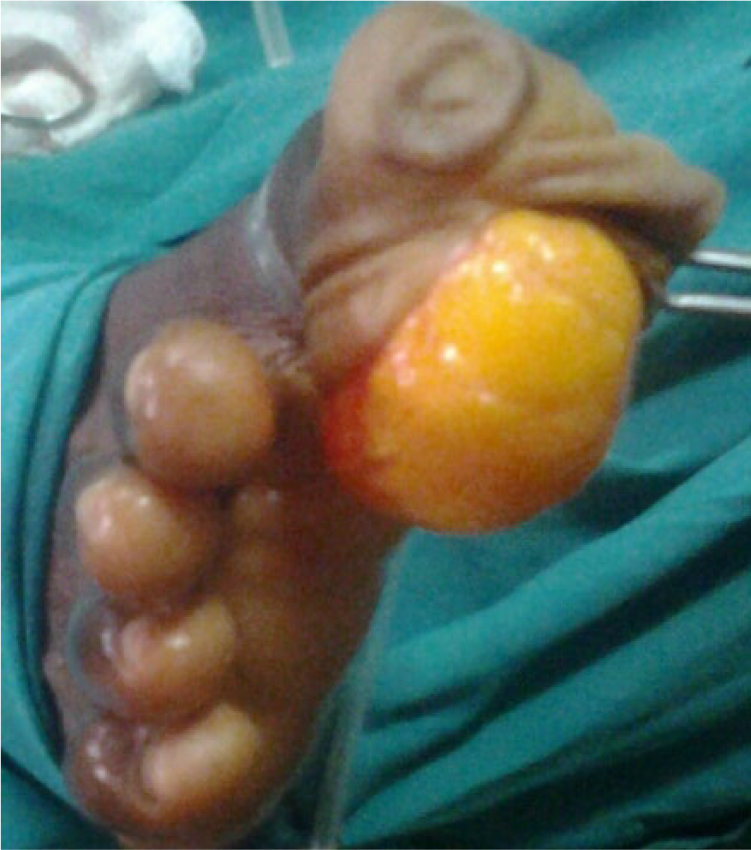
A 47-year-old man presented with two painless masses on his right great toe. The bigger mass was noticed 5 years before presentation, while the smaller distal mass was a year old. Masses had grown gradually in size and now affected his gait and prevented him from wearing shoes. On examination, he had two masses in the lateral side on the planter aspect of the right great toe (Figure 1). The proximal mass was bigger and was about 13 cm × 8 cm in size. It was globular, felt cystic but tense. It extended proximally into the web space and had caused separation of the great toe from the other toes. The distal mass was about 2.5 cm in diameter and was less tense. It slipped away when digital pressure was applied on its edge and was nearly completely reducible into the proximal bigger mass suggesting a common origin. They were both free from the skin and underlying tissue and transilluminated positively. He had no other masses on his body and no abnormal skin pigmentations. The neurovascular examination was normal and his inguinal lymph nodes were not enlarged.

Right great toe with masses.
A presumptive clinical diagnosis of lipoma was made to rule out neurofibromatosis and liposarcoma. Plain radiographs of the foot revealed soft tissue shadows and normal underlying bone. MRI could not be done because it was not available. He was managed with a marginal local excision using a longitudinal incision after a digital block with application of a tourniquet. A single, well-encapsulated multi-lobulated mass with typical visual features of lipoma extended to the web space (Figure 2). It was uniformly soft and free from the skin, underlying fascia, tendon sheath, and bone. The redundant planter skin was trimmed and the toe refashioned. He had an uneventful post-operative period. Histology showed mature fat cells and confirmed lipoma. He required no further treatment, and he has been followed up for 2 years without recurrence.
Intra-operative finding. A single, lobulated, well-encapsulated mass.
Discussion
Subcutaneous lipoma is rarely found in the planter aspect of the toes of adults. 1 Very few cases of massive lipoma of the toes have been reported. Most tumors of the toes are benign but there exists a wide range of diagnoses with atypical presentations making clinical diagnosis challenging. 2 Most are slow growing with asymptomatic evolution and are often associated with delay in diagnosis. 3
Subcutaneous lipoma typically presents as slow growing soft masses with clinical signs of lobulation and a positive slipping sign. Pressure applied on the edge of subcutaneous lipoma characteristically causes the tumor mass to slip away. 4 Some subcutaneous lipoma transilluminate positively. In the planter aspect of the toe of this patient, lobulation created an appearance of two separate masses. The distal mass slipped under pressure and could nearly completely reduce into the bigger proximal one. This suggested a common origin and a modification of the typical slipping sign. Compartmentalization of the subcutaneous fat layer in the planter aspect of the toe and foot by fibrous bands may be responsible for this modification. A dumb-bell lipoma in the planter aspect of the foot due to the peculiarities in the anatomy was reported. 5
Our patient toe was managed with marginal local excision after a presumptive clinical diagnosis like is done for most subcutaneous lipoma in other sites. MRI, recommended in the diagnosis of soft tissue tumors of the foot and toes, was not available in this center. MRI is particularly important in rapidly growing masses and those with atypical clinical findings that may suggest malignancy. 6 The use of MRI in the diagnosis of big soft tissue swellings of the foot is becoming a standard practice but access to MRI is still limited in some parts of the third world. MRI correctly diagnoses soft tissue tumors of the foot in 89% of case but it may be inconclusive in differentiating well-differentiated liposarcoma from benign lipoma.7,8 A T1-weighted MR image of lipoma shows lobulated, high signal intensity mass. Fat-saturated proton density-weighted MR image shows homogenous low signal intensity and there is no tumor mass enhancement after gadolinium enhancement on fat-saturated, T1-weighted image of lipoma. 9 Core needle biopsy may be needed for preoperative differentiation of lipoma from a well-differentiated liposarcoma in similar presentation.
The differential diagnosis in this patient includes plexiform neurofibromatosis and liposarcoma. A neurofibroma has a firm consistency, a limited mobility, and does not transilluminate positively. A liposarcoma has a relatively more rapid growth with a shorter history, is firm to hard in consistency, and may be attached to surrounding structures. Well-differentiated liposarcoma may be difficult to differentiate from lipoma. Our patient had a 5-year history of slow growing mass with modification of clinical features of a typical lipoma. With limited access to ancillary investigations, we chose an initially conservative surgical approach. The intra-operative finding of a well-encapsulated, lobulated, soft fatty mass that was free from adjacent structures further supported the diagnosis of lipoma. Local excision of lipoma is associated with recurrence rates of about 1%–2%. 10 The histology of the tumor specimen confirmed lipoma. A histology indicating a liposarcoma or other malignant lesion would require further surgical management with disarticulation of the toe and possible post-operative irradiation.
In conclusion, this size of lipoma is rare in the planter aspect of the toe and the diagnosis required a detailed interpretation of clinical signs. Understanding the modification of typical clinical signs of lipoma in the foot and toes may prove important when access to ancillary investigations is limited and when these investigations are inconclusive. Recognition of these modifications helped us reach a clinical diagnosis and to avoid an immediate amputation or disarticulation of the toe as was previously reported in massive lipoma of this size.11,12 Amputation of the great toe is associated with difficulty in walking requiring a re-training therapy.
Footnotes
Declaration of conflicting interests
The author(s) declared no potential conflicts of interest with respect to the research, authorship, and/or publication of this article.
Ethical approval
Our institution does not require ethical approval for reporting individual cases or case series.
Funding
The author(s) received no financial support for the research, authorship, and/or publication of this article.
Informed consent
Written informed consent was obtained from a legally authorized representative(s) for anonymized patient information to be published in this article.