
Abstract
Context:
Dysplastic pulmonary valve stenosis is a less common variety of valvular pulmonary stenosis. It is known to be part of Noonan syndrome. Bony hand anomalies in patients of pulmonary stenosis are very rare.
Case report:
A 50-year-old lady, with no significant past history, presented with slowly progressive breathlessness and fatigue, and had progressed from NYHA class 1 to 2 over 2 years. She had unilateral absent first metacarpal and diagnosed on workup to have dysplastic pulmonary valve stenosis and was treated with balloon valvuloplasty.
Conclusion:
Dysplastic pulmonary valve stenosis can rarely be associated with bony hand anomalies like absent first metacarpal.
Introduction
Valvular pulmonary stenosis is of three types: mobile dome shaped, bicuspid and dysplastic.
Dysplastic pulmonary stenosis is a less common variety with obstruction caused by myxomatous thickening of three separate but poor mobile leaflets without commissural fusion. It is associated with Noonan syndrome and hypertrophic cardiomyopathy. 1
Noonan syndrome shows features of short stature, webbed neck and broad-shaped chest. Bilateral elbow abnormalities are described but bony hand anomalies are not described in Noonan syndrome. Dysplastic pulmonary stenosis resembles dome-shaped pulmonary stenosis clinically, except for absence of ejection click. 2 Intervention is indicated at a gradient more than 40 mmHg. 3 Bony hand anomalies with dysplastic pulmonary stenosis is a very rare association and we believe has not been described in literature.
Case history
We report the case of a 50-year-old woman, who presented with a slowly progressive breathlessness and fatigue. She had progressed from an NYHA (New York Heart Association) class 1 to 2 over 2 years. Patient did not give any history of paroxysmal nocturnal dyspnoea, orthopnoea, chest pain, palpitations, syncope or oedema. Her obstetric history was significant for two uneventful pregnancies and deliveries, delivered in a primary health centre in a rural setup surprisingly with no complications. There was no significant family history.
On examination, the following results were obtained: pulse = 80 bpm, normal volume, regular; blood pressure (BP) = 110/70 mmHg; and both upper limbs jugular venous pressure (JVP) = 6 cm above sternal angle with prominent ‘a’ waves. There was no cyanosis, clubbing or growth retardation or evidence of peripheral oedema.
On abdominal examination, the liver was palpable and the liver span was 15 cm. There was absent left first metacarpal (Figure 1). No other skeletal deformity or facial dysmorphism was seen. Cardiovascular system (CVS) examination showed a systolic thrill in the second left intercostal space (ICS) parasternal area. On auscultation, S1 was normal and S2-single was soft. Ejection systolic murmur grade 4/6 intensity was heard in the left second ICS parasternal area radiating upward and to the left. There was no ejection click.

Absent first metacarpal.
Electrocardiogram (ECG) showed right axis deviation with right ventricular (RV) hypertrophy and right atrial (RA) enlargement. Chest X-ray showed normal pulmonary vascularity, mild prominence of main pulmonary artery and its left branch, and RV type of apex (Figure 2). Hand X-ray confirmed the absence of left first metacarpal (Figure 3). Transthoracic echo showed dilated RA with RV hypertrophy, tricuspid annular plane systolic excursion (TAPSE) of 18 mm and Tei index of 0.38. The pulmonary valve had thickened and immobile leaflets, absence of doming, hypoplastic valve annulus and minimal post-stenotic pulmonary artery dialation. The Doppler gradient across the valve was 60 mmHg (Figure 4). Cardiac catheterisation showed peak-to-peak gradient of 50 mmHg, hypoplastic pulmonary annulus and minimal post-stenotic dilatation of pulmonary artery. Patient underwent pulmonary balloon valvuloplasty. Immediately after procedure, the catheter-measured gradient was 30 mmHg. At follow up, after 2 months, she had relief of symptoms and the catheter-derived gradient had decreased to 20 mmHg and echo gradient to 30 mmHg. There was mild pulmonary regurgitation seen on follow up. The patient was followed monthly for 6 months after which she was lost to follow up.

Chest X-ray showed RA enlargement and RV type of apex.
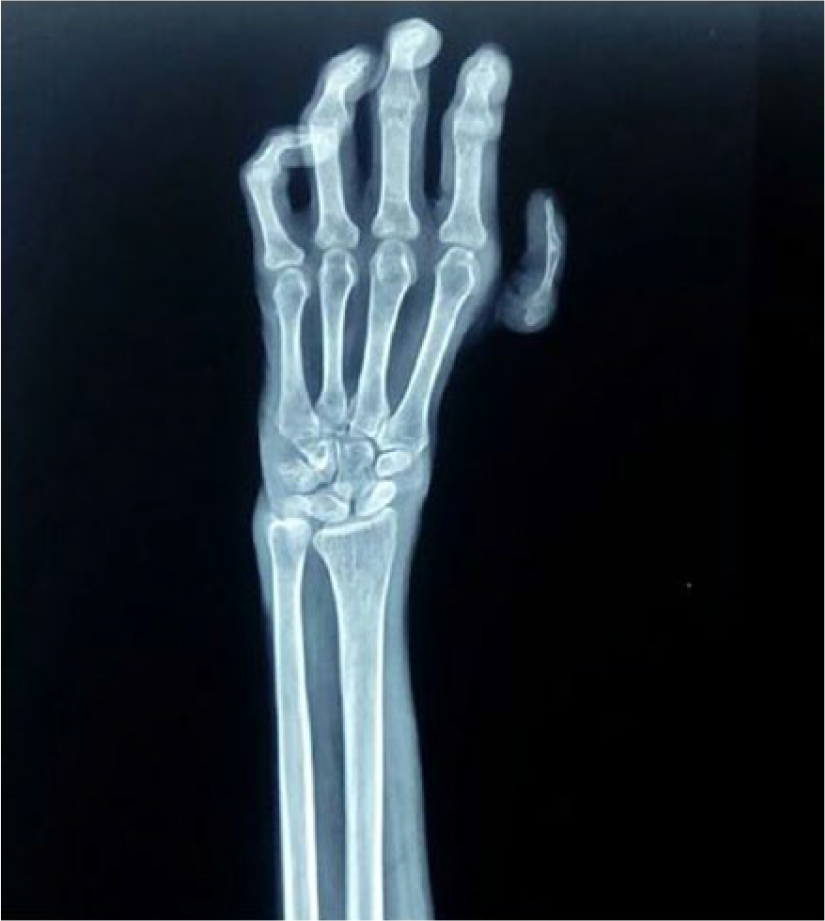
X-ray anteroposterior (AP) view of hand showing absent first metacarpal.

Doppler echo showing gradient of 60 mmHg through pulmonary valve.
Discussion
Dysplastic pulmonary stenosis is often seen associated with Noonan syndrome. It can present as late as fifth or sixth decade of life. Pulmonary stenosis can occur in isolation or be associated with bony hand anomalies such as elbow abnormalities, pectus excavatum or carinatum, as with Noonan syndrome; small chin, broad forehead, as with Williams syndrome and Alagille syndrome. The association of unilateral absent first metacarpal with pulmonary stenosis is quite a rare association and we believe has not been described in literature. These valves have a myxomatous morphology with poorly delineated lines of commissural fusion. As a result, the balloon valvuloplasty haemodynamics do not hold true as they do in typical form. Post-ballooning valve opening is unpredictable due to inherent morphology. Moreover, the valve annulus is smaller than doming valve variety.
These variations make the final outcome relatively less consistent. 4 Pre-procedure gradient less than 75 mmHg across pulmonary valve is the only significant factor associated with successful outcome. Immediate post-procedure gradient less than 60 mmHg is significantly associated with higher freedom from re-intervention rate. Thus, balloon valvuloplasty should be tried before consideration of surgery in such patients. 4 Bony hand anomalies usually direct clinicians to look for the presence of features of atrial septal defect (ASD) as part of Holt–Oram syndrome. But the possibility of pulmonary stenosis in such patients should always be kept in mind.
Thus, a careful cardiac examination and further evaluation with echocardiography are warranted in patients seen with absent first metacarpal as the diagnosis of pulmonary stenosis can be made even in the asymptomatic stage. If it is diagnosed, patient can be monitored and intervention can be done at appropriate indications to prevent further complications.
Footnotes
Declaration of conflicting interests
The author(s) declared no potential conflicts of interest with respect to the research, authorship and/or publication of this article.
Ethical approval
Our institution does not require ethical approval for reporting individual cases reports.
Funding
The author(s) received no financial support for the research, authorship and/or publication of this article.
Informed consent
Written informed consent was obtained from a legally authorised representative(s) for anonymized patient information to be published in this article.
Limitations of the case report
The pulmonary balloon valvuloplasty procedure images are unavailable due to loss of follow up of the patient. We regret the same. The main intention of this case report was to highlight the rare and as yet not described association of absent first metacarpal and dysplastic pulmonary stenosis.