
Abstract
Objective:
Venous structures of the transverse-sigmoid sinus region have been insufficiently studied by magnetic resonance venography, especially in the healthy Han Chinese population.
Methods:
Magnetic resonance venography data were reconstructed. The relevant parameters were recorded. A paired t-test was used to compare the diameters of the inferior petrous sinus at the origin and termination. An unpaired t-test, a chi-squared test or Fisher’s exact test was used to compare other data.
Results:
One hundred healthy participants were included. The average age was 36.6 ± 17.1 years, and the ratio of males to females was 1:1. The development of the transverse-sigmoid sinus had a right-sided predominance in 51% of patients. Statistical analysis revealed significant differences (p < 0.05) in the lengths of bilateral TSs, the lengths of bilateral partial TSs from the torcular to lateral tentorial sinus, the diameters between bilateral TSs at the origin and the lateral tentorial sinus, the diameters between bilateral TS-sigmoid sinus junctions and sigmoid sinus terminations, and the inferior petrous sinus diameters at the origin and termination. Statistical analysis revealed that the right lateral tentorial sinus was more likely to originate from the TS (p < 0.05). Statistical analysis revealed no significant differences (p < 0.05) between transverse-sigmoid sinus development and inferior petrous sinus continuity or between transverse-sigmoid sinus development and inferior petrous sinus continuity and bilateral inferior petrous sinus continuity.
Conclusions:
This study revealed that the right transverse-sigmoid sinus was predominantly larger in diameter, the torcular Herophili tended to deviate to the right, and the right lateral tentorial sinus tended to drain into the TS. The inferior petrous sinus at the origin was thicker than that at the termination, and the right inferior petrous sinus was thicker than the left inferior petrous sinus. transverse-sigmoid sinus development had no effect on inferior petrous sinus continuity, and there was no difference in inferior petrous sinus continuity between the left and right sides.
Introduction
The venous structures of the transverse-sigmoid sinus (TSS) region include the transverse sinus (TS), sigmoid sinus (SS), Labbé vein, superior petrous sinus (SPS), inferior petrous sinus (IPS), lateral tentorial sinus (LTS) and medial tentorial sinus (MTS). 1 These structures are important and drain vital intracranial structures. 2
Many intracranial diseases, such as tumors, dural arteriovenous fistulas, thrombosis and stenosis, can occur in the TSS region.3–5 Therefore, the anatomy of the TSS region warrants further study. Current available study methods include autopsy, digital subtraction angiography (DSA), computed tomographic angiography (CTA), and magnetic resonance venography (MRV).6–8 MRV is a noninvasive tool for reconstruction and is widely used to diagnose venous sinus disease.9,10 To date, studies on the venous structures of the TSS region have been insufficient, especially in the healthy Han Chinese population; therefore, we performed such an important study using 3.0 Tesla MRV.
Materials and methods
A retrospective MRV study of venous structures of the TSS region was performed in healthy Han Chinese participants during routine physical examinations that took place between October 2020 and October 2022. MRV examinations were performed using a 3.0 Tesla magnetic resonance scanner (Discovery 750, GE Healthcare, Milwaukee, WI, USA). This study was approved by the ethics committee of our institute.
Inclusion and exclusion criteria
MRV imaging was performed on a GE workstation (version 4.7) (GE Healthcare; Cytiva). During routine physical examinations, healthy participants were defined as individuals who had no intracranial disorders, such as tumors, traumatic brain injury, vascular diseases, venous sinus thrombosis, stenosis, occlusion or anomalies. Participants aged <18 years were excluded from our study because of a marked increase in venous flow and an underdeveloped downstream pathway of the SS after birth; consequently the TSS structures of adults are different from those of people younger than 18 years. 7
Software and tools used for postprocessing
The raw MRV data were sent to a GE workstation. Three-dimensional volume rendering procedures were used to reconstruct the raw data; then, the cutting tool was used to remove structures that interfered with the measurement and description. A distance-measuring tool was used to measure the vessel diameter. The two-click AVA tool was used to measure the curved length of a vessel. The degree tool was used to measure the angle between two vessels. Each parameter was measured three times, and the average value was used for analysis.
Measured parameters
The lengths of the TS, partial TS between the torcular and LTS origins, SS and IPS were measured. The diameters of the TS at the torcular and LTS origins, the TSS at the TS-SS junction, the SS at the termination (jugular bulb), the SPS at the origin, the IPS at the origin and termination, and the Labbé vein at its origin were measured. The angle between the TS and the vein of the Labbé was recorded (Figure 1). The development of the TSS, including the predominance, symmetry, and aplasia, was recorded (Figure 2). The development of the IPS, including continuity, discontinuity, and aphasia, was recorded, and continuity was defined as being well developed (Figure 3(a) and (b)). The origins of the LTS and MTS were recorded (Figure 3).

MRV-based measurement parameters. (a): No. 1 is the length of the transverse sinus, No. 2 is the length of the sigmoid sinus, and No. 3 is the diameter of the superior petrous sinus at its origin. (b): No. 4 is the length of the partial transverse sinus between the torcular Herophili and the vein of Labbé at its origin (lateral tentorial sinus). No. 5 is the diameter of the vein of Labbé at its origin. No. 6 is the diameter of the transverse sinus at the torcular Herophili. No. 7 is the diameter of the transverse sinus at the vein of Labbé at its origin (lateral tentorial sinus). No. 8 is the diameter of the junction of the transverse sinus and sigmoid sinus, and No. 9 is the diameter of the sigmoid sinus at its termination (jugular bulb). (c): No. 10 is the angle between the transverse sinus and the vein of Labbé. (d): No. 11 is the length of the inferior petrous sinus, No. 12 is the diameter of the inferior petrous sinus at its origin (cavernous sinus), and No. 13 is the diameter of the inferior petrous sinus at its termination (jugular bulb).

Development of the transverse-sigmoid sinus on MRV. (a): MRV showing the symmetry of the bilateral transverse-sigmoid sinuses. (b): MRV showing that the left transverse-sigmoid sinus (arrow) was slightly thicker than the right. (c): MRV showing that the left transverse-sigmoid sinus (arrow) was significantly thinner than the right. (d): MRV showing that the right transverse-sigmoid sinus (arrow) was aplastic.

Development of the inferior petrous sinus and locations of the tentorial sinus. (a): MRV showing that the left inferior petrous sinus was discontinuous and that the right inferior petrous sinus was continuous; the lateral tentorial sinuses originated from the transverse sinus, and the bilateral medial tentorial sinuses originated from the torcular sinus. (b): MRV showing that the bilateral inferior petrous sinuses were continuous, and the right medial tentorial sinus originated from the transverse sinus. (c): MRV showing that the bilateral lateral tentorial sinus originated from the transverse sinus far from the torcular Herophili, and the distances were different. (d): MRV showing that the right lateral tentorial sinus arose from the transverse sinus near the torcular Herophili.
Statistical analysis
Statistical assessments were performed using GraphPad Prism, version 9.5.1 (LLC, San Diego, CA, USA). Continuous variables are expressed as the mean ± standard deviation. A paired t‑test was used to compare the diameters of the IPS at cavernous sinus (CS) origin and at SS termination. An unpaired t-test was used to compare other measurement data between the bilateral sides. The chi-squared test or Fisher’s exact test was used to analyze count data. A p‑value < 0.05 was considered to indicate a statistically significant difference.
Results
General information
One hundred participants who met the inclusion criteria were enrolled in the study. The average age was 36.6 ± 17.1 years (range: 18–84 years), and the ratio of males to females was 1:1 (50/50). The detailed results are shown in Table 1.
Demographics of the subjects in this study.

Measured parameters
The development of TSS had a right-sided predominance in 51% (51/100) of patients. The length of the TS was 78.3 ± 10.2 mm on the left side and 72.9 ± 9.2 mm on the right side. The length of the SS was 39.8 ± 9.1 mm on the left side and 38.4 ± 8.7 mm on the right side. The diameter at the TS origin was 6.4 ± 2.8 mm on the left side and 7.9 ± 2.1 mm on the right side. The diameter at the termination of the SS was 5.9 ± 1.9 mm on the left side and 7.3 ± 1.9 mm on the right side. The diameter of the Labbé vein at the origin was 3.0 ± 0.9 mm on the left side and 3.1 ± 0.9 mm on the right side. The angle between the TS and the Labbé vein was 136.1 ± 15.9 degrees on the left side and 135.0 ± 19.4 degrees on the right side. The diameter of the SPS was 2.2 ± 0.8 mm on the left side and 2.1 ± 0.9 mm on the right side. The diameter of the IPS was 4.1 ± 1.2 mm at the right CS origin, 3.7 ± 1.1 mm at the right SS termination, 3.7 ± 1.0 mm at the left CS origin and 3.2 ± 0.9 mm at the left SS termination. The detailed data, including other parameters, are shown in Table 2. The locations of the LTS and MTS are shown in Table 3.
Summary of the measurement data used in this study.

CS: cavernous sinus; IPS: inferior petrous sinus; L: left; LTS: lateral tentorial sinus; MTS: medial tentorial sinus; R: right; Sig: sigmoid; SPS: superior petrous sinus; SS: sigmoid sinus; TS: transverse sinus; TSS: transverse-sigmoid sinus.
Indicate that paired tests were used. For other parameters, an unpaired test was used.
The locations of the tentorial sinus.

SS: sigmoid sinus; TS: transverse sinus; Stri. Sinus: straight sinus.
Fisher’s exact test and the Chi-square test were used.
Statistical results
Unpaired t-tests revealed significant differences in the lengths of the TSs, the lengths of the partial TSs from the torcular Herophili to the LTS, the diameters of the TSs at the origin, the TSs at the LTS, the TS-SS junctions and the SS termination between the left and right sides (p < 0.05). Paired t-tests revealed a significant difference in IPS diameter between the ipsilateral origin of the CS and the site of SS termination (p < 0.05). Unpaired t-tests revealed a significant difference between the diameters of the left and right IPSs at the origin and termination (p < 0.05). The results indicated that the right TSS was predominantly larger in diameter, the torcular Herophili tended to deviate to the right so that the right TS was shorter than the left TS, the IPS at the origin was thicker than that at the termination, and the right IPS was thicker than the left IPS (Table 2).
Fisher’s exact test revealed that the LTS originated from the TS or TS-SS junction (p < 0.05), indicating that the right LTS is more likely to originate from the TS than the left TS (Table 3). The relationship between TSS development and IPS continuity was analyzed by a chi-squared test, and no differences were found (p > 0.05). The difference in IPS continuity between the left and right sides was analyzed by Fisher’s exact test, and no differences were found (p > 0.05), indicating that TSS development had no effect on IPS continuity and that IPS continuity did not differ between the sides (Tables 4 and 5).
The relationship between TSS development and IPS continuity.

IPS: inferior petrous sinus; TSS: transverse-sigmoid sinus.
A Chi-squared test was used.
The relationship between the sides and IPS continuity.
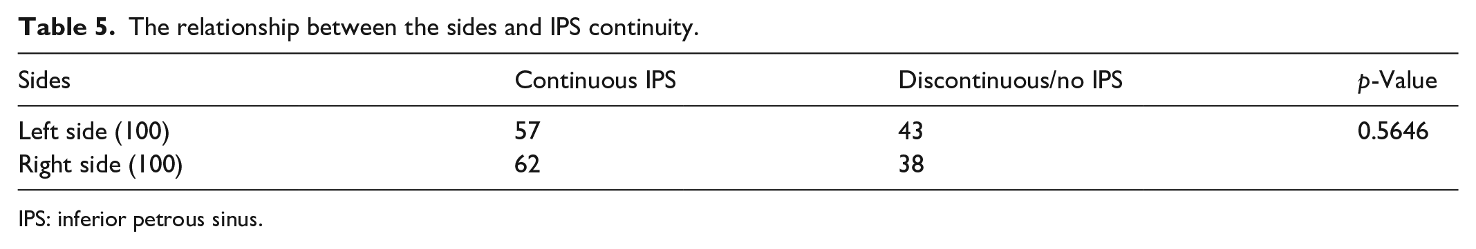
IPS: inferior petrous sinus.
Discussion
The TSS region is a complex anatomical region that mainly includes the TS, SS, vein of Labbé, SPS, and IPS. 11 The TS originates at the torcular Herophili and courses laterally to become the SS at the site immediately behind the petrous ridge, where the SPS originates. The vein of Labbé often originates from the TSS. The SS forms a gentle S-shape and terminates at the jugular bulb. The IPS passes through the petroclival fissure and connects to the cavernous sinus and basilar venous plexus at its upper end and the jugular bulb at its lower end. 1
These complex structures need continuous and further investigation. Currently, data for the Han Chinese population are insufficient.12,13 Catheter angiography remains the presumed gold standard for determining vascular anatomy; additionally, catheter angiography is invasive, and its luminal measurements are vulnerable to measurement errors. 9 MRV is a good noninvasive tool; it can offer three-dimensional reconstructions and does not require exposure to ionizing radiation or iodinated contrast media. MRV is a widely accepted modality for diagnosing cranial venous sinus disease.9,13 Therefore, our study used MRV to study the venous structures of the TSS region.
In general, the right TS is larger and receives most of the drainage from the superior sagittal sinus. The left TSS is usually smaller and predominantly receives drainage from the straight sinus. 1 Right-predominant TSS was the most common TSS in 50% of the TSSs; therefore, the right TSS was thicker than the left TSS for all of the different TSS locations.14,15 In our study, the diameters of the right TS at the origin and right SS at the termination were 7.9 ± 2.1 mm and 7.3 ± 1.9 mm, respectively, which were greater than those of the left TS. Due to the right-sided predominance of the TSS, the torcular Herophili often deviated to the right, which made the left TS longer than the right TS. Our study supported this finding. However, the SS length was similar in the left and right SS due to the fixed location.
The vein of Labbé is the largest anastomotic channel that crosses the temporal lobe between the Sylvian fissure and the TS.12,16–18 Its course and diameter may vary with race. In a study conducted in Portugal by Silva et al., 19 the vein of Labbé was predominant on the left side, the absence was 3% of the hemispheres, the mean angle between the trunk, and the TS was 111 degrees from the posterior TS, and the mean caliber was 3.2 mm. In a Chinese study by Han et al., 12 the diameter of the vein of Labbé was 2.8 mm. In our study, the diameter of the vein of Labbé was 3.0–3.1 mm. Similar to the above reports, no left-sided predominance was found, and the angle was 135°–136°. The difference in the angle between the TS and vein of Labbé from that reported by Silva et al. 19 may be due to differences in the study population.
The tentorium has a complex venous system that consists of internal tentorial veins, the LTS and the MTS. 20 The LTS is formed by the convergence of veins from the basal and lateral surfaces of the temporal and occipital lobes and courses laterally to drain into the TS or TS-SS junction. In our study, LTS drainage into the TS was more common than drainage into the TS-SS junction, especially on the right side. The MTS is formed by the convergence of veins from the superior surface of the cerebellum to the straight sinus or torcular Herophili. In this study, most of the MTSs drained into the straight sinus, but no difference was observed between the left and right sides. These findings have rarely been reported.
The IPS is an important structure, especially in the treatment of CS dural arteriovenous fistulas, and it plays an important role in venous drainage and the transvenous approach.21,22 Therefore, IPS development is important, and a continuous IPS can increase the convenience of catheterization. In a study by Gebarski et al., 23 bilateral IPSs were markedly asymmetric in 39% of patients, and in 8% of patients, the IPS was absent on at least one side. Our study investigated IPS development and revealed that the right IPS was predominant, but the IPS lengths were not different. Among all IPSs, 59.5% were continuous, and aplasia was present in 3.5% of the patients. These results confirmed the findings of Gebarski et al. 23
Conclusions
This study revealed that the right TSS was predominantly larger in diameter, the torcular Herophili tended to deviate to the right such that the right TS was shorter, the right LTS tended to be closer to the torcular Herophili, the IPS was thicker at its origin than at its termination, the right IPS was predominant, and the development of the left or right side of the TSS had no effect on IPS continuity. In addition, this study provided important data on the venous structures of the TSS region of the healthy Han Chinese population by using MRV, such as the length of the TS and SS and the diameters of the TS, SS, Labbé vein, IPS, and SPS.
Limitations
This work was a single-center retrospective study spanning 2 years that used preexisting MRV data; a sample size was not calculated, which may have led to an insufficient number of participants. Nevertheless, some interesting results were obtained, and this study was significant. Our study included only adult participants, and no participants under 18 years of age were involved. In the future, studies on the similarities and differences in venous structures of the TSS region between these two groups are warranted. This study focused only on the imaging and morphology of the TSS region, and the study of hemodynamics by 4D flow MRV, 24 invasive ultrafast miniature robot, 25 and other techniques is warranted.
Footnotes
Acknowledgements
None.
Author contributions
Zibo Zhou: data collection and writing—original draft; Jinlu Yu: study design, manuscript review and revision.
Availability of data and materials
All the raw data will be available upon request.
Declaration of conflicting interests
The author(s) declared no potential conflicts of interest with respect to the research, authorship, and/or publication of this article.
Funding
The authors received no financial support for the research, authorship, or publication of this article.
Ethical approval and consent to participate
This study was approved by the Ethics Committee of the First Hospital of Jilin University (No. 2022-KS-125). Written informed consent was waived because this was a retrospective study of preexisting MRV data.
Consent for publication
Written informed consent was waived because this was a retrospective study of preexisting MRV data.
Competing interests
The authors declare that they have no competing interests.