
Abstract
Endocrinologists are encountering patients with obesity-related complications such as metabolic syndrome (MetS) and type 2 diabetes mellitus (T2DM) on a daily basis. Nonalcoholic fatty liver disease (NAFLD) is a liver condition characterized by insulin resistance, hepatic steatosis and frequently T2DM. This is now the most common chronic liver condition in adults and is present in the majority of obese subjects. Liver fat accumulation may range from simple steatosis to severe steatohepatitis with hepatocyte necroinflammation (or nonalcoholic steatohepatitis [NASH]). Although the natural history is incompletely understood, NAFLD may lead to serious medical consequences ranging from cirrhosis and hepatocellular carcinoma to earlier onset of T2DM and cardiovascular disease (CVD). The diagnosis of NAFLD may be challenging because signs and symptoms are frequently absent or nonspecific, and thus easily missed. Liver aminotransferases may be helpful if elevated, but most times are normal in the presence of the disease. Liver imaging may assist in the diagnosis (ultrasound or MRI and spectroscopy) but a definitive diagnosis of NASH still requires a liver biopsy. This may change in the near future as novel biomarkers become available. Treatment of NAFLD includes aggressive management of associated cardiovascular risk factors and many times control of T2DM. Pioglitazone and vitamin E appear promising for patients with NASH, although long-term studies are unavailable. In summary, this review hopes to address the common clinical dilemmas that endocrinologists face in the diagnosis and management of NAFLD and increase awareness of a potentially serious medical condition.
Keywords
Introduction
Nonalcoholic fatty liver disease (NAFLD) has become the most common chronic liver disease in Western countries. It is a liver condition characterized by insulin resistance, hepatic steatosis and frequently prediabetes or T2DM. Liver fat accumulation may range from simple triglyceride accumulation to severe steatohepatitis with lobular necroinflammation and variable degrees of fibrosis (nonalcoholic steatohepatitis [NASH]), cirrhosis and even hepatocellular carcinoma [Bugianesi et al. 2007]. In a recent report by Musso and colleagues [Musso et al. 2010], it was estimated that NAFLD increases healthcare costs by 26% and that it will be the leading cause of liver transplantation by 2020. This study did not take into account the public health burden associated with NAFLD-related conditions, such as diabetes and cardiovascular disease (CVD) [Targher et al. 2010; Marchesini et al. 2003]. On a daily basis, endocrinologists see patients who are obese and have the metabolic syndrome (MetS); yet, most are unaware of NAFLD. There are several reasons why NAFLD has not become a more widely recognized problem: the diagnosis may be difficult, the natural history and clinical implications remain poorly understood, and pharmacological treatment is not well established, although this is likely to change in the near future. NAFLD is a disease that requires unique considerations and it is likely that endocrinologists will play a larger role in the future in screening and treating these patients.
Magnitude of the problem: prevalence and natural history
The precise prevalence of NAFLD remains unclear, depending on the methods used for screening, being lower when liver aminotransferases and/or liver ultrasound are used and higher with the gold-standard magnetic resonance spectroscopy (MRS). The prevalence of NAFLD in the industrialized countries is believed to be between 40% and 50%, with even higher prevalence rates in subjects with T2DM and as high as 90% in the morbidly obese [Chavez-Tapia et al. 2010; Musso et al. 2010; Pillai and Rinella, 2009]. In patients who have NAFLD, it is believed that about 40% may go on to develop NASH [Wieckowska et al. 2007; Browning et al. 2004b; Clark et al. 2002] although the true natural history of the disease is incompletely understood. The true prevalence of NASH in the general population is unknown because few studies have performed a liver biopsy in patients found to have NAFLD by liver aminotransferases or on liver imaging during routine screening [Leite et al. 2011; Williams et al. 2011]. However, it is clear that factors associated with disease progression include obesity and the cluster of factors associated with MetS, such as dyslipidemia, hypertension (HTN), insulin resistance and T2DM. For instance, in a recent analysis by our group, the presence of T2DM was associated with more insulin resistance and worse histology in patients with NASH [Ortiz-Lopez et al. 2010].
When comparing ethnicities, the Hispanic population has been reported to have a higher prevalence rate for NAFLD than the African American or White population [Williams et al. 2011; Neuschwander-Tetri et al. 2010; Mohanty et al. 2009; Browning et al. 2004b]. However, in these studies Hispanics had a higher prevalence of obesity, insulin resistance and T2DM, all established risk factors for NAFLD. A recent study in 152 subjects by Lomonaco and colleagues [Lomonaco et al. 2011a] has reported that Hispanics and Whites have similar severity of NASH if subjects are matched carefully for adiposity, and that previously described differences were more likely a reflection of the unfavorable metabolic risk of Hispanics. With the worldwide epidemic of obesity, the prevalence of NAFLD is increasing across the globe [Chitturi et al. 2011; Duseja, 2010]. Perhaps the most disturbing trend is the rise of NAFLD in the pediatric population, echoing the rise in childhood obesity [Mencin and Lavine, 2011; Schwimmer et al. 2006].
Simple steatosis can have a benign, nonprogressive course, but a number of studies suggest that approximately 30–40% of patients with NASH are at risk of developing fibrosis and potentially cirrhosis [Williams et al. 2011; Bugianesi et al. 2007; Ekstedt et al. 2006; Adams et al. 2005a; Browning et al. 2004a, 2004b; Fassio et al. 2004; Harrison et al. 2003]. Patients with NAFLD are also at an increased risk of end-stage liver disease, CVD and diabetes, which explain their overall increased mortality rate [Ekstedt et al. 2006; Adams et al. 2005b]. NASH can progress to cirrhosis in up to 5–15% of patients and is now recognized as the most common cause of cryptogenic cirrhosis [Caldwell, 2010; Bugianesi et al. 2007; Harrison, 2006]. Obesity and T2DM are present in a large portion of patients who develop cryptogenic cirrhosis, an association not seen in hepatitis-C-related cirrhosis or primary biliary cirrhosis. Both NASH and cryptogenic cirrhosis share many similar risk factors, including T2DM, obesity, and the MetS [Bugianesi et al. 2007; Adams et al. 2005a]. When compared with viral-associated cirrhosis, cirrhosis linked to NASH has a similar liver-related mortality, but a significantly higher CVD-related death rate [Musso et al. 2010].
Diagnosis of NAFLD: an endocrinologist’s challenge
The challenge endocrinologists face in the diagnosis of NAFLD is that signs and symptoms are frequently absent or nonspecific and thus easily missed. This requires a high degree of disease awareness. A complete history and physical examination are still useful tools that may offer clues about the disease. Some of the findings include general right upper quadrant pain, malaise, hepatomegaly or discomfort on exam, or evident signs of insulin resistance (i.e. acanthosis nigricans). While liver aminotransferases may be elevated (typically alanine aminotransferase [ALT] greater than aspartate aminotransferase [AST] levels), increases are usually mild to moderate and are normal in about two thirds of patients, making them an unreliable marker of NAFLD. Several medications (corticosteroids, HIV antiretroviral therapy, tamoxifen, others), viral hepatitis (i.e. hepatitis C genotype 3), autoimmune hepatitis and other conditions [Ali and Cusi, 2009; Vuppalanchi and Chalasani, 2009; Clark et al. 2003] should be ruled out. In addition, liver enzymes may even be normal in end-stage liver cirrhosis. Thus, the presence of elevated liver aminotransferases or fatty liver on imaging should prompt the physician to evaluate the patient for excessive alcohol intake and/or a number of liver-related medical illnesses (such as viral hepatitis, hemochromatosis, autoimmune liver disease, α-1 antitrypsin deficiency or Wilson’s disease) before the diagnosis of NAFLD be made.
Noninvasive assessments of NAFLD
Typically the first test used in the evaluation of patients with suspected fatty liver (usually by history and elevated AST/ALT) is an ultrasound (US). Its advantages are that it is rather inexpensive, noninvasive, widely available, and there is no radiation exposure. Steatosis on a liver US appears as hyperechogenic when compared with the spleen or kidney. Other useful features on US include beyond increased parenchymal echogenicity are hepatic and portal vein blurring, far gain attenuation of the diaphragm, gallbladder blurring [Mazhar et al. 2009; Liang et al. 2007]. Liver US can detect changes in parenchyma but does not provide accurate quantification of the amount of fat present and does not allow the severity of histological disease to be established. The disadvantages are that it is highly operator dependent and it is less accurate in patients who have a large body mass. Also, there is a decrease in sensitivity when the liver fat content is less than 20–30% leaving many patients undiagnosed. Computed tomography (CT) is another possible imaging modality but it also fails to precisely quantify the degree of steatosis [Saadeh et al. 2002]. A fatty liver will have a characteristic decrease in liver attenuation compared with the spleen, making the liver appear darker than the spleen.
Magnetic resonance spectroscopy (MRS), the current gold-standard technique for diagnosing fatty liver, provides more sensitivity and reproducibility for determining the amount of liver fat. In a multiethnic group of 345 subjects without any risk factors for NAFLD (lean, no or low alcohol consumption, normal plasma glucose, no history of liver disease and normal liver aminotransferases) the median liver fat content was 1.9% with a 95% percentile of 5.6%. Therefore, the diagnosis of NAFLD is considered when the liver fat content by MRS is >5.6% (equivalent to 55.6 mg/g of liver tissue) [Szczepaniak et al. 2005]. MRS has been shown to have a good correlation with the amount of liver fat estimated by liver biopsy in small studies [Szczepaniak et al. 1999; Longo et al. 1995] and in our own experience [Lomonaco et al. 2011a; Belfort et al. 2006]. The disadvantages of MRS are cost and that it is only available at few academic centers. A few of the less commonly used tests include assessment of fibrosis using the BARD score, the NAFLD fibrosis score, or the fibroscan [Dowman et al. 2011; Musso et al. 2010; Wong et al. 2010]. These tests, however, have not been fully evaluated for widespread clinical use.
Liver biopsy
A liver biopsy is the only way to confirm the diagnosis of NASH and grade the severity of steatohepatitis and stage fibrosis. It is usually performed under US guidance and is generally a safe and well-tolerated procedure in experienced hands. The invasive nature of the test and the lack of established pharmacological treatments make it an option rarely chosen by physicians in patients with NAFLD. At the current time, it is best indicated for the diagnosis of NASH in patients with clinical risk factors (i.e. severe obesity, T2DM) and markedly elevated liver aminotransferases (i.e. greater than threefold the upper limit of normal [ULN]), when other causes of liver disease have been excluded or if a treatment decision will be made based on the results.
Future noninvasive diagnosis of NAFLD and NASH
The fact that at least 70–80% of obese subjects have NAFLD, and many with NASH, highlights the need for a noninvasive diagnosis. Current efforts include the use of a combination of clinical parameters (body mass index [BMI], presence of diabetes, HTN) and plasma biochemical measurements frequently associated with the disease (such as plasma ALT, bilirubin, glucose, triglycerides, other) [Angulo et al. 2007; Wieckowska et al. 2007; Poynard et al. 2006; Ratziu et al. 2006], use of imaging by means of transient elastography [Gaia et al. 2011; Wong et al. 2010; Friedrich-Rust et al. 2008] or the use of new plasma biomarkers of NASH [Pagadala et al. 2009; Wieckowska et al. 2007].
The most promising biomarker in NASH is measurement of plasma caspase-cleaved cytokeratin-18 (CK-18) fragment levels [Feldstein et al. 2009]. CK-18 is a major intermediate filament protein in the liver. In NASH there is significant caspase activation and hepatocyte cell death by apoptosis. It is believed that the increased caspase activity can be measured in plasma from the spillover of caspase-cleaved CK-18 fragments into the bloodstream. A number of studies have demonstrated significant elevation of this protein in NASH when compared to controls with a fatty liver, but not steatohepatitis [Feldstein et al. 2009; Yilmaz et al. 2009; Younossi et al. 2008; Diab et al. 2008]. In our hands, CK-18 fragments are clearly elevated in patients with NAFLD compared with those without a fatty liver and also when comparing those with ‘benign’ steatosis versus patients with NASH [Cusi et al. unpublished]. However, it had less accuracy in necroinflammation grading or fibrosis staging within subjects with NASH. Future studies will confirm the role of CK-18 and other emerging plasma biomarkers in the management of patients with NASH.
Metabolic consequences of NAFLD
NAFLD and T2DM
The link between NAFLD and type 2 diabetes.
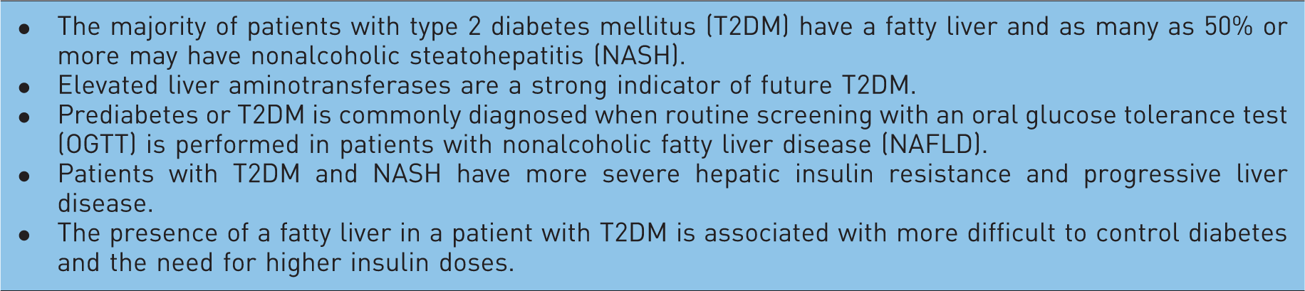
In our experience, about 70% of patients with NASH have disordered glucose metabolism, either impaired fasting glucose, impaired glucose tolerance or T2DM, when systematically screened with an oral glucose tolerance test (OGTT) [Ortiz-Lopez et al. 2010]. Earlier screening and diagnosis for T2DM in NAFLD patients may allow for early intervention and prevention of diabetes complications. This is important because the presence of fatty liver in T2DM is associated with more difficult to control diabetes and higher insulin requirements [Ryysy et al. 2000]. In addition, patients with T2DM and NASH have more severe hepatic insulin resistance and progressive liver disease [Cusi, 2009a]. Also advanced fibrosis is associated with obesity, insulin resistance, hepatocyte lipotoxicity, hyperinsulinemia, and abnormal glucose metabolism [Bataller et al. 2011; Neuschwander-Tetri et al. 2010].
NAFLD and CVD
The association of NAFLD with MetS and obesity has led to the connection between NAFLD and the development and progression of CVD (Figure 1). Both adult and children patients with NAFLD typically meet the criteria for the MetS (HTN, abdominal obesity, atherogenic dyslipidemia, insulin resistance or glucose intolerance) and thus have multiple risk factors for CVD. As summarized in Table 2, patients with NAFLD/NASH more frequently have T2DM, more severe dyslipidemia, more insulin resistance, worse subclinical inflammation (i.e. high-sensitivity C-reactive protein [hsCRP], interleukin 6 [IL-6], tumor necrosis factor alpha [TNF-α]) and may be affected by myocardial lipotoxicity.
Dysfunctional, insulin-resistant adipose tissue is common in overweight and obese subjects and leads to excessive free fatty acids (FFAs) in the liver. This promotes triglyceride accumulation, hepatocyte lipotoxicity with necrosis, inflammation and eventual fibrosis. The metabolic consequences are dyslipidemia, hyperglycemia, hyperinsulinemia and subclinical inflammation, all leading to premature cardiovascular disease (CVD). NAFLD, nonalcoholic fatty liver disease; NASH, nonalcoholic steatohepatitis; HDL-C, high-density lipoprotein cholesterol; LDL-C, low-density lipoprotein cholesterol. Risk factors for cardiovascular disease in nonalcoholic fatty liver disease.

Dyslipidemia in NAFLD is characterized by an increase in very-low-density lipoprotein (VLDL) secretion that leads to elevated plasma triglycerides (TG) and low high-density lipoprotein cholesterol (HDL-C) [Adiels et al. 2006]. There is an association between the higher secretion of apolipoprotein B particles and the increase in the atherogenicity of VLDL secreted by the liver [Adiels et al. 2008]. Moreover, decreased lipoprotein lipase clearance promotes postprandial lipemia and is another contributor to vascular damage. Hypertriglyceridemia in NAFLD also leads to small, dense LDL-C and an added risk of atherogenesis. Patients with elevated ALT and high plasma TG and cholesterol have >80% chance of having NAFLD [Browning, 2006].
Dysfunctional fat releases excessive amounts of free fatty acids (FFAs) leading to ectopic fat deposition in tissues that are poorly adapted to TG accumulation such as muscle, liver, and pancreatic β-cells [Cusi, 2010]. In 187 middle-aged obese patients with biopsy-proven NASH, compared with well-matched obese controls without NAFLD, we observed that the severity of adipose tissue insulin resistance kept a close association with metabolic and histological damage in patients with NASH [Lomonaco et al. 2011b]. We also observed that hepatic insulin resistance and steatosis correlated closely with the severity of insulin resistance in adipose tissue [Ortiz-Lopez et al. 2011]. This was even as both groups had similar BMI and total adiposity as measured by dual X-ray absorptiometry (DXA), suggesting that it is not the total amount of fat but its degree of dysfunction and insulin resistance that account for the development of NASH.
Dysfunctional adipocytes secrete many inflammatory cytokines previously believed to be produced only by macrophages (i.e. TNF-α, IL-6, resistin, monocyte chemoattractant protein-1 [MCP-1], plasminogen activator inhibitor-1 [PAI-1], visfatin, angiotensinogen, retinol-binding protein-4 [RBP-4], etc.) [Gregor and Hotamisligil, 2007; Shoelson et al. 2006]. It is now accepted that adipokines promote insulin resistance by inhibiting key insulin signaling steps in liver and muscle, and are actively involved in the recruitment and ‘activation’ of local macrophages that play a role in the development of adipose tissue insulin resistance, increased plasma FFAs and ultimately lipotoxicity [Cusi, 2011; Muhlhausler and Smith, 2009]. Dysfunctional adipocyte function is also characterized by reduced plasma adiponectin levels as reported in NASH, and its increase during pioglitazone treatment closely associated with histological improvement [Gastaldelli et al. 2010]. There is a close relationship between the state of subclinical inflammation, insulin resistance and atherogenesis in obesity and likely this contributes to the CVD of patients with NAFLD
A chronic increase in plasma FFA levels is especially harmful to the heart and vascular beds, and it is now accepted that it plays an active role in the development of CVD [Cusi, 2009b; McGavock et al. 2006]. MRS has been used to show an association between hepatic and cardiac lipid accumulation [Reingold et al. 2005]. Myocardial TG content is elevated in subjects with either glucose intolerance or T2DM [Perseghin et al. 2008; Kankaanpaa et al. 2006]. It is believed that the prevalence of coronary, cerebrovascular, and peripheral vascular disease is higher among patients with NAFLD than those without NAFLD [Targher et al. 2010]. We have observed that just a mild elevation in plasma FFA to levels observed in T2DM for 48–72 hours by means of a lipid infusion is sufficient to increase blood pressure and induces the production of markers of systemic inflammation (i.e. soluble intercellular adhesion molecule [ICAM] and vascular adhesion molecule [VCAM], endothelin [ET]-1) in lean healthy subjects [Kashyap et al. 2008; Tay et al. 2006]. Moreover, we have recently expanded these observations by showing that a 48-hour increase in plasma FFA concentration also increases soluble E-Selectin (sE-Selectin), myeloperoxidase (MPO), and total plasminogen activator inhibitor-1 (tPAI-1), indicators of a procoagulant state and associated with abnormal vascular reactivity [Mathew et al. 2010]. Patients with NAFLD have impaired flow-mediated vasodilatation and increased carotid-artery intima-media thickness [Targher et al. 2010]. However, although many lines of evidence suggest that patients with NAFLD are at higher risk of CVD, this hypothesis requires more rigorous evaluation and confirmation in large, controlled studies.
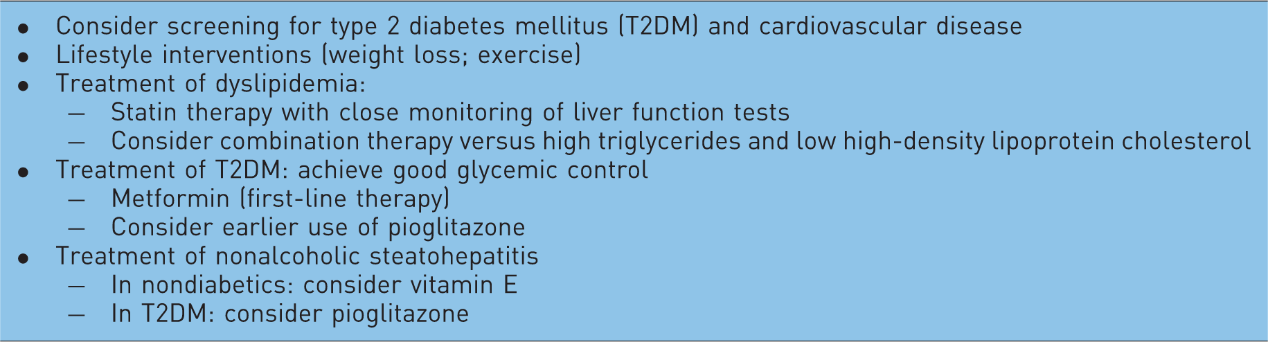
Management of NAFLD: practical considerations
Management guidelines for patients with nonalcoholic fatty liver disease.
Lifestyle intervention: diet and exercise
As awareness of the serious risks associated with NAFLD is increasing, there has been a greater effort to screen and implement combined lifestyle and pharmacological interventions. The most rational approach to weight reduction involves lifestyle modifications that incorporate diet and exercise [Cusi, 2009c]. Both, have proven effective to prevent T2DM [The Diabetes Prevention Program Research Group, 2005] and CVD [Fogelholm, 2010]. Many studies indicate that lifestyle [Haufe et al. 2011; Lazo et al. 2010; Kantartzis et al. 2009; Kirk et al. 2009; Viljanen et al. 2009] intervention may normalize liver aminotransferases and improve hepatic steatosis measured either by US [Sreenivasa Baba et al. 2006; Suzuki et al. 2005; Hickman et al. 2004; Kugelmas et al. 2003; Okita et al. 2001; Ueno et al. 1997; Andersen et al. 1991; Palmer and Schaffner, 1990] or MRS [Cowin et al. 2008; Larson-Meyer et al. 2008, 2006; Schafer et al. 2007; Thamer et al. 2007; Thomas et al. 2006; Petersen et al. 2005; Tamura et al. 2005; Westerbacka et al. 2005; Tiikkainen et al. 2003]. However, most of them are limited by small study size and short duration. Owing to the invasive nature of liver biopsy, numerous studies have used biochemical improvement as the primary endpoint but rather relied on surrogate markers, such as liver aminotransferases or imaging (US, CT, MRS). Histological improvement is proportional to the degree of total body weight loss. At least a total body weight loss of around 3–5% is necessary to improve liver steatosis, but a greater weight loss (7–10%) appears to be needed for improvements in necroinflammation in adult patients with NASH.
Because weight loss is challenging, approaches including medications or surgical procedures have been used in NAFLD. Medications such as orlistat [Harrison et al. 2009] or sibutramine [Zelber-Sagi et al. 2006] have not proved to be better than placebo if a comparable weight loss is achieved, suggesting that any benefit is strictly related to their potential to assist with weight loss. It is now accepted that bariatric surgery is associated with marked improvement or resolution of diabetes, HTN, and dyslipidemia [Chavez-Tapia et al. 2010; Pillai and Rinella, 2009]. In general, both Roux-en-Y gastric bypass (RYGB) and laparoscopic adjustable gastric banding (LAGB) have shown improvement in steatosis and a reduction in inflammation and ballooning. Fibrosis has been more inconsistent, with some studies reporting an increase in fibrosis [Csendes et al. 2006; Mathurin et al. 2009]. Procedures with a malabsorptive component, such as RYGB, lead to a greater weight loss and metabolic benefit than LAGB. However, LAGB appears to be becoming the procedure of choice in many centers as there is a clear trend favoring less invasive techniques (i.e. LAGB), although weight loss tends to be less. The best bariatric surgery for NASH is not known, as there are a number of limitations from the literature. Most studies are retrospective or uncontrolled and suffer from selection bias, poor standardization of presurgery and postsurgery dietary and follow-up procedures, and variable time of postsurgical follow up (from weeks to years). Of note, bariatric surgery as a method of weight loss is attractive as it has shown reductions in long-term mortality, but this has not been investigated specifically in patients with NAFLD [Sjostrom et al. 2007].
Treatment of dyslipidemia
As mentioned earlier, since NAFLD is strongly associated with the MetS and carries an elevated risk for CVD, patients would benefit from intensive medical intervention to control dyslipidemia. Asymptomatic elevations in AST or ALT three times the ULN have been reported with all statins in the general population. A threefold elevation of AST or ALT is seen in 1% of patients receiving initial and intermediate doses of statins and in 2–3% of patients at the maximal dose [McKenney et al. 2006]. The prescription of 3-hydroxy-3-methylglutaryl coenzyme A (HMG-CoA) reductase inhibitors, or statins, to patients with NAFLD is controversial, at best, due to an apparent risk of statin-related hepatotoxicity. However, several studies have now reported that statins can be safely used in patients with liver disease [Zamor and Russo, 2011; Argo et al. 2008; Riley et al. 2008; Ekstedt et al. 2007; Lewis et al. 2007; Chalasani, 2005]. Recent guidelines from the National Lipid Association (NLA) [McKenney et al. 2006], based at least in part on the assessment performed by the Expert Liver Panel [Cohen et al. 2006], concluded that statins could be given safely to patients with NAFLD or NASH. One should consider starting low-dose statin therapy, possibly of the more potent statins, and slowly titrate up with close monitoring of liver tests as needed to reach lipid targets. If an isolated asymptomatic aminotransferase level is found during a routine evaluation (up to three times the ULN), the NLA guidelines suggest that there is no need to discontinue the statin immediately but to have the test repeated. If still elevated, other causes should be ruled out and clinical judgment should be allowed to decide about continuing the statin or not. The appropriate frequency of monitoring liver function tests is unclear, but it is considered as appropriate to measure aminotransferase levels before starting therapy, 12 weeks after initiating therapy or after a dose increase, and periodically thereafter. However, the NLA did not believe that routine monitoring of liver aminotransferases was supported by the available evidence and suggested that the FDA reconsider this strategy. The statin should be discontinued at the first evidence of significant liver injury, the cause investigated and the patient referred to a hepatologist.
Because the most common dyslipidemia in NAFLD is an elevated plasma TG and a low HDL-C level, combination therapy with a lipid-lowering agent that specifically targets these defects is frequently needed. Fibrates can ameliorate atherogenic dyslipidemia in the MetS [Belfort et al. 2010] and improve dyslipidemia in NAFLD [Fernandez-Miranda et al. 2008; Basaranoglu et al. 1999]. Fenofibrate is preferred in combination therapy as it does not increase the levels of the statin [Bergman et al. 2004], in contrast to a twofold to threefold increase with gemfibrozil [Backman et al. 2000]. Based on results from recent clinical trials, combination therapy with fenofibrate should be targeted exclusively at patients with plasma TG concentrations greater than 200 mg/dl and a low HDL-C [Ginsberg et al. 2010; Keech et al. 2005; Tenenbaum et al. 2005]. Recently (25 May 2011), the AIM-HIGH study, a randomized, multicenter clinical trial sponsored by the National Heart, Lung and Blood Institute (NHLBI) in patients with low HDL-C and high TG, was discontinued due to the lack of efficacy in reducing CVD while examining the role of long-acting niacin (Niaspan®) in patients with a history of established CVD and well controlled LDL-C by simvastatin. There were 249 primary outcome events (15%) in the simvastatin plus placebo arm and 262 (15%) in the Niaspan plus simvastatin (p = 0.561). There were a total of 28 ischemic strokes (1.6%) in the Niaspan plus simvastatin arm and a total of 12 such events (0.7%) reported in the simvastatin arm.
Treatment of T2DM
Because insulin resistance is common in NAFLD, there has been significant interest in diabetes drugs with this mechanism of action, both for metformin and thiazolidinediones (TZDs). Metformin is a biguanide that ameliorates insulin resistance primarily at the level of the liver and to a lesser extent skeletal muscle [Cusi et al. 1996]. While several small trials have shown it can be used safely and that it reduces aminotransferase levels in NAFLD [Loomba et al. 2009; Duseja et al. 2007; Nair et al. 2004; Uygun et al. 2004; Marchesini et al. 2001], it should not be expected to improve histology [Haukeland et al. 2009; Loomba et al. 2009; Bugianesi et al. 2005]. In children and adolescents (aged 8–17 years), neither vitamin E nor metformin for 96 weeks significantly reduced plasma ALT levels (primary endpoint) or steatosis, lobular inflammation or fibrosis on individual scores, although the combined overall NAFLD Activity Score (NAS; NAS = combined steatosis, lobular inflammation, and ballooning) improved modestly with vitamin E compared with placebo [Lavine et al. 2011]. While the effect of metformin is rather modest to improve NASH, it remains as first-line therapy to treat hyperglycemia in T2DM and may have a modest beneficial effects on lipids and subclinical inflammation in this population [Cusi and DeFronzo, 1998].
Pioglitazone and rosiglitazone both belong to the thiazolidinedione (TZD) class of drugs that operate as ligands for the peroxisomal proliferators-activated receptor-γ(PPARγ)a class of nuclear transcription factors that are very abundant in adipose tissue. They are insulin-sensitizing agents approved for the treatment of T2DM only, but also effective in halting the progression of prediabetes to diabetes [DeFronzo et al. 2011; The DREAM (Diabetes REduction Assessment with ramipril and rosiglitazone Medication) Trial Investigators, 2006]. Pioglitazone, but not rosiglitazone [Ratziu et al. 2010, 2008], has proven to be the most useful drug for the treatment of NASH. Therefore, a strong consideration should be given to its earlier use in this population. Other oral agents (sulfonylureas, insulin, dipeptidyl peptidase [DPP] IV inhibitors) may also be used safely in NAFLD. Incretin mimetics, such as exenatide (Byetta®) or liraglutide (Victoza®), have generated significant interest as they promote weight loss and activation of hepatic GLP-1 signaling causes reduction of hepatic steatosis in rodents [Ding et al. 2006]. In patients with well-controlled T2DM, 6 months of exenatide twice daily has been reported to lead to an around 20% reduction in hepatic steatosis by MRS [Orsi et al. 2009]. However, it does not have a significant effect on reversing NASH [Kenny et al. 2010].
Treatment of NASH
Only two pharmacological interventions are currently promising in NASH: pioglitazone and vitamin E. TZDs exert positive changes on adipocytes (i.e. restoring adipocyte insulin sensitivity, increasing plasma adiponectin levels, reducing excessive lipolysis and plasma FFA levels, among others) and improve hepatic and peripheral (muscle) insulin sensitivity. The first proof-of-concept controlled trial [Belfort et al. 2006] in patients with NASH and either with IGT or T2DM, demonstrated that pioglitazone significantly lowered liver aminotransferases, increased plasma adiponectin levels and improved adipose tissue, liver and muscle insulin sensitivity. Liver steatosis, ballooning necrosis and inflammation histology scores improved significantly with pioglitazone (combined necroinflammation score was reduced by 85%). Liver fibrosis improved versus baseline but did not reach statistical significance when compared with placebo. The NAFLD activity score improved with pioglitazone in 73% compared with 24% of placebo-treated patients. Since this early report, two studies have expanded this observation to subjects without diabetes [Sanyal et al. 2010; Aithal et al. 2008], the largest being the PIVENS trial [Sanyal et al. 2010], with histological improvement in steatosis and inflammation but not fibrosis after 2 years of pioglitazone treatment. Unfortunately these studies have been of relative short duration (6–24 months) and await confirmation about their long-term benefit. It should also be kept in mind that pioglitazone is only approved for the treatment of patients with T2DM. Therefore, at the present time, physicians should consider its use primarily for patients with NASH that also have T2DM after careful consideration of the treatment options and integrated proper lifestyle intervention. Finally, vitamin E at doses of 400 units twice daily was equally effective in patients without diabetes and NASH [Sanyal et al. 2010] and should be considered as another viable and inexpensive choice for patients with NASH.
Summary
NAFLD is the most common chronic liver condition in adults and is present in the majority of obese subjects that endocrinologists see in their daily practice. It may lead to serious medical consequences ranging from cryptogenic cirrhosis to hepatocellular carcinoma as well as T2DM and CVD. The diagnosis of NAFLD is challenging and liver aminotransferases may be helpful if elevated, but if normal the clinician must still suspect the presence of the disease based on patient’s metabolic profile. Liver ultrasound may be of assistance in the diagnosis (MRI and spectroscopy is still a research tool) but a definitive diagnosis of NASH often requires ruling out other liver conditions and eventually a liver biopsy. Noninvasive approaches combining the clinical profile (obesity, T2DM, HTN, dyslipidemia) and novel biomarkers will change the management of the disease in the near future. Treatment of NAFLD includes lifestyle intervention and aggressive management of cardiovascular risk factors. Pioglitazone and vitamin E are currently the best pharmacological options for patients with NASH, although long-term studies are needed. Endocrinologists will likely be more often consulted and involved in the management of patients with NAFLD in the future.
Footnotes
Acknowledgements
Dr Kenneth Cusi is supported by the American Diabetes Association, the Burroughs Wellcome Fund, the Veterans Affairs Medical Research Fund, and the National Center for Research Resources (award number UL 1RR025767).
Conflict of interest statement
The authors declare no conflicts of interest in preparing this article.
Disclaimer
The content is solely the responsibility of the authors and does not necessarily represent the official views of the National Center for Research Resources of the National Institutes of Health.