
Abstract
The simultaneous occurrence of pregnancy and multiple myeloma (MM) is rare. The challenge of diagnosing MM during pregnancy is demonstrated in the case presented here. Despite the rarity of concurrent MM and pregnancy, this possibility should be considered in patients with signs and symptoms that may be attributed to MM so as not to delay the diagnosis and decision about pregnancy continuation and initiation of an appropriate and safe therapy to the mother and fetus. Treating physicians should be aware of the potential effects of MM therapies on the fetus and pregnancy outcomes.
Introduction
Multiple myeloma (MM) is a neoplastic proliferation of plasma cells in the bone marrow, accounting for 1% of all cancers and approximately 10% of all hematological malignancies. 1 The median age at MM diagnosis is 67–70 years and only 3% of the patients are diagnosed before 40 years of age. 2 Moreover, MM is approximately 1.5 times more prevalent in men compared with women. 3 Therefore, the simultaneous occurrence of pregnancy and MM is rare. The first case describing MM diagnosis during a pregnancy was published in 1965. 4 Between 1965 and 2020, 44 cases of MM, diagnosed around or during pregnancy, were reported in the literature.2,4–33 The most common early symptoms of MM are bone pain (58% of patients), fatigue (32%), and recurrent infections.34,35 End-target organ damage according to the CRAB criteria includes hypercalcemia (28%), renal failure (48%), anemia (73%), and lytic bone lesions (20–25%). 35 Some of these symptoms may also be attributed to the normal course of pregnancy. The challenge of diagnosing MM during pregnancy is demonstrated in the case presented here. The patient had signs that were primarily ascribed to her pregnancy and therefore the investigation was initially directed toward pregnancy-related disorders, such as iron deficiency, preeclampsia, or primary kidney disorder. The patient provided written consent for publishing this case report. The case report followed the CARE guidelines. 36
Case report
A 40-year-old previously healthy woman, gravida 12, para 10, was admitted for an investigation due to elevated blood pressure (180/115 mmHg) and acute renal failure (serum creatinine 1.5 mg/dL, previously normal) at gestational week 16 of index pregnancy. The patient was asymptomatic. Although she had a history of gestational hypertension during her three most recent pregnancies, she was not followed and was never treated for it. Notable laboratory findings included normocytic anemia, hemoglobin 9.4 g/dL, platelets 272 K/µL, and white blood cells 7.69 K/µL. There was no evidence of iron deficiency (iron 70 µg/dL, transferrin 277 mg/dL, ferritin 151.2 mg/mL). Serum albumin, lactate dehydrogenase, and electrolytes were within normal range. Total urine protein collected over 24 h was 1.3 g.
Therapy with labetalol 200 mg was started, and the patient was followed at the pregnancy high-risk unit in collaboration with the nephrology unit. During this period, blood pressure remained high and stable and renal function deteriorated to 2.06 mg/dL during week 22.
A kidney core needle biopsy, performed at week 23 + 4, showed 25 normocellular glomeruli without signs of active glomerular disease. No evidence of glomerular capillary wall thickening was observed. The interstitium showed focally prominent infiltration by leukocytes with multiple eosinophils and lymphocytes. Masson trichrome stain highlighted about 20% fibrosis. The tubules demonstrated multiple areas of tubulitis while the vessels showed no significant changes.
The morphological picture was compatible with tubulointerstitial disease with a prominent acute inflammatory component.
Immunostaining for cytomegalovirus, herpes simplex virus, and polyomavirus was negative. Congo red staining was negative for amyloid material. Immunofluorescence staining for IgA, IgG, IgM, C3, C4, C1q, kappa, lambda, fibrinogen, and albumin deposits was also negative. Scanning electron microscopy showed two normocellular glomeruli with prominent thinning of the glomerular basement membrane, and tubules with signs of epithelial cell damage.
The ultrastructural findings were consistent with thin membrane disease associated with tubular damage.
At 25 weeks of pregnancy, serum electrophoresis and immunofixation showed an unquantifiable IgG kappa monoclonal band. Serum IgG was 582 mg/dL (normal 700–1600), IgA 43 mg/dL (normal 70–500), and IgM 22 mg/dL (normal 40–280). Serum free light chain assay demonstrated high levels of kappa light chain 3120 mg/L (normal 3.3–19.4), preserved lambda 7.3 mg/L (normal 5.7–26.3), and a significantly skewed κ/λ ratio of 427.7 (0.26–1.65). Repeated 24-h urine protein collection demonstrated 2.21 g of urine protein, of which 1.91 g were Bence Jones protein. Bone marrow (BM) biopsy revealed infiltration by 70–80% kappa light chain-restricted plasma cells. Congo red stain was negative for amyloid. Fluorescent in situ hybridization (FISH) testing of plasma cells showed del13q and t(11;14). Whole-body magnetic resonance imaging (MRI) revealed lack of homogeneity along the spine vertebrae with multiple ‘salt and pepper’ foci. Focal lesions, 8 mm in diameter, were observed in the D8 vertebra and in the left pedicle of the D3 vertebra. Focal lesions, 17 mm in diameter, were observed in the left ilium wing and several bilateral tiny lesions were noted in the iliac bones. The BM along the thighs was inhomogeneous with no evidence for focal lesions (Figure 1).

Whole-body MRI performed at 25 weeks of pregnancy. (a) 17 × 13 mm focal lesion in the left ileum wing, and tiny lesions in the ileum bones; (b) heterogenic bone marrow along the thighs which is particularly pronounced in the right distal thigh.
The findings obtained from the MRI scan, BM biopsy, and the monoclonal proteins observed in the blood and urine all led to the diagnosis of oligosecretory IgG kappa MM, Revised International Staging System (R-ISS) 2, with predominantly kappa light chain secretory disease.
Upon the diagnosis of active MM with anemia, renal and skeletal injuries, there was a clear indication for treatment initiation. Considering that the patient did not wish to terminate the pregnancy, together with the advanced gestational age, therapy with high-dose dexamethasone (HDD) 40 mg/d was initiated.
HDD was administered on days 1–4, 9–12, and 17–20. After one treatment cycle there was no response: serum-free kappa remained stable at 2760 mg/L and serum creatinine was unchanged. Therefore, weekly cyclophosphamide (CTX) 500 mg/m2 was added to HDD on gestational week 27, resulting in partial response with a reduction in serum-free kappa to as low as 1540 mg/L. Creatinine improved to 1.21 mg/dL. The patient signed an informed consent for this treatment.
Therapy was continued during gestational weeks 27–34 with no further reductions in serum-free kappa light chain and in serum creatinine. Fetal development continued uneventfully, and fetal growth was appropriate for its gestational age.
On gestational week 35, increased creatinine level (1.27 mg/dL) and blood pressure (140/90 mmHg) were observed.
Due to the bony lesions in the pelvis observed by MRI, which increased the concern for a vaginal delivery, an elective caesarian section was performed on gestational week 36. A healthy female baby, weighing 1940 g, was delivered with an Apgar score of 9 and 10 at 1 and 5 min, respectively.
A day after the delivery, treatment with bortezomib, lenalidomide, and dexamethasone was started. Five 28-day cycles were given resulting in very good partial response, with normalization of κ/λ ratio at the fourth cycle. Creatinine remained stable at 1.3 mg/dL. The patient proceeded with high-dose melphalan (200 mg/m2) followed by autologous stem cell transplantation (ASCT) performed 8 months from diagnosis.
BM biopsy at day 87 post-ASCT was normocellular for age with polyclonal plasma cells comprising up to 5% of the BM cell population. The patient achieved stringent complete response (sCR) 3 months post-ASCT, and lenalidomide maintenance therapy was started and continues to date with stable sCR.
Discussion
The incidence of MM during pregnancy is uncommon. To date, 45 cases (including the present case) of MM, diagnosed around or during pregnancy, have been reported in the literature (Tables 1 and 2).2,4–33 These cases included patients aged 21–43 years diagnosed before conception, during pregnancy, and up to 3 months after delivery. Eight patients were diagnosed before they had conceived:6,7,13,15,25,29,31,32 five of them had active MM when they conceived,7,25,29,31,32 one had a known monoclonal gammopathy of undetermined significance (MGUS) about 6 years before pregnancy, stable MGUS/smoldering MM during pregnancy and progressive active MM in the postpartum period. 6 Two patients were in remission but the disease had relapsed during the pregnancy.13,15 Thirty-three patients were diagnosed during pregnancy: 7 during the first trimester,6,9,17,19 15 (including the present case) during the second trimester,2,4,5,8–11,13,16,22,26,33 and 11 during the third trimester. Five patients were diagnosed in the postpartum period.13,21,27,28 FISH and cytogenetic analysis were reported in a minority of patients, and no published data were available for analysis.
Summary of case reports on multiple myeloma in pregnancy (including the present report).

ASCT, autologous stem cell transplantation; CMOP, cyclophosphamide, melphalan, vincristine, prednisone; CTD, cyclophosphamide, thalidomide, dexamethasone; CTX, cyclophosphamide; DHI, double hemibody irradiation; ESHAP, etoposide, cisplatin, cytarabine, methylprednisolone; GA, gestational age; HDD, high-dose dexamethasone; HDM, high-dose melphalan; HPC, hematopoietic progenitor cell; ICD, idarubucin, cyclophosphamide, dexamethasone; ID, idarubucin, dexamethasone; IFN, interferon alpha; ISS, International Staging System; IVE, ifosfamide, epirubicin, etoposide; MGUS, monoclonal gammopathy of undetermined significance; MM, multiple myeloma; MP, melphalan, prednisone; MUC-1, Mucin 1, cell surface associated; NR, not reported; PBSC, peripheral blood stem cells; RD, lenalidomide, dexamethasone; rh-GCSF, recombinant human granulocyte colony-stimulating factor; RT, radiotherapy; TD, thalidomide, dexamethasone; VAD, bortezomib, doxorubicin, dexamethasone; VCD, bortezomib, cyclophosphamide, dexamethasone; VD, bortezomib, dexamethasone; VEGF, vascular endothelial growth factor; VRD, bortezomib, lenalidomide, dexamethasone; VTD, bortezomib, thalidomide, dexamethasone.
Chemotherapy was stopped 3–4 weeks before delivery to avoid hematological toxicity in the newborn.
Summary of the demographic and clinical characteristics of cases of multiple myeloma in pregnancy (including the present report).
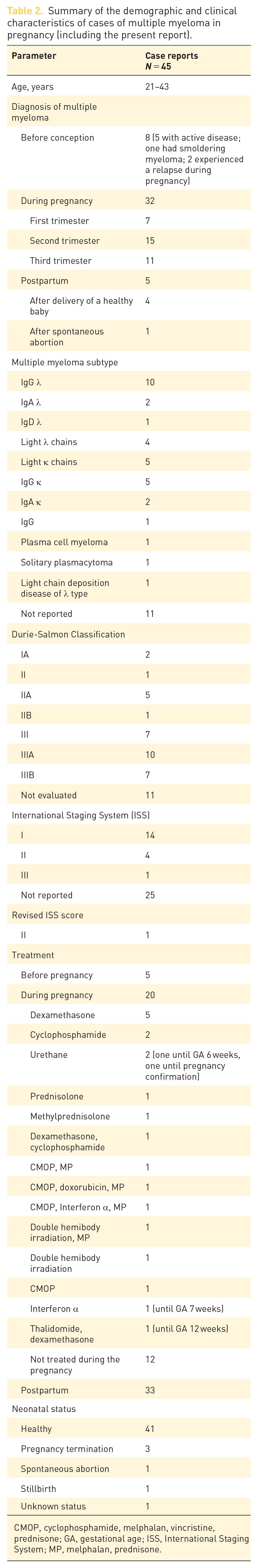
CMOP, cyclophosphamide, melphalan, vincristine, prednisone; GA, gestational age; ISS, International Staging System; MP, melphalan, prednisone.
As summarized in Table 2, all five patients with an active disease were treated with an anti-myeloma agent before pregnancy;7,25,29,31,32 four of them were treated with these agents during the first trimester until the pregnancy was confirmed.7,25,28,31 Sixteen patients received treatment for multiple myeloma during their pregnancy: seven with steroids only2,6,10,11,16 and the remaining nine patients with various therapies.4,9,29 Twelve patients were not treated during their pregnancy, most of them due to their request not to be treated while pregnant. Thirty-two of 45 patients were treated with anti-myeloma therapy after delivery or pregnancy termination.2,5–8,10,11,13–20,22–24,26,27,29,31–33
Forty-one women gave birth to 42 healthy babies, including a pair of twins. One patient had progressive disease and died after delivery. One newborn had an Apgar score of 5, which was followed by an uncomplicated neonatal course, 12 and another had seizures at birth that were related to difficulties with calcium regulation, but was healthy at follow-up. 23 Three pregnancies were terminated.8,13,22 One women had a spontaneous abortion, after which MM was diagnosed; the women gave birth to a healthy baby 5 years later, despite a relapse of the disease during the pregnancy. 13
Due to the low prevalence of MM in young adults, the initial investigation of the underlying causes of the patient’s symptoms – hypertension and acute renal failure – was directed toward pregnancy-related disorders, such as iron deficiency, preeclampsia, or primary kidney disorder, delaying the initial diagnosis. Once diagnosis was determined, treatment was initiated in order to delay disease progression and further damage to target organs while maintaining the safety of the fetus. The therapy regimen included CTX and HDD which enabled the completion of the pregnancy and the delivery of a healthy baby.
Treating physicians should be aware of potential effects of MM therapies on the fetus and pregnancy outcomes. Table 3 summarizes the limited evidence on the safety of the major classes of MM therapies during pregnancy. Most anti-MM agents have shown embryo lethality or teratogenicity when given during pregnancy in animal studies. Despite the widespread use of corticosteroids in MM protocols, 37 high-dose corticosteroids confer a slightly higher risk for fetal malformations in the first trimester, and a higher risk for obstetric complications in the second or third trimesters.38–41 Prednisolone is the preferred steroid as it is metabolized by the placenta with only 10% of maternal dose reaching the fetus. 42 In the case described here, the patient received HDD and CTX. CTX was also administered to two other pregnant patients – one in the second trimester and one in the third – but did not affect the development of the fetus.4,29 Low-dose CTX administered to a patient with Burkitt’s lymphoma during the third trimester showed good response with delivery of a normal baby, suggesting that low-dose intravenous CTX therapy may not be hazardous to the fetus during late pregnancy. 43
Classification of multiple myeloma therapy by fetal risks.

ADCC, antibody-dependent cell-mediated cytotoxicity; ADCP, antibody-dependent cellular phagocytosis; CDC, complement-dependent cytotoxicity; CTX, cyclophosphamide; FDA, Food and Drug Administration.
Chemical Hazards Emergency Medical Management. FDA Pregnancy Categories. US Department of Health and Human Services. Available from: https://chemm.nlm.nih.gov/pregnancycategories.htm.
Australian categorisation system for prescribing medicines in pregnancy. Australian Government Department of Health. Therapeutic Goods Administration. Available from: https://www.tga.gov.au/australian-categorisation-system-prescribing-medicines-pregnancy.
In conclusion, despite the rarity of concurrent MM and pregnancy, this possibility should be considered in patients with signs and symptoms that may be attributed to MM so as not to delay the diagnosis and decision about pregnancy continuation and initiation of appropriate, safe, and efficient therapy.
Footnotes
Author contributions
Conflict of interest statement
The authors declared no potential conflicts of interest with respect to the research, authorship, and/or publication of this article.
Funding
The authors received no financial support for the research, authorship, and/or publication of this article.