
Abstract
Objective
To review the history of chondrocyte transplantation, new approaches to treatment of human chondral and osteochondral defects, animal experiments, the choice of chondrocytes for transplantation, immunological features of human and animal chondrocytes and factors which could influence results of chondrocyte transplantation.
Design
As the material for review served numerous papers collected during our many years’ chondrocyte studies supplemented by PubMed search.
Results
Autologous chondrocytes, expanded in culture, were successfully used to repair damaged human articular cartilage. Numerous modifications of the original procedure benefited from a better understanding of factors influencing chondrocyte differentiation. Immunological studies suggested that survival of allogeneic transplants of bioengineered human neocartilage may depend on both passive and active mechanisms of immune evasion. Human chondrocytes with deleted expression of MHC class I molecules produced cartilage which, after transplantation into monkey articular cartilage was attacked by NK cells.
Conclusions
Immune response against human chondrocytes requires further investigation. It is already established that allogeneic chondrocytes are safe for treating chondral defects but not for healing osteochondral defects. Full reconstruction of cartilage defects by restoring anatomically identical hyaline cartilage seems not feasible.
Keywords
Introduction
It took Cheops 20 years to build the pyramid. The construction of the Parthenon lasted 15 years. The Colosseum took about 10 years to build. The Eiffel Tower was constructed in 2 years, 2 months, and 5 days. Collagenase from Clostridium histolyticum was identified by DeBellis et al. 1 in 1954, and it took 11 years to use it for isolating chondrocytes from mature cartilage.2,3 The first human ACI (autologous) was performed in October 1987, 4 which is 19 years since the first allogeneic osteochondral chondrocyte implantation in a rabbit model in 1968 5 but the paper with the first 23 patients operated on with ACI was published only in 1994.4,6 The safe use of allogeneic chondrocytes for treating chondral defects was first proposed in rats in 2000, 7 but it was only verified in a large animal study more than 20 years later.8,9 Why, after 60 years of studies, is there still no generally recognized procedure for articular cartilage reconstruction using chondrocytes? The present review will not answer this question but may draw attention to various problems involved in chondrocyte transplantation due to the metabolic specificities of chondrocytes or the way articular cartilage is nourished during joint movement. The particular issues are presented under the following headings:
Intramuscular Auto- and Allogeneic Chondrocyte Transplants: Cartilage as the Privileged Tissue
Early Studies on Chondrocyte Transplants into Articular Cartilage in Rabbits
Autologous Transplants of Chondrocytes in Humans
Cartilage Constructs and Expansion of Chondrocyte for Transplantation in Humans
Transplants of Allogeneic and Xenogeneic Articular Chondrocytes into Rabbit Articular Cartilage Defects
Syn- and Allogeneic Transplants of Chondrocytes in Rodents
Evaluation of Auto- and Allogeneic Rabbit and Rat Chondrocyte Transplants
Chondrocyte Transplants in Large Animals
Synovium, Immunoresponsive Cells, and Nutrition of Transplants
Nutrition of Cartilage: Influence of Oxygen
Rat Chondrocyte Tissue-Specific Antigen
Mesenchymal Cells and Cartilage Formation
Chondrocytes and Natural Killer (NK) Cells
Transplants of Human Allogeneic Chondrocytes in Humans and Genetically Modified Animals
Fas (CD95) Expression on Chondrocytes
Alloreactivity and Immunosuppressive Properties of Human Chondrocytes
The Possible Source of Allogeneic Chondrocytes for Transplantation
Is Reconstruction of True Articular Cartilage Possible?
Research Suggestion: How to Treat Osteochondral Defects?
Conclusions
Intramuscular Auto- and Allogeneic Chondrocyte Transplants: Cartilage as the Privileged Tissue
Successful, long-term allogeneic transplantation of cartilage has been reported in both humans and animals, leading to the concept that chondrocytes in such transplants are protected by extracellular matrix (ECM). 10 This concept was experimentally verified in two laboratories. Smith 3 transplanted fresh or cryopreserved allogeneic chondrocytes into defects of cancellous bone, did not observe an immune response, and concluded that it may depend on the rapid formation of a matrix protecting chondrocytes from contact with immunocompetent cells. Moskalewski and Kawiak 11 and Moskalewski et al. 12 intramuscularly transplanted autologous and allogeneic chondrocytes isolated from rabbit nasal septum cartilage and fragments of nasal septum autologous and allogeneic cartilage. In both cases, chondrocytes produced cartilage, but that formed by allogeneic cells was heavily infiltrated by lymphocytes and macrophages, suggesting that the recipients of chondrocytes were sensitized before a sufficient amount of matrix was formed to offer protection. Fragments of nasal allogeneic cartilage transplanted alone did not evoke an immune response. When similar fragments were transplanted together with isolated chondrocytes, infiltrates were frequently formed in the vicinity of the allogeneic fragments. Thus, isolated allogeneic chondrocytes evoked an immune response and presumably sensitized the animals against allogeneic cartilage fragments.
Similar results obtained Heyner 13 in a rat model. Some hosts were sensitized by skin allografts or injections of allogeneic lymph node and spleen cells. Chondrocytes were transplanted into the tip of the tongue for 30 days. Chondrocytes reformed cartilage both in nonsensitized and sensitized hosts, but in the latter, grafts were surrounded by a peripheral infiltrate of host mononuclear cells. Intact allogeneic cartilage grafts in nonsensitized hosts showed no evidence of resorption; in sensitized hosts, grafts were surrounded by lymphocytic infiltrate. Heyner 13 concluded that the matrix around the chondrocytes prevents the rejection of allografts of intact cartilage in nonsensitized hosts and that the privilege afforded such grafts is not a result of the lack of antigenicity of the cells.
These results raised the question of whether the prevention of chondrocyte recipient sensitization by short-lasting immunosuppression could give them time to produce a sufficient amount of interstitial substance to prevent rejection after the withdrawal of treatment. Indeed, isolated mouse epiphyseal or rib chondrocytes formed cartilage in syngeneic or allogeneic intramuscular transplants, but the latter became destroyed by infiltrating immune cells. Short-term immunosuppressive treatment decreased specific humoral antigraft immunity and prevented the development of cellular antichondrocyte immunity. Moreover, cartilage produced by allogeneic chondrocytes became surrounded by perichondrium, suggesting that the effect of immunosuppression could be long-lasting or even permanent.14,15
Książek and Moskalewski 16 transplanted chondrocytes isolated from the epiphyses of 5-day-old mice after sex determination. Syngeneic chondrocytes transplanted between animals of the same sex produced cartilage in which endochondral bone formation occurred in older transplants. Similarly behaved transplants of syngeneic female chondrocytes to males. Chondrocytes in syngeneic transplants from males to females across the H-Y (male-specific histocompatibility antigen) barrier formed cartilage infiltrated and resorbed by mononuclear cells. Similar changes occurred in allogeneic chondrocyte transplants across a weak (non-H-2; histocompatibility-2) barrier. In these transplants, which did not evoke an immune response (fully compatible system, transplants across the H-Y barrier in a nonrejector strain), chondrocytes reconstructed cartilage that hypertrophied, calcified, and was finally replaced by bone. When cartilage was surrounded by mononuclear infiltration, bone formation did not occur or was delayed, regardless of the degree of antigenic difference.
Romaniuk et al. 17 studied the rejection of cartilage formed by transplanted allogeneic rat epiphyseal chondrocytes using a panel of different monoclonal antibodies. One week after transplantation, the grafts were surrounded by numerous class II MHC (major histocompatibility complex) positive cells, CD4+ cells, and ED1+ monocytes/macrophages. In older transplants, there was a significant increase in the number of CD8+ cells. They accumulated close to the transplants, and some of them penetrated the cartilage matrix, suggesting that they might be involved in chondrocyte killing. At the time of transplantation, chondrocytes were endowed with RT1.D (Rat Histocompatibility 1 antigen) class II antigen but not with RT1.B class II molecule. However, after 1 week, the number of RT1.B positive chondrocytes increased during cartilage rejection. After 3 months, the cartilage was almost completely destroyed, and the intensity of infiltration was markedly decreased.
In a recent review, Garrity et al. 18 essentially supported Bacsich and Wyburn’s 10 concept of the protective function of cartilage matrix in an allogeneic environment and enriched it by pointing out that the average pore size of cartilage ECM is 6 nm in diameter, 19 while a lymphocyte has an average diameter of 6 to 7 μm. 20 Thus, lymphocytes cannot penetrate into cartilage and attack chondrocytes. It seems probable that in allogeneic intramuscular transplants, the formation of ECM by chondrocytes was too slow to form an impermeable barrier against the attack of infiltrating cells. To complicate matters further, infiltrations also appeared at fragments of allogeneic cartilage transplanted into muscle in animals sensitized by allogeneic skin 13 or chondrocyte transplants, 12 indicating that the protection offered by the matrix is not always sufficient.
Early Studies on Chondrocyte Transplants Into Articular Cartilage in Rabbits
Chesterman and Smith 5 transplanted allogeneic chondrocytes suspended in rabbit serum into articular cartilage defects and found the formation of cartilage by transplanted cells. However, at the bone marrow-cartilage interface, fibrous tissue formed, and the healing of the defect was not satisfactory. Interestingly, while skin transplants between strains of rabbits used in the experiment were rejected, the produced cartilage was free of infiltrations. Bentley and Greer 21 pipetted allogeneic epiphyseal or articular chondrocytes into deep articular cartilage defects and found that epiphyseal chondrocytes reconstructed the growth plate with the formation of endochondral bone, while articular chondrocytes formed clumps attacked by infiltrating cells. Freed et al. 22 transplanted articular allogeneic chondrocytes cultured and transplanted in a polyglycolic acid (PGA) scaffold into full-thickness defects without violating the subchondral bone. After six months, joint repair was better in cell-PGA allografts than in PGA alone. Infiltrations were absent on the histological slides. In this early period of isolated chondrocyte transplantation, comparisons of cryopreserved and nonfrozen chondrocytes demonstrated that both types could be successfully used for cartilage reconstruction.3,5,23,24
Autologous Transplants of Chondrocytes in Humans
Interest in chondrocyte transplantation was boosted by Brittberg et al. 4 publication on human cartilage repair by autologous chondrocyte implantation (ACI). The study was accompanied by experiments with autologous chondrocyte transplants in rabbits. 25 Chondrocytes were enzymatically liberated from a minor load-bearing area of articular cartilage, expanded in cell culture, and implanted into the cartilage defect. Soon, modifications of the procedure appeared, consisting of the transplantation of chondrocytes incorporated into atelocollagen. 26 Evaluation of the results of large full-thickness cartilage lesions demonstrated that the clinical and functional outcomes remain high even 10 to 20 years after the implantation.27,28 According to a 2014 survey, ACI has been performed in more than 30,000 patients worldwide since 1987. 29 Also, in the opinion of the working group of the German Society of Orthopaedics and Trauma, ACI is an established and well-accepted procedure for the treatment of localized full-thickness cartilage defects of the knee. 30 Overall, with a mean follow-up of 11.4 years, ACI demonstrated successful outcomes in 82% of patients. 31 Bentley et al. 32 compared the results of articular cartilage defects treatment with ACI and mosaicplasty in 58 and 42 patients, respectively. The mean follow-up was 19 months. Functional assessment with the modified Cincinnati and Stanmore scores and objective clinical assessment demonstrated that 88% of patients had excellent or good results after ACI compared with 69% after mosaicplasty. Thus, the study has shown significant superiority of ACI over mosaicplasty for the repair of articular defects in the knee. Carey et al. 33 identified through manual chart review patients who underwent ACI as treatment for unsalvageable osteochondritis dissecans (OCD) between 1990 and 2005. The median follow-up duration was 19 years. Most patients reached their preinjury activity level and would undergo ACI again if in the same situation. Thus, ACI for OCD provides a durable treatment option.
The plasticity, differentiation potential of chondrocytes, and involvement of growth factors were exhaustively reviewed by Tallheden et al. 34 Technical advances, such as the incorporation of chondrocytes into a suitable matrix before implantation, may further improve ACI results.29,35
Matrix-induced autologous chondrocyte implantation (MACI) is a third-generation ACI product in which the patient’s previously harvested chondrocytes are expanded in culture and seeded onto a collagen scaffold.27,36
Recently, many surgeons have favored the use of particulated articular cartilage for chondral and osteochondral repair, such as CAIS (Cartilage Autograft Implantation System), particulated autologous cartilage in a scaffold DeNovo NT, a tissue graft product made by Zimmer Biomet for the treatment mainly of knee and ankle defects, juvenile human allograft cartilage embedded in fibrin glue, autologous cartilage chips—with and without concomitant bone grafting, and autologous cartilage chips embedded in a clot consisting of platelet-poor plasma (PPP) and platelet-rich plasma (PRP). All these methods offer encouraging clinical results and cost reduction but require large randomized controlled studies, as recently reviewed in this journal. 37 Moreover, numerous companies have developed various forms of autologous chondrocyte preparations for direct clinical application. These products are listed and characterized in a recent review. 38 For example, CartiLife® is a fourth-generation ACI in which donor tissue is collected from the rib cartilage of a patient and then cultured to generate pellets that contain cartilage cells and extracellular substrates. Finally, pellet-type chondrocyte tissue is implanted into the defective area without a scaffold. 39
In attempts to shorten the typical ACI procedure to a one-stage cell transplantation, allogeneic mesenchymal stem cells (MSCs) were used to stimulate chondrons (chondrocytes with their pericellular matrix), enzymatically isolated from fragments of cartilage debrided during surgery. The procedure, called “Instant MSC Product accompanying Autologous Chondron Transplantation” (IMPACT), was used for the treatment of cartilage defects in 35 patients and resulted in statistically significant improvement in clinical outcomes. The repair tissue displayed hyaline-like features with a high concentration of proteoglycans and type II collagen. DNA analysis demonstrated that the regenerated tissue contained only the patient’s DNA. Thus, MSCs acted rather as “signaling cells” than differentiating stem cells. 40 Korpershoek et al. 41 presented a study protocol titled “Efficacy of one-stage cartilage repair using allogeneic mesenchymal stromal cells and autologous chondron transplantation (IMPACT) compared with nonsurgical treatment for focal articular cartilage lesions of the knee: study protocol for a crossover randomized controlled trial.” In the trial, sixty patients will be randomized to receive nonsurgical care or IMPACT. The authors expect to develop a health economic model to estimate the incremental cost-effectiveness ratio of IMPACT versus nonsurgical treatment in terms of costs per quality-adjusted life year over a 5-year time horizon. The study is designed to evaluate the efficacy of IMPACT compared with nonsurgical care. The trial was registered in 2019. 41
Just recently appeared the paper by Niemeyer et al. 42 who in the study involving 100 patients found hydrogel-based ACI to be a safe and effective treatment option for patients with large knee cartilage defects with sustained efficacy up to 5 years. No remarkable adverse events or safety issues were noted.
Cartilage Constructs and Expansion of Chondrocytes for Transplantation in Humans
Scaffolds formed from an esterified derivative of hyaluronic acid called HYAFF® 11 served as the basis for development of Hyalograft® C, a tissue-engineered graft consisting of autologous chondrocytes expanded in culture. Within the cohort of 141 patients with average follow-up time of 38 months, 91.5% of patients improved according to the International Knee Documentation Committee subjective evaluation, moreover 76% and 88% of patients had no pain and mobility problems, respectively, assessed by the EuroQol-EQ5D measure. Authors conclude that the positive clinical results indicate that Hyalograft® C is a safe and effective therapeutic option for the treatment of articular cartilage lesions. 43
Adkisson et al. 44 found that juvenile articular chondrocytes cultured in a serum-free system produce a sufficient number of growth factors to guide the production of hyaline cartilage in vitro. Chondrocytes from adult articular cartilage failed to produce neo-cartilage when grown under identical conditions. The authors speculate that the onset of puberty is associated with an age-related decrease in autocrine-mediated cell signaling, whereby articular chondrocytes lose their capacity to proliferate and maintain cartilage homeostasis.
A new possibility for the formation of cell sheets suitable for transplantation was introduced by Okano et al. 45 Commercial polystyrene culture dishes were covered with polymer acting as a temperature switch, creating hydrophilic surfaces below 32 °C and hydrophobic surfaces above 32 °C. Sheets of liver and endothelial cells could be detached from the PIPAAm-grafted surface simply by reducing the temperature below the polymer transition temperature, eliminating the need for proteolytic enzymes for cell detachment and preserving their organization within the sheet. This technique was adopted for chondrocyte transplantation, first in rabbit tests. 46
The advantages offered by bioengineered and layered chondrocyte sheets were presented by Sato et al.47,48 Mitani et al. 49 studied the expression of SOX9, collagen type II, integrin α10, and fibronectin genes in chondrocytes from triple-layered sheets produced in temperature-responsive culture dishes and found that it was significantly increased compared with those in conventional monolayer culture. Moreover, synovial tissue obtained from the same joint and serving as the chondrocyte nutritive supply source could accelerate the preparation of chondrocyte sheets. Transplantable tissue could be obtained on average in 10.5 days. 50 Autologous chondrocyte sheet transplants were used by Sato et al. 51 for the treatment of knee osteoarthritis (OA) in eight patients and found the therapy effective.
Tissue engineering strategies aimed at the repair of articular cartilage injury were presented in a recent review and, in the opinion of the review authors, hold promise for solving cartilage reconstruction problems. 52 Another approach was proposed by Park et al., 53 who described the chondrogenic differentiation of fetal cartilage-derived progenitor cells (FCPCs) to adult cells using a three-dimensional spheroid culture method based on silica nanopatterning techniques. Acevedo Rua et al. 54 proposed to use nasal chondrocyte–based tissue-engineered cartilage (N-TEC) for treatment of degenerative cartilage defects in humans. First, they established that N-TEC maintained cartilaginous properties when exposed in vitro to inflammatory stimuli found in osteoarthritic joints and favorably altered the inflammatory profile of cells from osteoarthritic joints. N-TEC survived after ectopic engraftment in mouse models reproducing a human osteochondral OA tissue environment, and in sheep articular cartilage defects that mimic degenerative settings. The safety of autologous N-TEC for the treatment of osteoarthritic cartilage defects was verified by transplantation into the knees of two patients with advanced OA who were otherwise considered for unicondylar knee arthroplasty. No adverse reactions were recorded, and patients reported reduced pain as well as improved joint function and life quality 14 months after surgery. Authors suggests that N-TEC can directly contribute to cartilage repair in osteoarthritic joints. 54 Lehoczky et al. 55 aimed at implementation of autologous NCs in a single-stage minimally invasive technique for the repair of small to middle-size focal cartilage lesions. Human nasoseptal cartilage chondrocytes (NCs) isolated by short collagenase digestion were able to proliferate and produce histochemically detectable cartilage matrix within an injectable, swiftly cross-linked polyethylene glycol (PEG) gel enriched with human platelet lysate (hPL). NC-hPL-PEG gels integrated with osteochondral tissues obtained from allogenic knee joints after subcutaneous implantation into nude mice. In view of the positive results of the in vitro and in vivo tests authors believe that the envisioned therapeutic strategy may obtain the approval of the modified PEG gel as medical device or together with nasoseptal chondrocytes as a “combination medical device–advanced therapy medicinal product. 55 Mumme et al. 56 studied relevance of engineered cartilage maturation in a randomized multicenter trial for articular cartilage repair using autologous nasal chondrocytes (NCs). Clinical outcome of in vitro–matured NC-tissue-engineered cartilage (N-TEC) was tested versus undifferentiated NC-cell-activated matrices (N-CAM) in focal full- thickness knee cartilage defects. Isolated NCs were expanded in a monolayer for 2 weeks and seeded on the porous side of a collagen membrane. NCs were cultured in the membrane for either 14 days to manufacture N-TEC or for 2 days to manufacture N-CAM. Before implantation, N-CAM contained cells with limited chondrocytic morphology and ECM staining for glycosaminoglycans and type 2 collagen. N-TEC, which underwent a longer NC culture time, was phenotypically, structurally, and functionally more like hyaline cartilage than N-CAM. The overall Knee Injury Osteoarthritis Outcome Score (KOOS) increased with clinical relevance in both groups compared with preoperative values. KOOS was higher at 24 months for N-TEC than for N-CAM. N-TEC, but not N-CAM, was similarly effective in patients with larger defects. In author’s opinion results of the trial validate the clinical efficacy of NC- based grafts for articular cartilage repair and supports the clinical relevance of engineering mature tissues, even for patients with more challenging cartilage defects. 56
The problem of obtaining a sufficient number of chondrocytes for transplantation was addressed using a hollow-fiber bioreactor (Quantum). Tests with chondrocytes isolated from femoral condyles from patients undergoing total knee replacement surgery demonstrated that the rapid reactor upscale expansion did not alter the chondrocyte phenotype when compared with chondrocytes expanded in plastic vessels. Therefore, the use of a bioreactor provides an attractive method for rapidly manufacturing chondrocytes for clinical use (Figure 1). 57
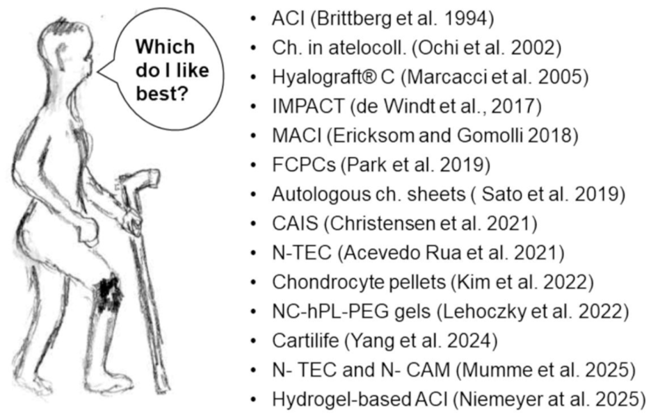
Various possibilities of articular cartilage defects treatment.
Transplants of Allogeneic and Xenogeneic Articular Chondrocytes Into Rabbit Articular Cartilage Defects
Rabbits are frequently used in studies on cartilage reconstruction, testing various ways of chondrocyte administration. During evaluation of rabbit articular chondrocyte transplants deserves particular attention spontaneous healing of cartilage lesions described by Shapiro et al. 58 The authors drilled full-thickness 3 mm diameter articular cartilage defects in New Zealand white rabbits and studied their spontaneous healing. In the first few days, fibrinous arcades were established across the defect, serving to orient mesenchymal cell ingrowth along the long axes. The first evidence of cartilage matrix appeared in 10 days. At 2 weeks, cartilage matrix was present immediately below the surface of collagenous tissue rich in flattened fibrocartilaginous cells. At 3 weeks, nearly all defects had a layer of cartilage. At 24 weeks, both the tidemark and the compact lamellar subchondral bone plate had been re-established. The authors concluded “Many rabbits had excellent histological repair in the absence of any specific treatment.” So, it seems that in rabbits, chondrocytes implanted into damaged cartilage only accelerated (or possibly disturbed) the healing of defects, which, in due time, would heal spontaneously.
The following text presents numerous technical approaches aimed at improving the treatment of articular cartilage defects. Thus, allogeneic chondrocytes were transplanted into chondral defects as sheets detached by trypsin and covered with a periosteal flap. After 12 weeks, the transplant formed tissue with hyaline cartilage. 59 Chondrocytes cultured in temperature-responsive culture dishes, collected with PVDF (polyvinylidene fluoride) membrane, were placed on the other cell sheet to produce multilayered cell sheets floating in the culture medium. Layered chondrocyte sheets were able to maintain the cartilaginous phenotype, could be attached to the sites of cartilage damage, and serve as a barrier to prevent the loss of proteoglycan from these sites. 46 Boopalan et al. 60 transplanted auto- and allogeneic cultured-to-confluence chondrocytes as cell suspension into articular cartilage defects covered with a periosteal flap and observed regeneration of hyaline/mixed hyaline cartilage. No significant difference in the performance between autologous and allogeneic transplants was observed.
Osteochondral defects were implanted with allogeneic chondrocytes cultured in two-layered collagen 61 or in collagen gel.62 -64 The latter authors observed a high percentage of apoptotic cells in the formed cartilage. Chondrocytes were also placed in agarose gel, 65 in lactic acid polymer fleece, 66 in alginate gel, 67 or in type II collagen gel scaffold. 68 Nagai et al. 69 kept chondrocytes in a static culture for 7 days, then in the rotational or static mold formed plates which were inserted into the defects to integrate with the host cartilage. The lower portion of the repair tissue contained hypertrophic chondrocytes that were remodeling the subchondral bone. Ito et al. 70 transplanted allogeneic synovial cells with or without allogeneic chondrocyte sheets produced in temperature-responsive culture dishes in various ratios and obtained the best cartilage formation with synovial 12 x 106 cells and layered chondrocyte sheets with 1.7 x 10⁶ cells. Lin et al. 71 transplanted chondrocytes cultured in vitro for 7 days and modeled into microspheres in hyaluronan/collagen II (HA/Col II) carrier. Man et al. 72 transplanted chondrocytes in chitosan hydrogel-demineralized bone matrix hybrid scaffold. Tani et al. 73 transplanted chondrocytes cultured with synovium from knee joints, prepared in three-layered sheets; some sheets remained nonvitrified, others were vitrified and thawed, with no difference in performance observed. Jakutowicz et al. 74 transplanted auto- and allogeneic chondrocytes cultured on polyethersulfone (PES) membranes. Cartilage was formed in both auto- and allogeneic transplants.
In all the above-quoted reports, chondrocytes produced cartilage matrix, and no signs of rejection were noted. These findings contrast with the study by Arzi et al., 75 in which allogeneic neocartilage constructs were rejected in patella cartilage defects while in the trochlea they integrated with the underlying bone. The rejection at the patella site was explained by the proximity of the transplant to synovium containing immunoreactive cells. Xenogeneic (bovine chondrocytes) constructs evoked a strong response at both locations.
Syn- and Allogeneic Transplants of Chondrocytes in Rodents
Moskalewski et al. 76 transplanted syngeneic chondrocytes isolated from the epiphyses of newborn rats and suspended in hyaluronic acid intramuscularly or into defects in knee joints reaching bone underneath the cartilage. In intramuscular transplants, chondrocytes produced cartilage which ossified, while in articular cartilage transplants, chondrocytes in the superficial layer formed cartilage, and in the deep layer, hypertrophy, matrix calcification, and bone formation occurred. Thus, either the intra-articular environment inhibited the maturation of chondrocytes, or articular chondrocytes present among the transplanted cells accumulated close to the joint lumen and reconstructed articular cartilage.
Other experiment were performed14,77 to find out whether short-lasting immunosuppression would allow allogeneic chondrocytes to produce a sufficient amount of matrix to resist rejection. Chondrocytes from epiphyseal cartilage of CFW/LI mice transplanted intramuscularly into C57BI/10 mice reconstructed cartilage, which became resorbed by infiltrating cells. Immunosuppression with cyclosporin and cladribine (2-chlorodeoxyadenosine) suppressed the formation of infiltrations and allowed the formation of bone trabeculae covered by osteoblasts.14,77 In articular cartilage transplants, allogeneic chondrocytes also formed cartilage, but immunosuppression only slightly decreased the intensity of infiltrations and did not prevent cartilage resorption. 78 Thus, strong immunosuppressive treatment of rats with allogeneic chondrocyte transplants was more effective in inhibiting the general immunological response than the reaction in the joint. 79 Moreover, the attack of immune cells proceeded from the direction of the bone marrow, while the cartilage surface remained free of infiltrations. This suggested that the immunization occurred via the bone marrow and not via the joint cavity. Following this observation, a barrier from bone cement was formed between the bone marrow cavity and transplanted allogeneic chondrocytes. Reconstructed cartilage had a similar appearance to the normal cartilage surrounding the transplant, and there were no signs of cartilage resorption. 7 Since articular cartilage nutrition takes place via the synovial fluid and not via the bone marrow,80,81 authors concluded that allogeneic chondrocytes could serve as a good material for articular cartilage repair if a proper barrier was constructed. Subsequent studies demonstrated a contribution of solutes from bone marrow cavity for maintaining of articular cartilage—the problem is discussed in the Synovium, Immunoresponsive Cells, and Nutrition of Transplants section.
Noguchi et al. 82 transplanted syngeneic and allogeneic rat articular chondrocytes cultured for 2 weeks into full-thickness cylindrical osteochondral defects in the femoral articular surface. During the 12 weeks, all eight defects with syngeneic transplants had healed successfully, compared with only four of eight defects with transplanted allogeneic chondrocytes with delayed subchondral bone formation. At 26 and 52 weeks, there was no significant difference in the success rate between the isografted and allografted groups. At four weeks, inflammatory cell infiltration at the periphery of the defects was slight in the isografted knees, somewhat conspicuous in the allografted ones. At 26 weeks, there was no inflammatory cell infiltration.
Moskalewski et al. 83 produced syngeneic and allogeneic transplants into osteochondral defects in the articular cartilage of naïve rats or syngeneic transplants in rats sensitized with allogeneic chondrocytes injected intramuscularly. Syngeneic transplants produced cartilage without signs of rejection. CD8+ lymphocytes and macrophages accumulated in the vicinity of cartilage produced by similar transplants in animals previously sensitized with intramuscular transplants of allogeneic chondrocytes. CD8+ cells penetrated into the peripheral part of the cartilage, while macrophages advanced much more deeply. The synovium from rats with osteochondral transplants of allogeneic chondrocytes or syngeneic chondrocytes after sensitization contained macrophages and CD8+ cells. The rejection of cartilage formed by syngeneic chondrocyte transplants in sensitized animals argued in favor of chondrocyte-specific antigen expression.
Takaku et al. 84 used luciferase-expressing transgenic Lewis rats and cultured luciferase-labeled chondrocytes and synovial cells to produce sheets on temperature-sensitive dishes. The sheets were transplanted into rat knee joints with osteochondral defects. Both chondrocytes and synovial cells survived for an extended period of time at the transplanted site, without signs of inflammation, thereby confirming the safety and utility of cell sheets in promoting cartilage regeneration.
Takatori et al. 85 used male Wistar rats with early arthritis induced by intra-articular administration of monoiodoacetic acid (MIA) as recipients of chondrocyte sheets produced in culture from articular chondrocytes of male Lewis rats. Transplantation of cell sheets alleviated pain and decreased cartilage degeneration. Macroscopic observations showed the absence of inflammatory symptoms in the knee, such as swelling or reddening.
Takao et al. 86 seeded expandable limb-bud mesenchymal cells (ExpLBM) derived from human-induced pluripotent stem cells into dishes for the initiation of chondrocyte induction. Then the cells were transferred into temperature-sensitive dishes to produce chondrocyte sheets. The sheets were transplanted into defects in the knee articular cartilage of immunodeficient X-SCID (X-linked severe combined immunodeficiency) rats. Implanted material was positive for glycosaminoglycans, aggrecan protein (ACAN), and collagen type II. The authors suggest that the validation of the efficacy of transplantation is required in larger animals that more closely resemble humans.
Evaluation of Auto- and Allogeneic Rabbit and Rat Chondrocyte Transplants
Since rabbit allogeneic chondrocytes injected intramuscularly or transplanted in a plasma clot sensitized recipients, resulting in an immune attack on the reformed cartilage,11 -13 the question arises why in the 19 reports on transplants of allogeneic chondrocytes into articular cartilage defects presented in the review, only in 2 of them21,75 was there an immune response in the form of lymphocytic infiltrations.
Contrary to that, cartilage produced by allogeneic chondrocytes in rats evoked strong immunological reaction in form of lymphocytic infiltrations which occurrence could not be prevented by immunosuppression.78,79
In most of the above quoted papers, chondrocytes were implanted in various types of scaffolds. It is well documented that cartilage matrix prevents contact of allogeneic chondrocytes with the immune system (reviewed by Garrity et al. 18 ), but it remains unknown how much matrix is needed for protection. Thus, it is possible that chondrocytes transplanted in scaffolds are able to produce it in sufficient amounts to obtain protection before sensitization could occur, and that may explain the success of many transplants. There is, however, also a possibility that the performance of rabbit and rat osteochondral transplants depends on the differences in the content of bone marrow in long bones in both species. In the bones of the limbs of 4- to 6-month-old rabbits the marrow cavity was predominantly composed of adipose cells, with the proximal femur retaining a small amount of red marrow. 87 In 4-month-old Wistar rats hematopoietic tissue covered 21% at the femoral epiphyses and about 70% at diaphysis. 88 It leads to the conclusion that for the full evaluation of the immunological reaction toward chondrocyte transplant it is also necessary to study the immunological status of the site of transplantation.
Chondrocyte Transplants in Large Animals
Studies on large animals are not numerous. Mainil-Varlet et al. 89 cultured allogeneic chondrocytes in a bioreactor, resulting in the formation of cartilage-like constructs measuring 700 μm to 1 mm in thickness after 3-4 weeks. Constructs were transplanted into superficial and full-thickness (FT) articular defects in the patellar groove of the femoral condyle of Yucatan minipigs. After 24 weeks, in some FT endochondral bone formation and in some inflammatory responses were observed.
Ebihara et al. 90 used allogeneic articular chondrocytes for the preparation of multilayered sheets in temperature-responsive culture dishes. Sheets were implanted into the chondral defects of minipigs and produced mainly adequate cartilage repair. There were no signs of inflammation.
Abe et al. 9 used induced pluripotent cynomolgus monkey stem cells (cyiPSCs) for the formation of allogeneic cartilage organoids (cyiPS-Cart). cyiPSCs expressed pluripotency markers, while cyiPS-Cart expressed chondrocyte markers. Chondral defects with implanted allogeneic cartilage organoids were filled with cartilaginous tissue both at 4 and 17 weeks after surgery, survived, and integrated with articular cartilage. There was no lymphocyte accumulation around chondral defects transplanted with allogeneic cyiPS-Cart.
Krauze et al. 91 transplanted allogeneic chondrocytes from porcine xiphoid cartilage into a sternal incision wound. Implanted chondrocytes formed cartilage without significant side effects. The authors conclude that allogeneic chondrocytes may accelerate wound healing after median sternotomy.
Synovium, Immunoresponsive Cells, and Nutrition of Transplants
Synovium contains bone marrow-derived macrophages and occasionally dendritic cells.92 -94 The influenza virus deposited in the joint space induced a strong systemic antibody response of IgG, IgA, and IgM classes. This response was significantly higher (P < 0.05) than that of control subjects immunized subcutaneously. 95 Moskalewski et al. 83 produced syngeneic and allogeneic transplants into osteochondral defects in the articular cartilage of naïve rats or syngeneic transplants in rats sensitized with allogeneic chondrocytes injected intramuscularly. Syngeneic transplants produced cartilage without signs of rejection. CD8+ lymphocytes and macrophages accumulated in the vicinity of cartilage produced by similar transplants in animals previously sensitized with intramuscular transplants of allogeneic chondrocytes. CD8+ cells penetrated into the peripheral part of the cartilage, while macrophages advanced much more deeply. The synovium also contained macrophages and CD8+ cells. Thus, synovium could probably process chondrocyte antigens, serving as the first link necessary for sensitization via the joint cavity, but chondral cartilage transplants do not contact synovium, making this way of sensitization improbable. Synovium is also mainly responsible for the nutrition of articular cartilage.80,81 Arkill and Winlove 96 found, however, that calcified cartilage in the cartilage-subchondral bone area is permeable to small solutes and that subchondral circulation may make a significant contribution to the nutrition of deep cartilage. Wang et al. 97 studied the role of synovial fluid versus subchondral bone marrow as the source of nutrients and found that the former is the dominant source of sustenance for adult cartilage structure and function. When the only nutrition source was the bone marrow cavity, cartilage degenerated.
Nutrition of Articular Cartilage: Influence of Oxygen
For successful cartilage transplants, it is important to ensure satisfactory nutrition for transplanted chondrocytes forming cartilage. While the influence of axial compression on solute transport into cartilage was recognized already in 1959 by McCutchen, 98 the importance of sliding for cartilage nutrition was realized only recently. Mass transfer by sliding was found to be considerably higher compared with uniaxial compression, 99 a fact important for the construction of transplants in articular cartilage defects which should have contact with the opposite cartilage surface to benefit from sliding movement. This raises the question of which of the numerous ways of chondrocyte transplantation would be optimal for chondrocyte nutrition. Moreover, since chondrocytes are predominantly anaerobic cell types, there must be an effective efflux from the cell of the large quantities of lactic acid produced if intracellular pH is to be maintained, 100 and this process may also be influenced by the form of chondrocyte transplant. Studies on rodents or other small animals would not be adequate due to differences in cartilage dimensions between these animals and human cartilage.
An important aspect of chondrocyte physiology is their adaptation to hypoxia. Combining hypoxia with spheroid culture markedly increased the expressions of cartilage-specific collagen II and aggrecan at protein and mRNA levels. 101 Spheroids generated by human articular chondrocytes in hypoxia showed significantly increased glycosaminoglycan synthesis and up-regulated the expression of transcription factor SOX9, ACAN, COL2A1, COMP (cartilage oligomeric matrix protein), and zinc finger protein SNAI1 genes compared with those obtained under normoxic conditions. 102
Mennan et al. 103 compared articular chondrocytes cultured in ~21% (normoxic chondrocytes, NC) and 2% (hypoxic chondrocytes, HC) oxygen in both monolayer and three-dimensional (3D) pellet culture and also with freshly isolated chondrocytes (FC). The markers CD166 and CD151, indicative of chondrogenic de-differentiation, were significantly higher on NC compared with HC and lowest on FC. NC also produced the highest levels of CD106 and showed the greatest levels of IDO expression (indoleamine 2,3-dioxygenase, a known mediator of immune evasion, [see Munn et al. 104 ] following interferon-γ stimulation, indicating immunomodulatory potential. Thus, hypoxic conditions encourage retention of a chondrogenic phenotype with some immunomodulatory potential, whereas normoxia promotes dedifferentiation of chondrocytes toward an MSC phenotype with loss of chondrogenic potency but enhanced immunomodulatory capacity. These observations suggest that chondrocytes cultured in hypoxia would be more suitable for transplants than normoxic cells, but the culture of cells at low oxygen concentration is expensive and troublesome. It remains unknown how long it would take chondrocytes cultured in normoxia to return to the typical joint cavity environment after transplantation and whether this period of adjustment would have a significant influence on the final results.
Rat Chondrocyte Tissue-Specific Antigen
The observation that syngeneic chondrocytes of naïve rats stimulate T cell reactivity both in the in vitro proliferation and cytotoxic assays led to the suggestion that chondrocytes may express a tissue-specific antigen (reviewed in more detail by Lance, 105 Moskalewski 106 and Moskalewski et al. 107 ). The presence of such antigen(s) was then demonstrated by transplantation experiments. Cartilage produced in 2-week-old intramuscular transplants in rats by syngeneic chondrocytes did not display any signs of rejection, but cartilage formed by similar transplants in animals presensitized with allogeneic chondrocytes was infiltrated and rejected. 108 Rat chondrocytes intramuscularly transplanted into rabbits produced cartilage, which within 2 weeks deteriorated and was invaded by macrophages and fibroblasts. Sera of rabbits which received three consecutive transplants of chondrocytes contained high titers of antichondrocyte cytotoxic antibodies which could be removed by absorption with rat splenocytes. The absorbed serum lysed chondrocytes but not fibroblasts, endotheliocytes, or thymocytes. Western blot with chondrocyte lysates detected an antigen with Mr ~ 74 kD. 109 The antigen was subsequently identified as sialylated transmembrane protein Tmp21 (21 kDa transmembrane-trafficking protein) belonging to the p24 protein family. 110 These proteins participate in the traffic between the endoplasmic reticulum and Golgi complex and in some cells appear also in the membrane of secretory granules and plasmalemma. Antibodies reacting with p24 antigen could also be produced in rats immunized with surface chondrocyte proteins extracted from cultured chondrocytes and suspended in incomplete Freund’s adjuvant. 111 Expression of Tmp21 decreased in cultured chondrocytes concomitantly with the decline of collagen type II and aggrecan and the rise of collagen type I and versican expression. 112 Thus, it may be considered as a rat chondrocyte differentiation antigen. As far as we know, no attempts were undertaken to identify it in chondrocytes of other species, but there are data suggesting that it is absent in human chondrocytes. Yamaga et al. 113 produced five monoclonal antibodies against human articular chondrocytes and found that they all reacted with chondrocytes in frozen human articular cartilage. When tested against 29 different noncartilaginous tissues, each of these antibodies had distinct reactivity patterns, suggesting that each reacted with different antigens. Thus, all chondrocyte antigens detected with produced antibodies were shared by one or another human tissue.
Summarizing, rat chondrocytes express Tmp21, which may be considered as a specific rat chondrocyte differentiation antigen with immunogenic properties, but it is rather a rat biological specificity without implications for human chondrocyte transplantation.
Mesenchymal Cells and Cartilage Formation
Hunziker et al. 114 produced partial-thickness defects in the articular cartilage of adult rabbits and Yucatan minipigs and studied the effects of chondroitinase ABC or trypsin, fibrin clots, and mitogenic growth factors on the healing process. The chondroitinase treatment removed a superficial layer of proteoglycans from the surface of the defect, and the fibrin clot formed a scaffold for the migration of synovial mesenchymal cells. At 48 weeks, the entire cavity of the defect remained filled with fibrous connective tissue, but cartilage did not form. The idea of the differentiation of articular cartilage chondrocytes from bone marrow stem cells was critically discussed by Somoza et al., 115 who stressed that “MSCs have an intrinsic differentiation program reminiscent of endochondral bone formation, which they follow after exposure to specific reagents as a part of current differentiation protocols.” Thus, production of chondrocytes from bone marrow stem cells requires further studies to obtain material suitable for articular cartilage reconstruction. Ryan et al. 116 used allo-MSCs from DA or LEW rats to study chondrocyte differentiation. MSCs expanded in culture, expressed low levels of MHC class I, and lacked expression of CD45, MHC class II, CD80, and CD86. After 3-week culture, the cells suspended in alginate and stimulated with transforming growth factor TGFβ3 and bone morphogenetic protein BMP-2 differentiated to a chondrogenic phenotype and expressed MHC class I and MHC class II. Differentiated MSCs induced proliferation of allogeneic lymphocytes, whereas undifferentiated MSCs did not. Three-dimensional chondrogenic differentiation induced in an alginate matrix with TGFβ3 and BMP-2 served to immobilize and potentially protect MSCs at the site of implantation. Allogeneic undifferentiated MSC alginate subcutaneous implants attracted low numbers of infiltrating mononuclear cells, whereas allogeneic differentiated MSCs were heavily infiltrated. Thus, allogeneic cells induced a systemic allogeneic lymphocyte response, not prevented by encapsulation in alginate. Aldrich et al. 117 in a recent review argued that MSCs act as signaling and modulatory cells, exerting their influence through trophic and immune mediation rather than as a cell replacement therapy, and stressed that understanding the distribution and persistence of transplanted MSCs is necessary to fully realize their potential in cartilage regeneration techniques and tissue engineering applications.
Chondrocytes and Natural Killer (NK) Cells
Rejection of isolated chondrocyte grafts may be also mediated by the natural cytotoxicity mechanisms as normal chondrocytes were found susceptible to lysis by the natural killer (NK) cells. This is a universal mechanism since susceptibility to lysis by NK cells was found to be an attribute of chondrocytes originating from different types of cartilage from humans, pigs, rats, and mice, 15 ,118 -120 and this phenomenon appears to be associated with specific chondrocyte phenotype. 121 Recognition and lysis of porcine costal chondrocytes by activated NK cells may include participation of stimulatory natural cytotoxicity receptors (NCR) family such as NKp30, NKp44, and NKp46 receptors as well as NKG2D stimulatory molecule 120 ; however, expression of MIC-A or MIC-B target structures on chondrocytes have not been confirmed, so far. Nevertheless, expression of NKp44 ligand (NKp44L) was confirmed in normal human articular chondrocytes. 122 Interestingly, normal human articular chondrocytes constitutively express LLT1/CLEC2D molecule being a ligand to human inhibitory NKR-P1 (CD161) NK cell receptor. 123 This may imply that there also exist some mechanisms protecting chondrocytes from NK cell lysis. However, this protection does not work in case of activated NK cells. 123
The significance of natural cytotoxicity in chondrocyte transplantation may be further illustrated by a recent study showing that cartilage formed in osteochondral defects in monkey knee joints following transplantation of B2M−/− cyiPSCs lacking β2-microglobulin, a light chain of MHC class I molecules, was specifically infiltrated by NK cells. 124 This is consistent with the “missing self” theory that expression of MHC class I plays a protective role against NK cells via interaction with inhibitory Killer Immunoglobulin-like Receptors (KIR).125,126
Transplants of Human Allogeneic Chondrocytes in Humans and Genetically Modified Animals
Dhollander et al. 127 produced an alginate scaffold with human mature donor allogeneic chondrocytes for the treatment of chondral and osteochondral knee lesions. Statistically significant clinical improvement became apparent after 6 and 12 months, but the correlation between clinical outcome and magnetic resonance imaging (MRI) findings was poor. Immune reactions were not mentioned.
Tompkins et al. 128 summarized the use of chondral fragments obtained from juvenile chondrocytes by DeNovo NT. The material before transplantation was kept frozen in storage medium and implanted with fibrin adhesive. It has been used since its introduction in 2007 in more than 5000 patients. The literature to date documenting results with this technique is sparse and consists of case reports, usually optimistic. Immune reactions were not discussed.
Perrier-Groult et al. 129 transplanted human articular chondrocytes expressing human leukocyte antigens HLA A2+ or HLA A2- in agarose hydrogels into humanized mice subcutaneous pouches. After 4 weeks, chondrocytes produced matrix regardless of the HLA A2 phenotype. No sign of T-cell or macrophage infiltration was observed.
Toyoda et al. 130 transplanted polydactyly-derived chondrocyte sheets (PD sheets) produced in temperature-responsive culture dishes into the patellar groove of the femur of female Japanese white rabbits. Animals received immunosuppression with tacrolimus before and after transplantation. Results varied, with some sheets performing poorly and others excellently according to International Cartilage Repair Society (ICRS) scores. Microarray analysis identified 205 genes with a positive correlation with the ICRS score and 238 genes showing a negative correlation. The authors underline that they identified genes which may contribute to hyaline cartilage regeneration via PD sheet transplantation.
Takizawa et al. 131 used articular chondrocytes and synovial tissue from unwanted postoperative tissues of 71- to 78-year-old patients for the formation of layered chondrocyte or synoviocyte or chondrocyte + synovial sheets. They were transplanted into the patellar groove of the femur of immune-deficient rats (F334/NJcl-rnu/rnu). Chondrocyte sheets produced cartilage regeneration with hyaline cartilage and fibrocartilage; in the remaining groups, only fibrous tissue was formed.
Kondo et al. 132 used scaffold-free, human juvenile cartilage-derived chondrocyte sheets from routine surgical discards using thermo-responsive culture ware. The cells showed mesenchymal surface markers and lacked MHC class II expression. Chondrocytes transplanted into nude rat focal osteochondral defects formed neocartilage at 4 weeks, with type II collagen, aggrecan, and human vimentin.
Hamahashi et al. 133 used polydactyly-derived allogeneic chondrocyte cell-sheets prepared in temperature-responsive culture dishes for implantation into human osteoarthritic knee defects. After 12 months, defects contained cartilage with a distinct ECM. Immune response was not discussed and was absent in the enclosed illustrations. The authors conclude that polydactyly-derived allogeneic chondrocyte cell-sheets transplantation was effective in treating osteoarthritis.
Kutaish et al. 134 produced hyaline cartilage microtissues (Cartibeads) engineered from adult dedifferentiated chondrocytes by a three-step method: (1) expansion with high wingless-related integration site WNT, (2) starvation with low WNT, and (3) TGFβ3, BMP-2, and insulin-like growth factor IGF1 stimulation and Cartibeads formation. Cartibeads were transplanted into severe combined immunodeficient (SCID) mice and produced nontumorigenic hyaline-like cartilage. The authors stress that the use of Cartibeads enables standardized production of hyaline-cartilage microtissues from a small cartilage sample.
Fas (CD95) Expression on Chondrocytes
Analysis of isolated human articular chondrocytes demonstrated that 28% of them express Fas and are susceptible to Fas-induced apoptosis. Fas-positive cells were located mainly in the superficial and midzones of articular cartilage. 135 Mouse chondrocytes exposed to granulocyte colony-stimulating factor (G-CSF) expressed Fas ligand. G-CSF-treated tissue-engineered cartilage showed less infiltration by macrophages, with increased formation of cartilage after subcutaneous transplantation compared with nonexposed controls. The interactions between chondrocytes and macrophages may increase G-CSF secretion in macrophages and FasL on chondrocytes, which in turn induce the apoptosis of macrophages and suppress tissue reactions, promoting the maturation of tissue-engineered cartilage. 136
Alloreactivity and Immunosuppressive Properties of Human Chondrocytes
Jobanputra et al. 137 demonstrated that chondrocytes isolated from femoral heads cartilage constitutively express MHC class I molecules. Class II expression was induced by interferon (INFγ). Intercellular adhesion molecule 1 (ICAM1) was present on 94% to 99% of freshly isolated cells and declined with culture but could be induced by INFγ. Bocelli-Tyndall et al. 138 found that chondrocytes collected postmortem from the lateral femoral condyles of adult males cultured in control or TGFβ1, fibroblast growth factor—FGF2, and platelet-derived growth factor—PDGFbb-enriched medium (TFP) undergo dedifferentiation. Chondrocytes cultured in control medium exhibited a cell number-dependent anti-proliferative effect on peripheral blood mononuclear cells (PBMCs) proliferation driven by anti-CD3 antibody. Chondrocytes expanded (dedifferentiated) in TFP medium had a significantly lower anti-proliferative effect. Thus, human articular chondrocytes have a marked anti-proliferative effect on anti-CD3 stimulated PBMCs, which is reduced upon culture in medium inducing extensive cell dedifferentiation.
Nochi et al. 139 isolated juvenile chondrocytes from the Neocartilage disks supplied by ISTO Technologies and used them for reaction with purified CD4+ T cells activated with CD28 and CD3 cross-linking mAbs. Chondrocytes inhibited lymphocyte proliferation only in direct contact with lymphocytes. The reaction was not observed if chondrocytes were physically separated from lymphocytes. The results demonstrated the requirement for cell-to-cell contact in the chondrocyte-mediated inhibition of activated CD4+ T cell proliferation.
Nochi et al. 139 found also that chondrocytes from juvenile cartilage donors suppressed CD4+ T cell proliferative responses by direct action on the responding T cells. Adkisson et al. 140 found also that chondrocytes from juvenile cartilage donors are unable to stimulate allogeneic T cells in vitro, and they do not constitutively express cell surface molecules required for induction of T cell immunoresponsiveness. Moreover, juvenile chondrocytes expressed multiple molecules of inhibitory B7 family members (B7-DC, B7-H2, B7-H3, and B7-H4) and produced chondromodulin-1, identified as an inhibitor of T cell proliferation (see Setoguchi et al. 141 ), and indoleamine 2,3-dioxygenase, a known mediator of immune evasion (see Munn et al. 104 ). Thus, the survival of allogeneic transplants of bioengineered neocartilage may depend on both passive and active mechanisms of immune evasion.
Abe et al. 142 found that chondrocytes from knee osteoarthritic cartilage expressed MHC class I but not MHC class II molecules or B7-1/2-positive co-stimulatory molecules, failed to trigger an allogeneic PBMCs reaction, and did not induce apoptosis of allogeneic PBMCs. Thus, chondrocytes from osteoarthritic knees of older patients exhibited similar immunomodulatory properties in vitro to those in juveniles or adults.
Okutani et al. 124 deleted the expression of MHC class I molecules to reduce the immune reactions against chondrocytes. For this purpose, the B2M gene in cynomolgus monkey iPS cells (cyiPSCs) was deleted to obtain B2M−/− cyiPSCs using the CRISPR/Cas9 system. The transplantation of B2M−/− cyiPSCs in immunodeficient mice resulted in teratoma that contained cartilage, indicating that the lack of MHC class I molecules on the cell surface affects neither the pluripotency nor the chondrogenic differentiation capacity of cyiPSCs. By modifying the chondrogenic differentiation protocol for human iPSCs, authors succeeded at differentiating B2M+/+ and B2M−/− cyiPSCs toward chondrocytes followed by cartilage formation in vitro. An in vitro mixed lymphocyte reaction assay showed that neither B2M+/+ nor B2M−/− cyiPSC-derived cartilage cells stimulated the proliferation of PBMCs. On the contrary, osteochondral defects in monkey knee joints that received allogeneic transplantations of cyiPSC-derived cartilage showed an accumulation of leukocytes with more NK cells around B2M−/− cyiPSC-derived cartilage than B2M+/+ cartilage, suggesting complex mechanisms in the immune reaction of allogeneic cartilage transplanted in osteochondral defects in vivo.
Recently, Abe and Tsumaki
8
presented immunological problems involved in allogeneic chondrocyte transplantation and concluded that
Allogeneic iPSC-derived cartilage transplantation, a new therapeutic option, could be a good indication for chondral defects without an immune response. HLA type matching or iPSC lines in which HLA genes are edited can provide a solution to suppress the immune response in osteochondral defects.
The presented studies disclosed numerous important immunoreactive features of human chondrocytes. Our studies7,79 and those of Abe et al. 9 and Abe and Tsumaki 8 allow for the conclusion that allogeneic chondrocytes can be safely used for the treatment of chondral defects without connection with the bone marrow cavity.
There is, however, missing information concerning human chondrocyte antigenic properties. For example, the demonstration that mice male chondrocytes express the H-Y antigen 16 was not followed by a similar study with human chondrocytes, thus the matter remains terra incognita. Osiecka-Iwan et al. 143 found that sera from rats immunized with extract containing surface chondrocyte proteins reacted not only with rat chondrocyte-specific antigen (transmembrane Tmp21 protein belonging to the p24 protein family) but also with COMP. COMP is attached to the chondrocyte membrane by integrins, and its presence in chondrocyte extract is not surprising. The release of COMP from cartilage occurs in OA,144,145 and high serum levels of COMP are associated with OA progression.146 -148 Autoantibodies to COMP were also detected in patients with rheumatoid arthritis (RA) 149 and collapsing polychondritis. 150 Thus, it seems unfortunate that in numerous studies on chondrocytes differentiating from mesenchymal cells or re-differentiation of dedifferentiated chondrocytes, the presence/absence of this protein was not evaluated.
The Possible Source of Allogeneic Chondrocytes for Transplantation
Important information about the survival of articular chondrocytes taken from corpses and kept at 4 °C, 11 °C, 23 °C, and 35 °C was provided by Alibegović et al. 151 The fraction of viable chondrocytes was much higher in the samples stored at 11 °C and 23 °C than at 4 °C or 35 °C. The viability of chondrocytes stored at 11 °C was above 30% at the last measurement on day 63 and was best preserved in the superficial zone. Cells with chondroprogenitor properties can be isolated from human adult articular cartilage and used for agarose suspension cultures. 152 Moreover, subsequent studies demonstrated that the cells from the superficial layer of human articular cartilage have stem cell properties.34,153,154
In enzymatically treated cartilage explant culture, chondrocytes from the superficial zone had higher migratory activity than chondrocytes from the deep zone. 155 Thus, cadaveric cartilage with a predominance of living superficial chondrocytes could be a good source of chondrocytes for allogeneic transplants.
Olivos-Meza et al. 156 found that it is possible to isolate viable chondrocytes from cadaveric human donors from samples processed in the first 48 hours postmortem. There is no significant difference between the number of chondrocytes isolated from live or cadaveric donors. Cryopreservation of cadaveric primary chondrocytes does not alter the capability to form cartilage-like tissue.
In further studies, Olivos-Meza et al. 157 formulated criteria for the collection of chondrocytes from cadaveric donors as younger than 29 years with a BMI (body mass index) below 26 kg/m². The processing time to obtain the chondrocytes should be less than 60 hours. In view of Alibegović et al. 151 observations (quoted above), this time could be extended if the cadavers were cooled to the proper temperature, for example, 11 °C.
For cartilage reconstruction, infant or juvenile chondrocytes may be better. Adkisson et al. 44 found that proteoglycan content and collagen type II and type IX mRNAs in fresh juvenile chondrocytes were 100- and 700-fold higher, respectively, than in adult chondrocytes. The immunological status of juvenile chondrocytes is presented in the “Alloreactivity and Immunosuppressive Properties of Human Chondrocytes” paragraph. On average, juvenile articular cartilage contains 4-fold greater numbers of viable cells than adult articular cartilage. 128 Juvenile chondral fragments in culture produced more proteoglycans and collagen type II than mature chondral fragments. 158 Mortazavi et al. 159 found that the growth rate and matrix production by infant chondrocytes were significantly higher than those of adult chondrocytes. Recently, iPSCs have been proposed as a new source of allogeneic chondrocytes. 8 The use of pluripotent stem cells is, however, limited by their tumorigenic potential. 160
Is Reconstruction of True Articular Cartilage Possible?
Articular cartilage is stratified into superficial, deep, and transitional zones (or upper, middle, and lower zones). At the bottom of the lower zone, chondrocytes hypertrophy, express collagen type X, and the matrix calcifies. 161 The mineralization front, often called a tidemark, appears as a rough, continuous plane pockmarked by densely packed, roundish cavities left by the chondrocytes. 162 Calcified cartilage may serve as a link between articular cartilage and the underlying subchondral bone. 163 The particular layers of articular cartilage differ not only by shape and distribution of chondrocytes but also by content and composition of matrix components, particularly collagen fibrils. Studies of collagen fibril distribution in articular cartilage usually refer to the classical study of Beninghoff 164 based on gelatin-embedded sections, who described their extended in unloaded and arcade-like distribution in loaded articular cartilage. Classical Beninghoff’s observations were clarified and extended with modern techniques. Thus, electron microscopy studies showed that the collagen fibrils near the articular surface are predominantly parallel to the surface, while the fibrils in the deeper zones have a more random distribution with a tendency to be perpendicular to the surface. 165 Polarized light microscopy observations based on dynamic changes of the collagen network in articular cartilage during growth and maturation of pigs, 166 sheep, 167 and rabbits 168 suggested that during articular cartilage maturation, the nonorganized collagen fibril network slowly assumes classical Benninghoff architecture. Mansfield et al.169,170 used polarization-sensitive second harmonic generation (P-SHG) imaging to map collagen organization in articular cartilage and found that in the superficial zone, the fibrils are arranged predominantly parallel to the articular surface; in the deep zone, they are arranged perpendicular to the surface; while in the transitional zone closer to the deep zone, fibrils run perpendicularly to the surface and, approaching the superficial zone, gradually assume an arch-shaped form. Under tensile loading, the arrangement of fibrils undergoes re-organization with an increase in local organization in the superficial zone.
The collagen fibril network determines the mechanical properties of articular cartilage, but replication of this collagen network remains a challenge for articular cartilage tissue engineering efforts. 171 Moreover, the site-specific differences in articular cartilage properties in tension, compression, and friction, observed for example in the human tibiofemoral joint, demonstrate adaptations to different loading experienced by the surfaces 172 and further suggest that the reconstruction of articular cartilage from isolated chondrocytes may be particularly difficult.
Some attempts were directed at the reconstruction of zonal diversification. For example, Kim et al. 173 demonstrated that chondrocytes isolated from upper, middle, and lower zones of calves’ articular cartilage differed in growth kinetics and gene expression in monolayer and in matrix synthesis in 3D culture in photopolymerizing gels. After 3-week culture, each zone of multi-layered constructs had similar histologic findings to that of native articular cartilage, including the presence of collagen fibrils. Since, however, the particular zones were separated by the gel, reconstruction of the collagen fibril pattern characteristic of articular cartilage could not be evaluated. In another study the cartilaginous constructs formed from superficial zone chondrocytes exhibited less matrix growth and lower compressive properties than constructs from middle zone chondrocytes. Expression of superficial zone protein (SZP) was highest at the construct surfaces. 174 The problems with articular cartilage reconstruction were well demonstrated by Wakitani et al., 63 who transplanted articular chondrocytes into full-thickness large (6 x 3 x 3 mm) defects in rabbit knee articular cartilage. The defects were repaired with histologically apparent hyaline cartilage, but the grafted tissue did not remodel and differentiate into the morphological zones seen in normal articular cartilage. No tidemark or subchondral bony plate formed even 48 weeks after transplantation. Thus, it seems that chondrocytes transplanted into articular cartilage defects may effectively produce hyaline cartilage. However, reconstruction of true articular cartilage is, at present, not feasible.
The problems with harvesting a sufficient number of autologous articular chondrocytes for articular cartilage regeneration stimulated studies on the use of heterotopic chondrocytes of nasoseptal or costal origin (reviewed by El Sayed et al. 175 ). The authors conclude that it remains to be established whether heterotopic chondrocytes implanted into joint cartilage defects can fulfill regular articular cartilage functions. It seems rather doubtful in view of the specific arrangement of collagen fibrils in the latter.
The use of nonarticular chondrocytes may also cause other, still unrecognized problems. Tanabe et al. 176 studied cytokine expression by articular chondrocytes of infants, rheumatoid arthritis RA and OA patients and found that four cytokine mRNAs, interleukins IL6, IL8, IL11, and macrophage colony-stimulating factor (M-CSF), were detected in all human chondrocytes, regardless of source. IL10 and tumor necrosis factor (TNF) transcripts were found in at least 12 of the 14 human samples. IL13, G-CSF, granulocyte macrophage colony-stimulating factor (GM-CSF), and lymphotoxin alpha—LTα (formerly known as TNF-β) mRNAs were found more predominantly in infant samples and in samples from patients with RA compared with samples from patients with OA. Another group of cytokine mRNAs, IL1 (α, β), IL4, IL5, and IL7, were only weakly expressed in some human samples. IL2, IL3, and IFNγ transcripts were not found. Nonarticular chondrocytes may produce other cytokines, which could adversely affect cartilage performance.
Articular chondrocytes also produce several growth factors. McCutchen 98 formulated the theory of “weeping” joint lubrication, stating that the loading of articular cartilage during motion squeezes interstitial fluid with lubricating properties needed for proper joint function. The cartilage interstitial fluid (CIF) squeezed at a pressure of 3 bar from articular-epiphyseal cartilage complexes dissected from newborn rats contained basic FGF, IGF1, TGFβ1, BMP-7, M-CSF, G-CSF, and leukemia inhibitory factor (LIF). Synovial membrane exposed to CIF reacted by stimulated expression of hyaluronan synthase (HAS)1 and 2, lubricin, collagen I, versican, aggrecan, matrix metalloproteinases (MMPs)2 and 3, tissue inhibitors of metalloproteinases (TIMPs)1-3, interleukin IL6 and TGFβ1, and decreased expression of TNF and IL1β. 177 Crucial ligament fibroblasts exposed to CIF reacted by stimulation of the expression of HAS1, HAS2, aggrecan, lubricin, MMP3, TIMP3, and TGFβ1, while the expression of collagen type I, versican, MMP2, TIMP2, TNF, and IL1β was inhibited. 143 Production of growth factors by cartilage formed in articular cartilage defects has not yet been determined. In the case of small defects, the deficit of growth factors produced by chondrocytes would probably pass unnoticed, but in the case of large defects, the ability of transplanted chondrocytes to produce growth factors could be important for joint function. Moreover, production of growth factors by chondrocytes could depend on their age. Bonasia et al. 158 found, for example, that ECM production by juvenile chondrocytes is inhibited by adult chondrocytes. Also, Olivos-Meza et al. 156 observed that co-culture of primary and passaged chondrocytes enhances the histological quality of newly formed tissue compared with non-co-cultured cells. Both these results could depend on the differences in growth factor production by chondrocytes at various stages of maturation.
Research Suggestion—How to Treat Osteochondral Defects
As discussed in the paragraph “Sensitization by allogeneic chondrocyte transplants into osteochondral defects and nourishment of articular cartilage,” rejection of cartilage formed by allogeneic transplants in rat cartilage defects by immunocompetent cells came from the direction of the bone marrow, while the cartilage surface remained free of infiltrations. A mechanical barrier formed from bone cement between the bone marrow cavity and transplanted allogeneic chondrocytes prevented sensitization, and the reconstructed cartilage had a similar appearance to the normal cartilage surrounding the transplant without signs of resorption. 7 At the time of that publication, the passage of small solutes from the subchondral bone area to articular cartilage was not recognized. Now, the barrier should be constructed from permeable material, such as a dense collagen layer. In such conditions, allogeneic chondrocytes could form cartilage saved from contact with bone marrow cavity cells and with access to solutes from the bone marrow cavity. The osteal part of the defect could be filled with bone inducers, for example, in the form of shredded bone fragments or BMPs attached to collagen or another carrier. Pippenger et al. 178 found in tibial plateaus obtained from OA patients undergoing total knee arthroplasty that about 20% of the cells were positive for alkaline phosphatase, an early osteoblast/osteoprogenitor marker in the nonhematopoietic cell fraction. In the hematopoietic cell fraction, the majority of cells were of monocytic origin (>80%) displaying strong surface expression of CD14. Thus, the cell population in the osteal part of the osteochondral defect seems to be adequate to produce bone in response to osteoinductive stimuli.
Conclusions
Allogeneic chondrocytes may be used for chondral implants without apprehension of immunological problems. The treatment of osteochondral defects requires further studies. There are several papers describing various ways of chondrocyte transplantation and also a number of papers on the use of dedifferentiated-re-differentiated chondrocytes or special constructs with genetically modified cells with changed antigenic expression. In view of the considerable demand for articular cartilage treatment, it would be useful to have a testing system for comparison of various chondrocyte preparations. For example, which forms of chondrocytes used for transplantation offer better conditions for transplant nutrition? Would the use of hypoxic chondrocytes offer advantages compensating for the increased costs? There would be a role for some Orthopaedic Research Society to help create an adequate testing system. Short-term effects (such as survival of chondrocytes, synthesis of matrix components, removal of lactic acid) could presumably be evaluated in chondral transplants of human chondrocytes in pigs or sheep. There is, unfortunately, a lack of information on chondral xenogeneic transplants. The one osteochondral transplant of bovine chondrocytes in rabbits 75 evoked, as could be expected, a strong immune response. In chondral transplants, better protected from contact with the immune system, xenogeneic cartilage would presumably survive and could serve for short-term observations. Long-term observations would require immunosuppressive treatment of recipients.
Footnotes
Ethics Considerations
Ethical approval was not sought for the present study because it is a systematic review and does not directly involve patients.
Acknowledgment and Funding
The authors received no financial support for the research, authorship, and/or publication of this article.
Declaration of Conflicting Interests
The authors declared no potential conflicts of interest with respect to the research, authorship, and/or publication of this article.