
Abstract
Objective
Articular cartilage exists in a hypoxic environment, which motivates the use of hypoxia-simulating chemical agents to improve matrix production in cartilage tissue engineering. The aim of this study was to investigate whether dimethyloxalylglycine (DMOG), a HIF-1α stabilizer, would improve matrix production in 3-dimensional (3D) porcine synovial-derived mesenchymal stem cell (SYN-MSC) co-culture with chondrocytes.
Design
Pellet cultures and scaffold-based engineered cartilage were grown in vitro to determine the impact of chemically simulated hypoxia on 2 types of 3D cell culture. DMOG-treated groups were exposed to DMOG from day 14 to day 21 and grown up to 6 weeks with n = 3 per condition and time point.
Results
The addition of DMOG resulted in HIF-1α stabilization in the exterior of the engineered constructs, which resulted in increased regional type II collagen deposition, but the stabilization did not translate to overall increased extracellular matrix deposition. There was no increase in HIF-1α stabilization in the pellet cultures. DMOG treatment also negatively affected the mechanical competency of the engineered cartilage.
Conclusions
Despite previous studies that demonstrated the efficacy of DMOG, here, short-term treatment with DMOG did not have a uniformly positive impact on the chondrogenic capacity of SYN-MSCs in either pellet culture or in scaffold-based engineered cartilage, as evidenced by reduced matrix production. Such 3D constructs generally have a naturally occurring hypoxic center, which allows for the stabilization of HIF-1α in the interior tissue. Thus, short-term addition of DMOG may not further improve this in cartilage tissue engineered constructs.
Introduction
Articular cartilage is under 2 constant stresses, mechanical strain and hypoxia. In chronically hypoxic tissue such as articular cartilage, hypoxia supports cartilage development, survival, and function. 1 Mammalian tissue responds to and regulates the cellular response to low oxygen levels via mediation with hypoxia-inducible factor 1α (HIF-1α). 2 HIF-1α is a hypoxia-dependent constituent of the protein complex HIF, a transcription factor that upregulates gene targets important for chondrogenesis in mesenchymal stem cells. In normoxic cell culture conditions, hydroxylase inhibitor enzymes degrade HIF-1α. Previous research has shown that hypoxia enhances chondrogenic differentiation of mesenchymal stem cells (MSCs) when compared with ambient oxygen tension cell culture conditions.3,4
HIF-1α levels are controlled posttranslationally and are specifically regulated by 2 types of oxygen-sensing hydroxylases, prolyl hydroxylase domain-containing proteins (PHDs) and factor inhibiting HIF (FIH). 2 Furthermore, Murphy et al. compared chondrogenic differentiation of both murine and humane bone marrow MSCs under normoxia and hypoxia at various degrees (20%, 5%, and 1% O2) and found that chondrogenic differentiation took 4 days and increased the intensity of collage type II staining in the reduced oxygen groups compared to 1 week in the ambient oxygen group. 1
In an effort to generate tissue engineered cartilage (TEC), co-culture systems of chondrocytes and mesenchymal stem cells are often used. 5 Given the instability of chondrocyte phenotype and function in vitro, establishing a hypoxic environment via the stabilization of HIF-1α is a potential solution to the negative impact of in vitro oxygenation. 6 This study aimed to investigate the effects of chemically inducing hypoxia with the hydroxylase inhibitor dimethyloxalylglycine (DMOG) in engineered articular cartilage culture. This approach could potentially provide an effective and convenient alternative to cell culture in hypoxia chambers, which are expensive and sometimes technically challenging to use. DMOG is a competitive inhibitor of hydroxylase enzymes, whose presence has been shown to result in increased nuclear localization of HIF-1α proteins. 7 DMOG strongly binds to the 2-OG region of both hydroxylases, rendering these enzymes functionally inhibited. Previous studies found that hydroxylase inhibitors that employ mechanisms other than competitive inhibition of 2-OG regions result in increased nuclear localization of HIF-1α, but did not result in upregulation of its downstream gene targets. 7 However, a strong consideration against use of DMOG is that it is a pan-hydroxylase inhibitor and could affect other biological processes aside from those mediated by HIF-1α, which may limit its potential. 2 The current study addresses whether the positive aspects of the DMOG mechanism of action related to hypoxia improves matrix production in engineered cartilage.
Methods
Chondrocyte and SYN-MSC Cell Isolation
The chondrocytes and synovial (SYN)-MSCs used in both approaches, pellet and engineered construct culture, were from the same animal and isolation for each individual experiment that comprised the biological triplicate. Porcine articular chondrocytes were obtained via an overnight collagenase digestion (Worthington Biochemical) from juvenile porcine condyles (n = 3 separate animals; Animal Biotech). SYN-MSCs were isolated from the synovial membrane of the same animal via an 8-hour collagenase digestion. SYN-MSC and chondrocytes were expanded to passage 3 in growth media consisting of DMEM (Dulbecco’s modified Eagle’s medium) high glucose (Gibco, Invitrogen) with 10% fetal bovine serum (Hyclone) and 1% pen/strep (Lonza) as previously described. 8 Studies were completed in biological triplicates with n = 3 per condition and time point.
Pellet Study
SYN-MSC and chondrocyte co-cultures were centrifuged in a 15-mL conical tube at a concentration of 0.5 × 10⁶ cells and cultured in chondrogenic media with or without the hypoxic agent DMOG. The ratio of cell types was 4 SYN-MSCs to 1 chondrocyte to remain consistent with the tissue engineered cartilage. Media was changed 3 times per week. Pellets were harvested at days 14, 21, and 28 for data analysis (n = 3 per time point and condition). Control media was also changed daily from day 14 to day 21 to remain consistent with the DMOG media change frequency, described below.
Tissue Engineered Cartilage (TEC)
SYN-MSC and chondrocytes (ratio of 4 MSCs to 1 chondrocyte) were photoencapsulated in 1.5% methacrylated HA (MeHA) hydrogel disks (4 mm, 2 mm thickness) at a concentration of 40 million cells/mL of MeHA and cultured in chondrogenic media (DMEM, 1% ITS+Premix, 50 µg/mL L-proline, 0.1 µM dexamethasone, 0.9 mM sodium pyruvate, and antibiotics) supplemented with ascorbate (50 µg/mL) and 10 ng/mL transforming growth factor-β3 at 37°C and 5% CO2 as previously described. 8 Culture media was replaced twice per week and harvested at weeks 4 and 6 for evaluation (n = 3 per week and condition). Control media was also changed daily from day 14 to day 21 to remain consistent with the DMOG media change frequency, described below.
DMOG Treatment
Dimethyloxalylglycine (Sigma-Aldrich) was dissolved in dimethyl sulfoxide, filtered, aliquoted in 200 µM concentrations and stored at −20°C prior to use. Fresh aliquots were added to media and deemed “treatment media.” Treated media was changed daily at the same time from day 14 to day 21 of culture for the pellets and TECs. Nontreated pellets and TEC had media change daily from day 14 to day 21 of culture to remain consistent with media change frequency of DMOG treatment. Week 3 time point samples were harvested for HIF-1α stabilization analysis within 8 hours of treatment (n = 3 each for pellets and TECs). The time point of DMOG treatment initiation and the duration of treatment were based on a previous study. 7
Nuclear HIF-1α Co-Localization
To understand the impact of DMOG treatment on HIF-1α, immunofluorescent staining for HIF-1α was visualized and quantified in the tissues. This approach was utilized to assess whether differences were apparent in the center versus exterior of the tissues. Pellets and tissue engineered cartilage were sectioned using a CM3050 S Research Cryostat (Leica) at 5 µm and stained for HIF-1α using anti-HIF-1α and secondary antibodies (Fisher Scientific), and with DAPI for nuclear staining. Immunofluorescent images were collected on a laser scanning confocal microscope (FV1200, Olympus), HIF-1α appearing green, and DAPI blue. Images were collected at 20× from the center of the tissue, and from the exterior edge, through 100 µm into the tissues, for comparison of HIF-1α stabilization. Co-localization of overlapping DAPI and HIF-1α signal was analyzed by Image Pro Plus 7.0 (MediaCybernetics), and an increase in HIF-1α signal considered “stabilization” of the signal.
Histology
Sections from pellets and tissue engineered constructs (n = 3 per group) were stained with for Alcian blue (Kit, Richard-Allen Scientific) and hematoxylin and eosin to visualize proteoglycans. Because Alcian blue stain intensity correlates positively to sulfated GAG content only, this stain can optimally demonstrate proteoglycan content between groups while not being affected by the methacrylated hyaluronic acid hydrogel. 9 Picrosirius red was used to visualize collagen (Kit, Polysciences Inc.), 10 and anti-collagen type II antibodies (Millipore-Sigma) were used to visualize type II collagen
Biochemistry
Pellets were weighed wet, lyophilized for 48 hours, reweighed dry, and digested in 0.5 mg/mL Proteinase-K at 60°C overnight (n = 3). Wet weights were used to ratio biochemical data. The glycosaminoglycan (GAG) content was measured using the dimethylmethylene blue (DMMB; Sigma Chemicals) dye-binding assay.
Tissue engineered cartilage followed the same sample preparation (n = 3). In addition to glycosaminoglycans, the overall collagen content was assessed by measuring the orthohydroxyproline content via dimethylaminobenzaldehyde and chloramine-T assay. 11
Spectral Imaging Compositional Analysis
Spectral images were obtained to assess the relative distribution of specific components of the tissue. Tissue engineered cartilage constructs were sectioned at 30 µm thickness, and pellets sectioned at 5 µm thickness onto low-e microscope slides (Kevley Technologies; n = 3 per treatment and time points). Spectral imaging data were collected from pellet and TEC sections using a Spotlight 400 imaging spectrometer (Perkin Elmer) with a spectral resolution of 8 cm−1, a spatial resolution of 25 µm, and 2 co-added scans in the mid-IR range (MIR) of 800 to 2000 cm−1. Sample data were ratioed to a clean region on a low-e slide. Spectral images were then imported into ISys 5.0 software (Malvern Instruments) for processing. A Savitzky-Golay second derivative filter with 11 smoothing points was applied to the spectra to enhance underlying absorbance peaks. Peak height ratios of absorbance peaks were determined by ratioing the 1338 cm−1 collagen peak to the 1656 cm−1 Amide I total protein peak, and ratioing the 856 cm−1 proteoglycan peak to the Amide I peak. 12 The calculated peak height ratio provides a quantification for the relative amounts of collagen and proteoglycan present in the tissues.
Attenuated Total Reflection (ATR) Spectroscopy
Attenuated total reflection (ATR) spectral data were collected from pellets (n = 3 pellets per treatment and time point) to assess bulk collagen composition. Pellets were removed from the conical tubes, washed 3 times with phosphate-buffered saline, and immediately placed on the ATR diamond crystal. Ten spectra were collected per pellet, with data collection taking approximately 30 seconds per spectra, adding up to a total of 5 minutes to collect 10 spectra from each sample. Spectra were collected on a Nicolet iS5 FTIR spectrometer equipped with an iD7 ATR accessory (Thermo Scientific), from 600 to 4000 cm−1 using 32 co-added scans at a spectral resolution of 8 cm−1. Sample data were ratioed to an air background. A Savitzky-Golay second derivative filter with 11 smoothing points was applied to the spectra to enhance resolution of underlying peaks. Bulk collagen content was quantified by ratioing the 1338 cm−1 peak to the Amide II total protein peak. The Amide II absorbance peak is used here instead of the Amide I, as a significant water absorbance overlaps the Amide I absorbance.
Mechanical Assessment
Tissue engineered cartilage constructs were harvested for mechanical analysis at weeks 4 and 6 (n = 3 per treatment group and time point). All mechanical tests were performed on a Bose ElectroForce 3230 (Bose) using a 1000 g load cell as previously described. 8 The thickness and diameter of the constructs were measured prior to testing with a caliper. The unconfined equilibrium compressive modulus was derived from a stress relaxation test at 10% strain followed by 1000-second relaxation. After equilibration, the dynamic modulus was determined using 5 sinusoidal cycles of compression at 1 Hz (1% strain amplitude). The equilibrium modulus and dynamic modulus of each construct were calculated in a custom macro in Microsoft Excel.
Statistical Analysis
Results of outcome parameters were reported as means and standard deviations. Differences in condition (DMOG treated or untreated) and weeks were assessed by a 2-way ANOVA and Tukey’s HSD test, with P < 0.05 considered significant.
Results
Pellets: Nuclear HIF-1α Co-Localization and Signal Quantification
HIF-1α signal can be observed co-localized with nuclei in both DMOG-treated and untreated groups at day 21 (
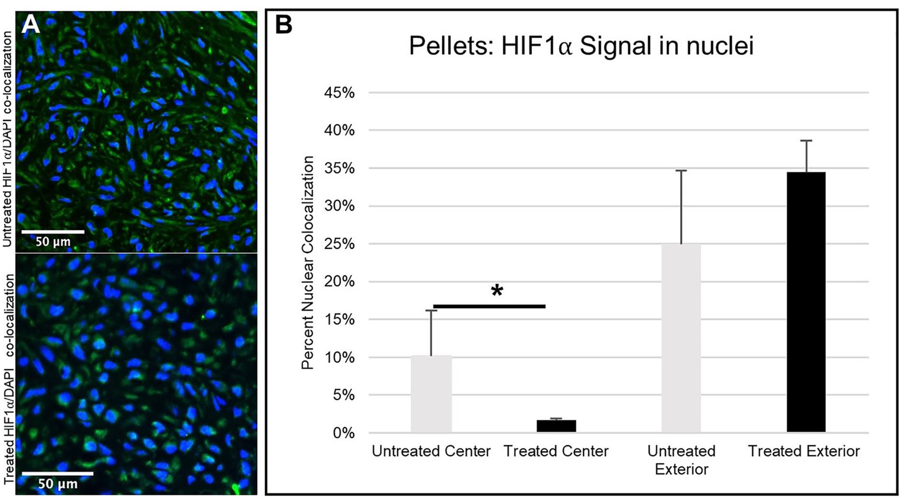
Immunofluorescence images of HIF-1α (green) co-localized with cell nuclei (blue) in (
Pellets: Qualitative Histology
The Alcian blue and picrosirius red stains demonstrated proteoglycan and collagen deposition, respectively, throughout the pellets for both the untreated and DMOG-treated pellets (
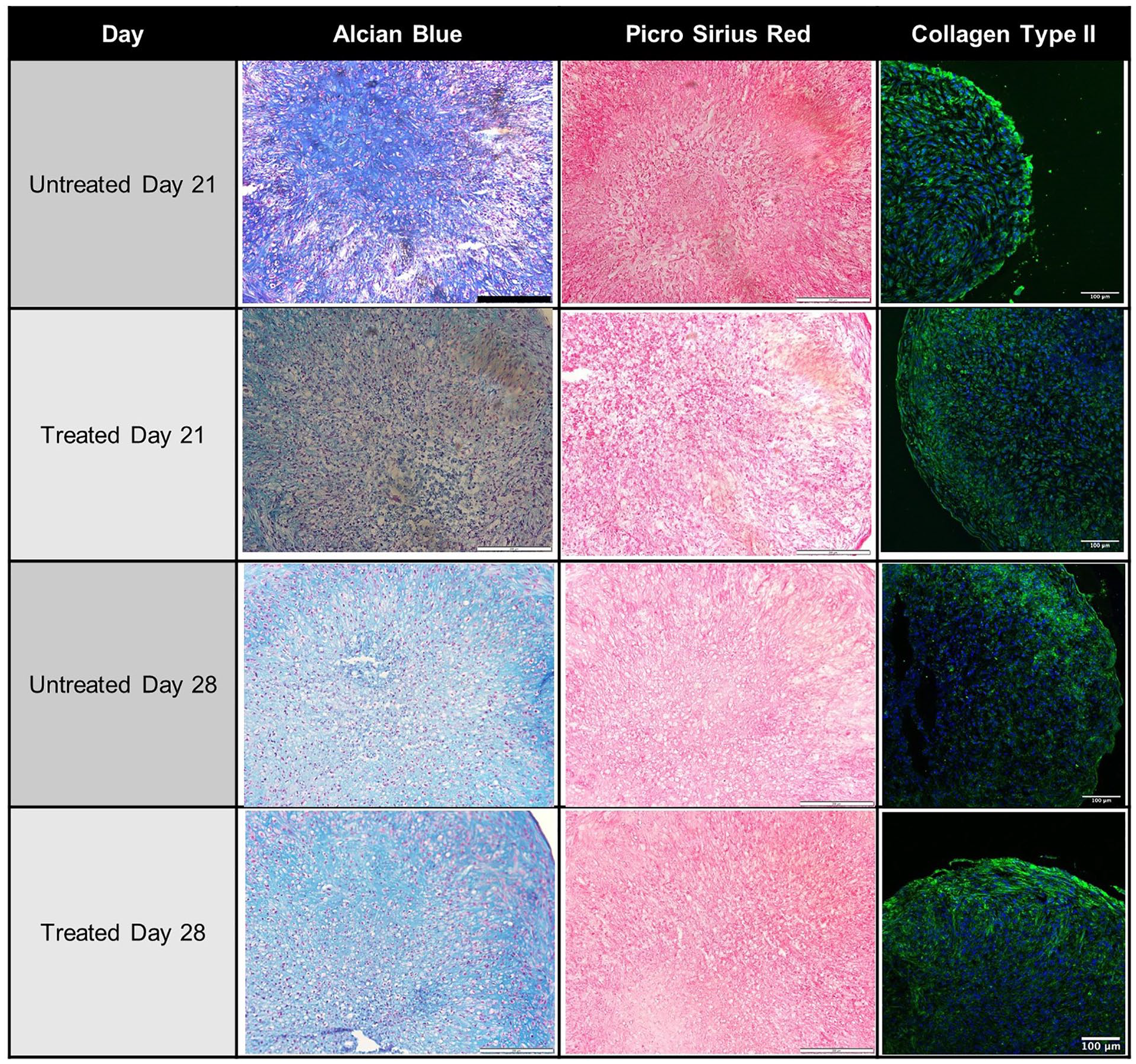
Representative histological images of each pellet culture group. Alcian blue was used to visualize deposited proteoglycans and picrosirius red to stain collagen. Scale bar for Alcian blue and picrosirius red images = 200 µm. Histological images shown are supplemented with corresponding images of immunofluorescent staining for type II collagen (green), with cell nuclei (blue) stained with DAPI. Scale bar for immunofluorescence images = 100 µm.
Pellets: Spatial Distribution and Quantification of Proteins
Spectral imaging was used to assess the spatial distribution of proteoglycan and collagen in pellets treated with or without DMOG. The spectral imaging method allows not only for the visual spatial distribution of proteins, but also the relative quantification of each protein. Proteoglycan was found distributed throughout the tissues (
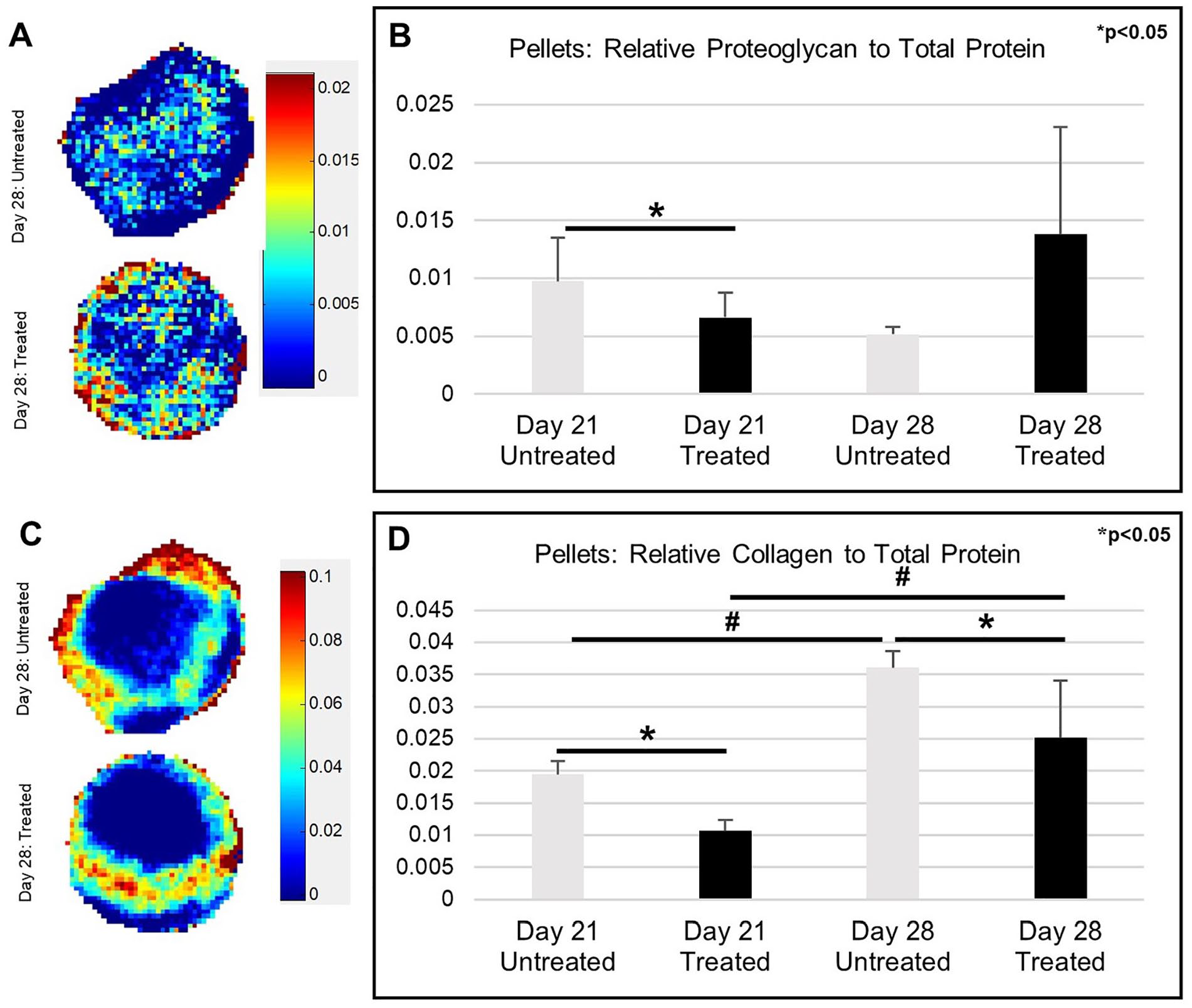
(
Pellets: Bulk Quantification of Collagen by ATR
The ATR technique was used to quantify differences in collagen content in the pellets, in lieu of biochemistry, which was not sensitive enough for the small amounts of tissue analyzed. The collagen peak in the untreated day 28 sample is both visibly clear (
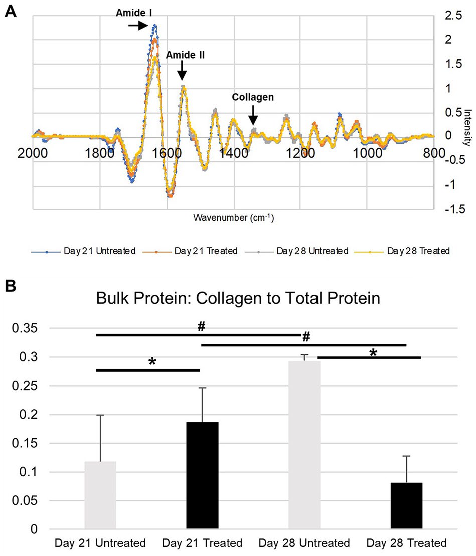
(
Pellets: Biochemical Assessment
Pellets were assessed for differences in proteoglycan content by the sGAG DMMB assay. DMOG did not impact bulk proteoglycan deposition at day 21 or day 28 when compared to the untreated groups (
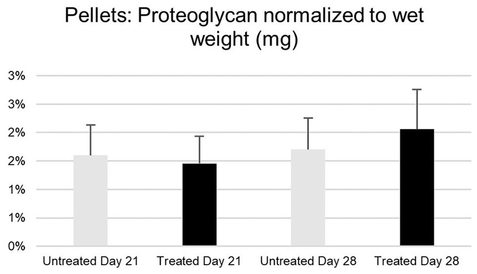
Quantification of bulk proteoglycan in cartilage pellets for DMOG-treated and untreated groups at days 21 and 28 (using the DMMB assay). No statistical differences among groups.
TECs: Nuclear HIF-1α Co-Localization and Signal Quantification
The co-localization of HIF-1α in the nuclei in Week 3 tissue engineered cartilage (
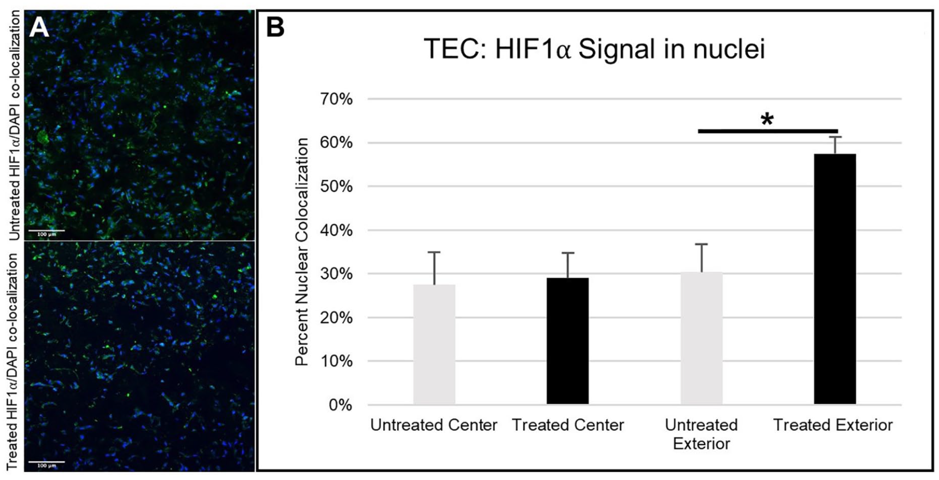
Immunofluorescence images of HIF-1α (green) co-localized with cell nuclei (blue) in (
TECs: Qualitative Histology
Tissue sections stained with Alcian blue (
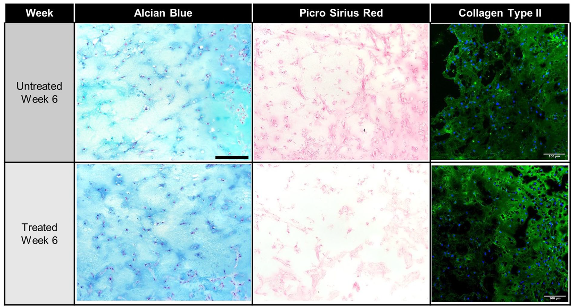
Representative histological images of the engineered cartilage construct groups at week 6. Alcian blue was used to visualize deposited proteoglycans and picrosirius red to stain collagen. Scale bar for Alcian blue and picrosirius red images = 200 µm. Histological images shown are supplemented with corresponding images of immunofluorescent staining for type II collagen (green), with cell nuclei (blue) stained with DAPI. Arrows indicate edge of construct. Scale bar for immunofluorescence images = 100 µm.
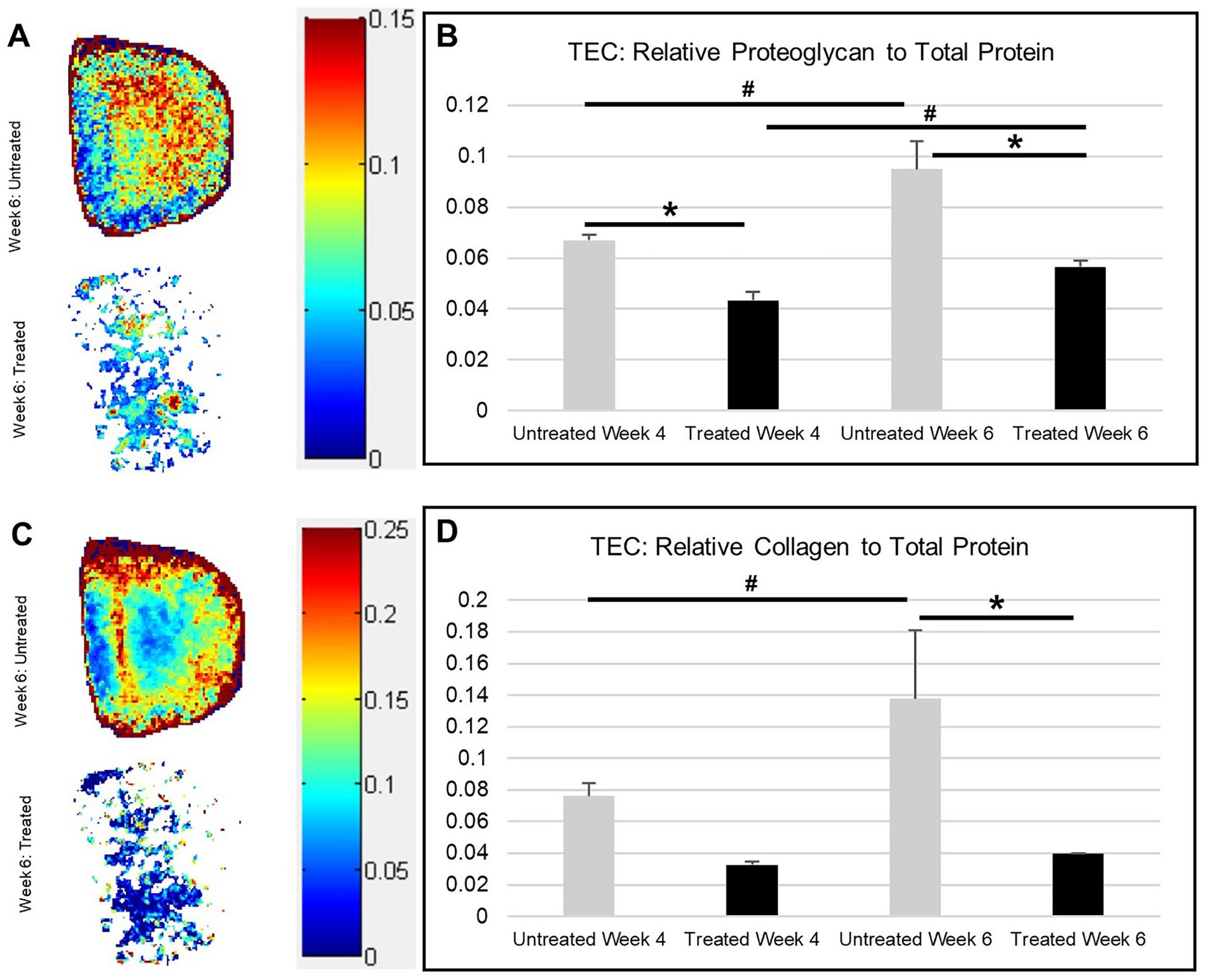
TECs: Spatial Distribution and Quantification of Proteins
Spectral imaging allowed for the evaluation of relative amounts and spatial distribution of protein within the DMOG-treated and untreated constructs (
(
TECs: Biochemical Assessment
Consistent with the spatial distribution analysis and quantification of proteins by spectral imaging, the biochemical results of proteoglycan (
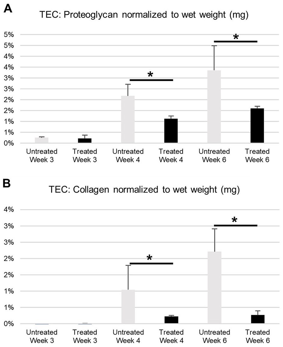
(
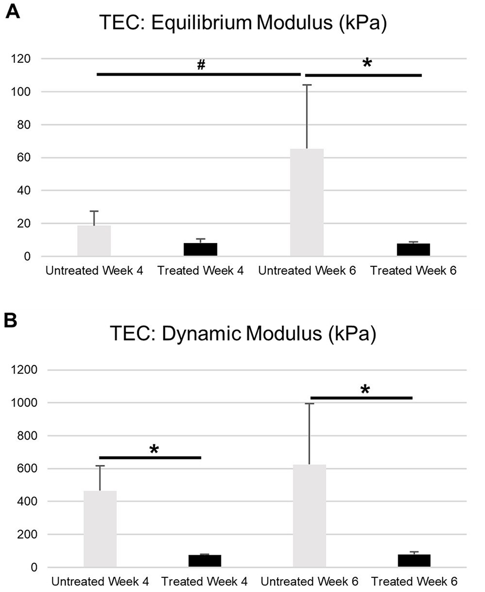
TECs: Mechanical Assessment
Deposited proteoglycan and collagen play a critical role in the compressive properties of cartilage. Thus, the statistical decrease in both equilibrium modulus (
(
Discussion
The motivation behind this study was to investigate the impact of a hypoxia simulating agent, DMOG, on the differentiation on SYN-MSCs in scaffold-free and scaffold-based tissue engineered cartilage. This study, which represents the first DMOG treatment for cartilage tissue engineering, found that DMOG had a negative effect on the cartilaginous extracellular matrix deposition despite an increase in HIF-1α stabilization in regions of the tissues.
Cartilage tissue was engineered using a co-culture system comprised of chondrocytes and synovial-derived mesenchymal stem cells. SYN-MSCs were chosen because of their in situ role as tissue-specific stem cells for chondrogenesis. 13 Recreating the native in vivo microenvironment in vitro can be advantageous for the expansion to yield functional SYN-MSCs, and several studies have demonstrated the benefit of MSC expansion in low oxygen tension.14,15 Furthermore, in situ oxygen tension presents as a gradient with a decrease in tension from the articular cartilage surface to the subchondral bone. SYN-MSCs do not experience the exact same oxygen tension as chondrocytes although the microenvironment in the synovium remains hypoxic. 16 This study investigated whether stabilizing HIF-1α in vitro using a chemical agent could be an alternative to hypoxia chambers by targeting the transcription factor degraded by oxygen exposure.
Dimethyloxalylglycine, cobalt chloride (CoCl2), and deferoxamine (DFO) have been shown to augment chondrogenic differentiation by mimicking hypoxic conditions required for chondrocyte growth. Cobalt chloride does so by replacing iron in prolyl hydroxylase 2 (PHD2), acting as a chelating agent to prevent the degradation of HIF-1α, a necessary transcription activator that is upregulated in hypoxia to promote ECM growth. Cobalt chloride has been shown to increase chondrogenic and osteogenic potential, as well as inhibit adipogenesis. 17 When 0.1 mM CoCl2, a concentration empirically determined to limit toxicity to cells, was added to Murine MSCs (C3H/10T1/2) for 24 or 48 hours before growth in chondrogenic medium, HIF-1α mRNA was significantly increased, and accompanied by a significant increase in aggrecan and type II collagen (Col2a1) at 48 hours of exposure, and a slight increase in Sox9 at 24 hours of exposure.
Deferoxamine (DFO or desferrioxamine) is a chelating agent initially designed to remove excess iron in the bloodstream and has been shown to have additional biological applications by synergizing with the action of TGF-β to induce chondrogenesis. 18 The mechanism of action of DFO is related to the sequestration of iron, which is necessary for the action factor inhibiting HIF (FIH) and PHD2, thereby amplifying the concentration of HIF-1α within the cell and simulating the effects of hypoxia. 7 With the addition of DFO and TGF-β to chondrocytes, Col2a1, Sox9, and aggrecan expression, as well as cell proliferation, were significantly increased, compared to the null control, DFO alone, and TGF-β alone. 18
A previous study using PC12 cells (rat adrenal phaeochromocytoma) grown in monolayer treated with DMOG at a concentration of 1 mM for 2 hours showed HIF stabilization by Western blot analysis. 19 The current study is the first to assess the effect of DMOG by consideration of spatial localization, and investigation of HIF-1α stabilization co-localized with nuclei. Co-localization with nuclei was an important step in this evaluation, as HIF-1α needs to be translocated into the nucleus to initiate transcription of target genes. Furthermore, co-localization within specific regions was representative of a spatially stabilized molecule, thereby confirming that hypoxia was achieved throughout the tissue and not only at the naturally hypoxic core. The exterior of the tissue engineered construct is not typically hypoxic, as is the construct center, and therefore the presence of HIF-1α in the exterior was enhanced by DMOG treatment. The results, a demonstration of increased HIF-1α stabilization in the exterior of tissue engineered cartilage constructs treated with DMOG, unfortunately did not translate to positive changes in the cartilaginous matrix. Accordingly, at the time points used in this study, DMOG likely did not improve chondrogenic differentiation in SYN-MSCs co-cultured with chondrocytes in pellets or in tissue engineered cartilage and therefore was not a suitable alternative to traditional hypoxia chambers.
The reduced cartilaginous extracellular matrix found in DMOG-treated pellets and treated TEC are indicative of a negative impact of DMOG. These results were not consistent with previous studies where low oxygen settings, such as chambers, were used, to enhance chondrogenesis.15,20,21 The extracellular matrix attenuation in the DMOG treated tissue engineered cartilage were subsequently reflected in the mechanical properties of the tissue, which is the ultimate determination of tissue function.
Previous research indicated that DMOG is effective at inducing HIF-1α nuclear localization and regulation of gene targets in 2D monolayers of human bone marrow mesenchymal stem cells. However, this research also showed that DMOG inhibits extracellular matrix (ECM) protein incorporation, ultimately yielding significantly reduced cartilage-like ECM with less collagen type II and glycosaminoglycans (GAGs) unless exposed only during later stages of chondrogenesis. 7 Here, samples were treated with DMOG from day 7 to day 14 of culture as previously described for the upregulation of chondrogenic genes. 7 Further exploration into dosing options may be a potential strategy for the use of DMOG for chondrogenesis.
Although here ECM deposition was impaired with DMOG treatment, Taheem et al. also found that DMOG lowers the extracellular deposition of unwanted collagen type X, reducing chondrocyte hypertrophy. 7 While genes involved in stem cell chondrogenesis were upregulated transcriptionally with DMOG exposure, extracellular matrix collagen type II and GAG content were lower than the control group. 7 Thus, results from this study were consistent with Taheem et al. in the truncated deposition of ECM proteins. Another interesting study by Jahangir et al. explored the angiogenic-osteogenic effects of using DMOG-containing 3D scaffolds. 22 Cell-seeded groups were embedded with adipose derived mesenchymal stem cells, and controls were implanted without embedded cells (cell-free). Treatment of DMOG produced a scaffold structure with rougher and thicker pore walls, as well as overall reduced porosity. Additionally, genes involved in osteogenesis and angiogenesis were transcriptionally regulated by the presence of DMOG, as measured by qualitative reverse transcriptase polymerase chain reaction. 22
One explanation for the results of the current study is that DMOG, which is a 2-oxoglutarate analog, can inhibit prolyl 4-hydroxylase, an enzyme involved in the synthesis of 4-hydroxyproline residues, which are crucial for stabilizing collagen triple helices in physiological conditions. 23 Although sustained exposure to DMOG may in fact inhibit incorporation of ECM proteins into hydrogel matrix, some hydrogel-embedded cultures have shown success using DMOG with short-term exposure. Sathy et al. found that research models that employ hydrogels containing DMOG, rather than sustained supplementation of the chemical in cell media, result in enhanced chondrogenesis with no long-term detriments to collagen production. 24 This can be attributed to 80% to 100% of the DMOG load being released from the hydrogel within 72 hours of encapsulation. While type II collagen deposition was reduced after 1 week of culture using this delivery mechanism, no long-term reductions were observed. 24 Thus, a novel strategy for DMOG incorporation could be useful to improve type II collagen deposition by chondrocytes, and the subsequent generation of functional cartilage.
Another potential path that may have affected DMOG efficacy was a possible competition with HIF-1α stabilization through natural hypoxia. It is well established that 3-dimensional cell culture generates a naturally occurring hypoxic core due to the lack of oxygen diffusion. 25 The HIF-1α co-localization study showed that HIF-1α was stabilized in both the treated and untreated groups, indicating that oxygen tension remained the same on the interior center and exterior of the pellets. In the tissue engineered constructs, which were larger than pellets at 2 mm thick and a circumference of 4 mm, DMOG did elevate HIF-1α stabilization in the exterior region. However, the interior center results indicated that the lower oxygen tension had already generated a hypoxic core regardless of DMOG supplementation. Untreated tissue engineered cartilage deposited greater proteoglycan and collagen than the DMOG-treated group, suggesting that the naturally occurring hypoxic environment was sufficient for robust cartilaginous output.
Furthermore, clinical use of tissue engineered cartilage embedded with DMOG must also take into consideration the potential side effect of DMOG diffusion to adjacent tissues. It is known, for example, that DMOG can act independently of the PHD/HIF pathway. Zhdanov et al. found that DMOG can inhibit cellular respiration in a concentration-dependent manner within minutes of exposure, significant at concentrations as low as 0.25 mM. 26 DMOG inhibition of cellular respiration halts the consumption of intracellular oxygen in hypoxic cell microenvironments, like those present in cartilage tissue, allowing for the reoxygenation of cells. This response persisted even after siRNA knockdown of HIF-1α and HIF-2α, demonstrating an alternative biochemical mechanism to that of HIF pathways. 26 Intracellular reoxygenation could disrupt the normal hypoxic physiology of chondrocytes and their progenitors and reduced cellular respiration could further alter cell functioning. Furthermore, research by Zhdanov et al. also demonstrated that prolonged cell exposure to DMOG resulted in mitochondrial structural changes beyond those induced by hypoxia, including increased size, altered chemical composition, and changes in proton gradients. 27 The long-term consequences of these changes remain under studied but are of clinical importance prior to adoption of DMOG-embedded hydrogels in implanted tissue engineered cartilage.
In conclusion, this study investigated the potential use of a hypoxia-simulating agent to improve the chondrogenic capacity of SYN-MSCs in engineered cartilage tissue by pellet formation and hydrogel-based scaffolds. The work herein demonstrated that sustained DMOG treatment is not a suitable alternative for traditional methods to control oxygen tension in vitro. However, the work opens the door to deeper investigations with DMOG, which could involve studies focused on varying concentrations and dosing time, or novel approaches for release to improve cartilage tissue engineering outcomes.
Footnotes
Acknowledgments and Funding
The author(s) disclosed receipt of the following financial support for the research, authorship, and/or publication of this article: Funded by NIH R01 AR056145.
Declaration of Conflicting Interests
The author(s) declared no potential conflicts of interest with respect to the research, authorship, and/or publication of this article.
Ethical Approval
Ethical approval was not sought for the present study because ethical approval was not required for this study.
Animal Welfare
Guidelines for humane animal treatment did not apply to the present study because no live animals were used to conduct the research described herein.