
Abstract
This study analyzed the morphological and biomechanical characteristics of perimeniscal capsule in knee joint thus establishing the roles of these tissues. A total of 10 human cadaver knees were used in this study. Medial meniscus and the adjacently surrounding joint capsules were harvested then sectioned both axially and coronally, followed by scanning electron microscopy analysis. The medial meniscus (anterior, middle, posterior) and the adjacent perimeniscal capsules (superior, peripheral) were biomechanically assessed to ascertain the tensile modulus. Among the perimeniscal capsules, the peripherally located capsules were morphologically different from the superiorly located capsules: The peripheral perimeniscal capsule was thicker and showed circumferentially oriented fibers whereas the superior perimeniscal capsule fibers were thinner and arranged in vertical orientation. The peripheral capsule also yielded significantly greater tensile modulus compared with the superior capsule biomechanically. We conclude that depending on its anatomical location, the perimeniscal capsule consists of fibers of varying orientations. This may be important in maintaining the circumferential hoop tension of the meniscus especially in the presence of circumferentially oriented and thick peripheral capsule fibers, which coincidentally have higher tensile modulus.
Introduction
The meniscus is the primary stabilizing structure of the knee joint. Its geometry and microstructure contribute to the essential meniscal functions such as load transfer, shock absorption, and full conformity resulting in minimum contact pressure.1-6 The framework of predominantly circumferentially oriented collagen fibers is subject to compression forces. The meniscus resists radial displacement by developing circumferential hoop tension.2,3,7-10 These fibers are not only connected to fewer radially aligned fibers that help prevent meniscus rupture but also tightly integrated with the surrounding capsule.2,3,7-10 Thus, when subjected to axial loading, this integrational network of fibers transfers these forces not only to the meniscus body itself but also to the surrounding capsular structures. Since the surrounding capsule is also indirectly subjected to axial compression, its structure should be robust enough to sustain constant and repeated axial loads. To date, the microstructure and the role of the meniscus itself have already been well described2,3,7-10; however, the anatomical and functional aspects of the adjacent capsule that surrounds the meniscus wall has not been established. Dugas et al. 11 emphasized the importance of the meniscocapsular junction structure, in which meniscocapsular separation occurs in the presence of increased tibiofemoral contact pressure. 11 It is plausible that the tissue of meniscocapsular junction could be histologically and biomechanically different from that of the surrounding capsule, thus enabling it to withstand higher level of stress. It is only apt to assume that in order for appropriate amount of stresses to be tolerated by different tissues at various anatomical sites, such as the peripheries of the meniscus (meniscus capsule) and the upper and lower capsule (joint capsule), these tissues should possess distinct properties specific to their function. However, this notion has not been previously tested.
Thus, the aim of the present study is to analyze the morphological and biomechanical characteristics of the perimeniscal capsule and evaluate its functional role in knee joint; specifically focusing on their anatomical arrangements and ability to cope with biomechanical stresses.
Materials and Methods
Tissue Specimen Collection
A total of 7 human cadavers (14 knees, mean age 80.7 years, age range 54-92 years) were used in this study. Of the 14 knees, those with histories of knee trauma, knee surgery, cartilage degeneration (grade IV) and meniscus tear were excluded, and the remaining 10 knees were selected for this study. Medial meniscus and the adjacently surrounding joint capsules were surgically isolated from cadavers. Using a sterile scalpel, the meniscus, meniscus capsule, and joint capsule were harvested from anterior, middle, and posterior aspects of each knee. The joint capsule was obtained 2 cm above the meniscus body. Each specimen was then sectioned in radial (coronal) and axial orientations ( Fig. 1 ). The coronal cross-sections allowed end-on imaging of the primary collagen network, whilst the axial cross-sections allowed imaging along the main axis of these fibers.
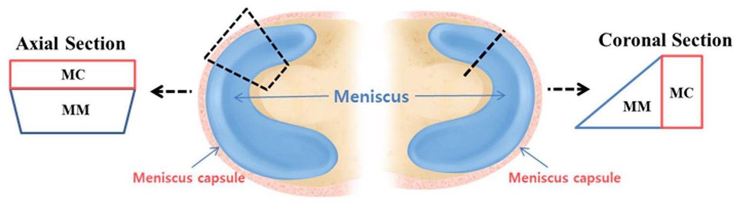
A schematic illustration showing the meniscus and perimeniscus capsule of interest in coronal and axial section. MM, medial meniscus; MC, meniscus capsule.
Histology of Constructs
The specimens were rinsed in distilled water for 1 to 2 days and were then dehydrated, and later embedded in paraffin wax. The paraffin blocks were sectioned from the meniscus capsule with a sliding microtome (RM2255, LEICA, Nussloch, Germany) in the thickness of 7 μm. For histological evaluation, sections were deparaffinized, rehydrated through a series of graded ethanol, and later stained. Axial sections were stained with Safranin-O for sulfated proteoglycans in the matrix as well as with Picro Sirius Red for collagens. The slides were first stained by Mayer’s hematoxylin and then stained using 0.1% Sirius red solution for 1 hour. After the stained slides were differentiated with 50% acetic acid, they underwent dehydration procedures with 95% and 100% alcohol. Finally, the samples were rinsed with xylene and mounted for image analysis.
Scanning Electron Microscopy
Medial capsules were studied under the scanning electron microscope in order to analyze the texture of the collagen fibrils. The samples were subdivided into anterior, middle, and posterior segments by uniform radial (coronal) and axial sections to allow us to permit topographic classification. The segments from different layers and regions of the meniscus capsule segments were kept in 10% NaOH solution at room temperature over a period of 5 to 6 days. Subsequently, the specimens were rinsed in distilled water for 1 to 2 days, and then they were saturated in 1% tannic acid for 4 to 5 hours. After rinsing the specimens in distilled water for 24 hours, they were counter-fixed in 1% OsO4 solution. The specimens were dehydrated in a series of graded concentrations of ethanol and then freeze-cracked with a razor blade in liquid nitrogen and finally critical-point dried using liquid CO2. The specimens were sputtered with gold and studied under a field emission scanning electron microscope (JSM-6700F, JEOL, Japan).
Mechanical Testing
Mechanical characterization of tensile properties of the specimens were determined on an H5KT (Tinius Olsen, Horsham, PA, USA) testing frame equipped with a 5000 N load cell. Each meniscus capsule, joint capsule and meniscus of the anterior, middle, and posterior segments were tested accordingly. Each specimen was clamped in a material testing machine such that the axis of tension was perpendicular to the simulated tear. After preloading to 0.1 N, the testing was performed at a grip-to-grip distance of 5 mm and a strain rate of 1 mm/min. An offset strain of 0.2% in the stress-strain curves was used to determine the yield stress.
Statistical Analysis
All quantitative data sets are expressed as mean ± standard deviation. GraphPad Prism 6 (GraphPad, San Diego, CA, USA) was used to produce graphic images. One-way analysis of variance using Tukey multiple comparison and unpaired t tests were performed to assess whether there were statistically significant differences in the results of different data sets, with a value of P < 0.05 being considered significantly different.
Results
Histological Analysis of Meniscus and Capsules
In Safranin-O staining, meniscus showed abundant glycosaminoglycan contents, whereas both meniscus capsule and joint capsule revealed no evident glycosaminoglycan staining. ( Fig. 2A ). In comparison between meniscus capsule and joint capsule, meniscus capsule has shown significantly distinct collagen fiber orientation from joint capsule in Picro Sirius Red staining ( Fig. 2B ).
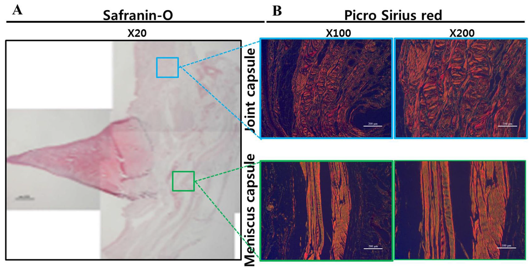
Histological analysis of medial meniscus and capsules (meniscus capsule and joint capsule) in Safranin-O staining (
Morphological Analysis by Scanning Electron Microscopy
When comparing the axial sections of meniscus capsule and joint capsule, the peripheral meniscus capsule showed multiple thick and rosary-shaped collagen fibers in circumferential orientation; ( Fig. 3A , C , and E ), whereas the upper joint capsule revealed thinner collagen fibers arrayed in vertical orientation ( Fig. 3B , D , and F ). When the diameter of the collagen fiber was measured and compared, no significant difference was noted in its diameter according to the anatomic position in both meniscus capsule and joint capsule ( Fig. 4 ).
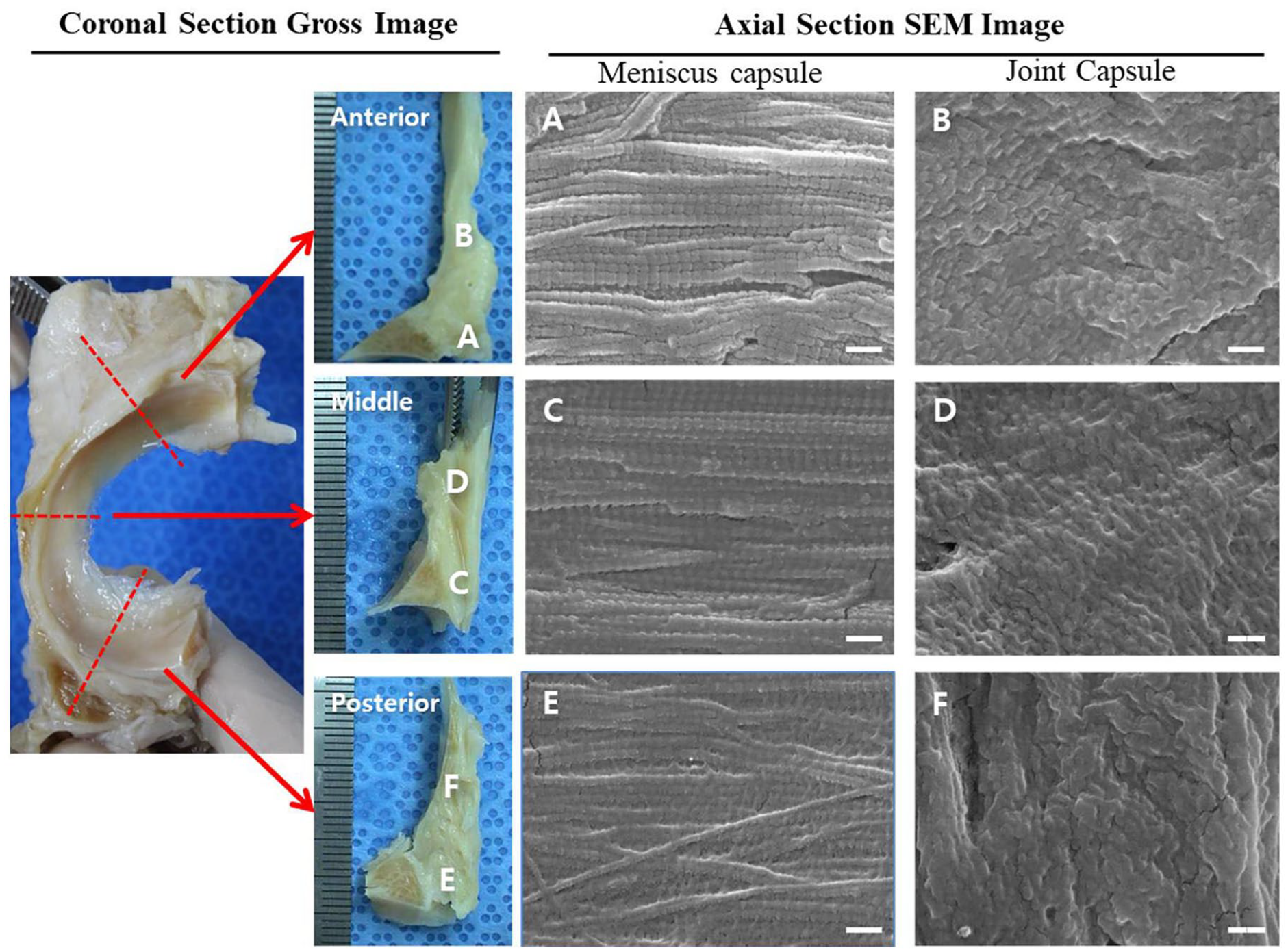
Morphological analysis of meniscus capsule and joint capsule by scanning electron microscope (×60,000, scale bar 200 nm). (
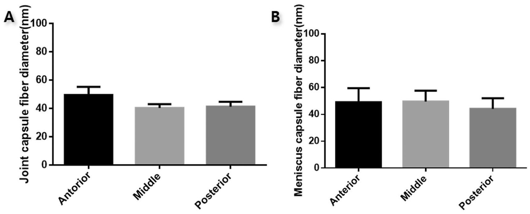
Comparison of the mean collagen fiber diameter according to the anatomical position in meniscus capsule and joint capsule. (
In morphological comparison between medial meniscus and meniscus capsule, both medial meniscus and meniscus capsule showed nearly identical collagen fiber orientation in coronal section indicating morphological similarity between the 2 entities ( Fig. 5 ).
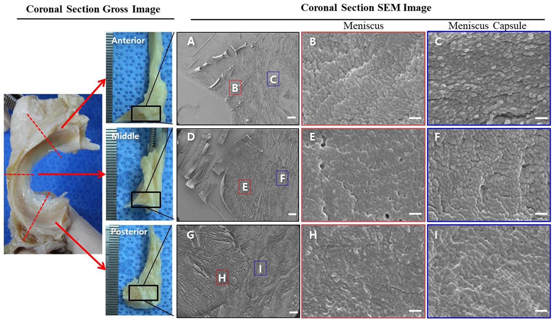
Morphological comparison of medial meniscus and corresponding meniscus capsule using scanning electron microscope. (
Biomechanical Analysis
Circumferential tensile modulus was measured and compared among the meniscus, meniscus capsule, and joint capsule. Meniscus capsule showed significantly greater circumferential tensile modulus than joint capsule in the anterior, middle, and posterior regions (15.20 ± 5.57 vs. 3.27 ± 1.29 kPa in anterior, P < 0.01; 19.80 ± 7.19 vs. 1.58 ± 0.79 kPa in middle, P < 0.001; 21.55 ± 8.80 vs. 5.49 ± 2.54 kPa in posterior, P < 0.05) Similarly, meniscus revealed significantly greater circumferential tensile modulus than meniscus capsule in the anterior, middle, and posterior regions (177.22 ± 77.35 vs. 15.20 ± 5.57 kPa in anterior, P < 0.0001; 229.92 ± 87.53 vs. 19.80 ± 7.19 kPa in middle, P < 0.0001; 109.53 ± 39.90 vs. 21.55 ± 8.80 kPa in posterior, P < 0.0001). Overall, meniscus capsule has shown about 4.2% to 30.0% circumferential tensile modulus of the corresponding adjacent meniscus ( Fig. 6 ).
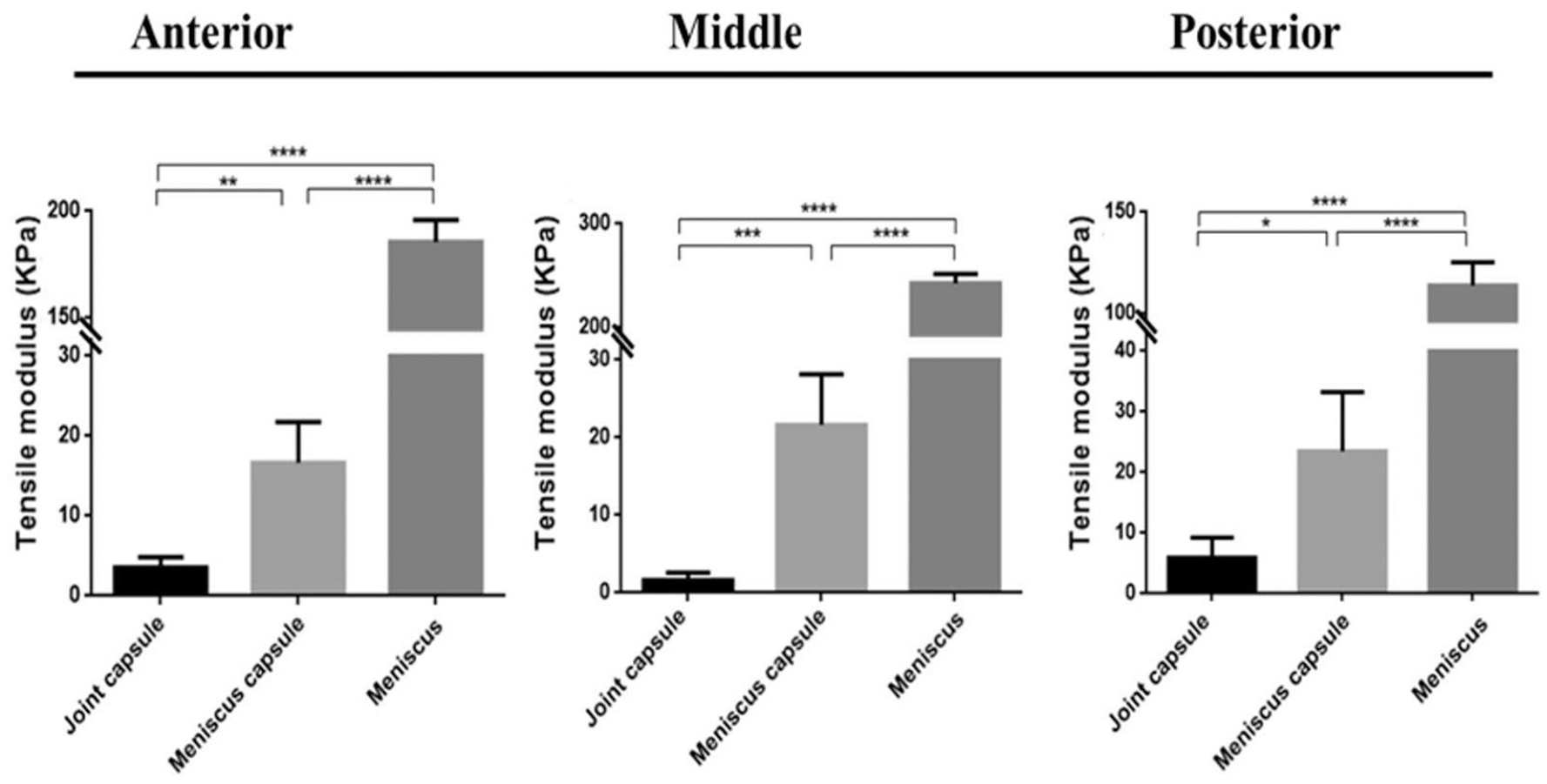
Comparison between the circumferential tensile modulus of the meniscus, meniscus capsule, and joint capsule in anterior, middle, and posterior regions. *P < 0.05, **P < 0.01, ***P < 0.001, ****P < 0.0001.
Discussion
The present study demonstrates that the perimeniscal capsule contains fibers of varying orientations based on its anatomical position. The peripheral perimeniscal capsule was thicker and showed circumferentially oriented fibers whereas the upper perimeniscal capsule fibers were thinner and arranged in vertical orientation. The distinct fiber orientation of the perimeniscal capsule might be due to the need to adapt to the different kinds of stresses subjected at the different anatomical sites. Since the meniscus capsule is subjected to radial hoop stresses, the morphological changes have been induced remodeling to circumferential orientation to effectively sustain the hoop stresses. In the same manner, the joint capsule is subjected to vertical tensile force and thus vertically oriented to efficiently resist the vertical tensile force.
Meniscus extrusion is frequently observed in patients with meniscus root tear or meniscal transplantation and is frequently accompanied with knee osteoarthritis.12-17 Because meniscus extrusion decreases meniscal coverage of the tibial plateau and poorly distributes load bearing in knee joint, meniscal extrusion is one of the strongest predictors for the progression of osteoarthritis and should be given clinical significance.17,18 Even with the great deal of technology in repair techniques, meniscus root repair does not reduce the meniscus extrusion and does not prevent the progression of arthrosis completely. 19 In other words, restoration of the meniscal continuity itself does not reduce the meniscal extrusion perfectly. In fact, meniscus extrusion is also positively correlated with the meniscal degeneration even though there are no definite loss in its continuity. 13 Thus, some other structures besides the meniscus itself, most likely the adjacent capsule, may play an additive role in resisting the hoop tension and preventing meniscal extrusion. In this study, the meniscus capsule, which circumferentially covers the meniscus, was observed to sustain the hoop stress in part by showing about 4.2% to 30.0% circumferential tensile modulus of the corresponding adjacent meniscus. Also, the meniscus capsule was thick and circumferentially oriented showing morphological similarity with the meniscus itself that presumed to effectively sustain the hoop stress. Based on our findings, it seems plausible that in treating the cases with meniscus extrusion with compromised hoop tension, the perimeniscal capsule as well as the meniscus should be considered as a complex to maintain the hoop tension and restore the normal knee biomechanics.
Whether perimeniscal capsule is made by nature or acquired by repetitive axial weight is unknown from this study. Wolff’s law states that, when continuous and repetitive stimuli are applied, the morphological structure is adapted and remodeled in response to the loads placed on it.20,21 Thus, when the meniscus is subject to repetitive axial stress in a knee joint, the peripherally adjacent surrounding capsule that wraps the meniscus should be adapted and remodeled to sustain the radial hoop stress. At the same time, since the radial outward stress may stretch the joint capsule in vertical direction, the upper and lower adjacent capsule should be adapted and remodeled in response to the vertical tensile force ( Fig. 7 ). Distinct collagen fiber orientation of the perimeniscal capsule according to its anatomical position may be presumed to be an acquired change because of a variable biomechanical environment in this study.
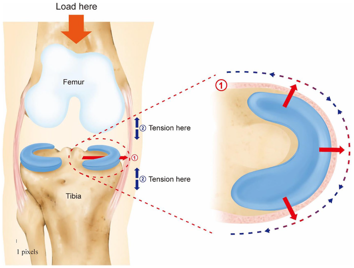
Schematic diagram demonstrating meniscus Wolff’s law. When the meniscus is subject to repetitive axial stress in a knee joint, the peripherally adjacent surrounding capsule that wraps the meniscus should be adapted and remodeled to sustain the radial hoop stress (small red arrow). Meanwhile, since the radial outward stress may stretch the joint capsule in vertical direction, the upper and lower adjacent capsule should be able to adapt and remodel in response to the vertical tensile force (small blue arrow).
Limitations should be addressed regarding this study. First, although the morphological structure of the meniscus and the peri-meniscus capsule was analyzed based on macroscopic dissection of the meniscus and investigated from scanning electron microscopy in this study, our findings do not present the native anatomy of the structures thoroughly. More enhanced imaging techniques, such as 3-dimensional microscopy, 8 may aid in more precise morphological evaluation of the perimeniscal capsule. Second, a rather small number of cadavers with old ages were used for this study. Yet, in the perspective that all the specimens resulted in homogenous characteristics in the morphology and similar trends in the biomechanical analysis, the number of the cadaver specimens seemed to be enough to reach our conclusion.
Conclusions
The perimeniscal capsule tends to have distinct fiber orientation according to its anatomical position. The peripheral perimeniscal capsule is thicker, circumferentially oriented, and shows greater tensile modules whereas the upper perimeniscal capsule fibers are thinner and vertically oriented.
Footnotes
Acknowledgments and Funding
The author(s) disclosed receipt of the following financial support for the research, authorship, and/or publication of this article: This work was supported by the Ministry of Education of the Republic of Korea and the National Research Foundation of Korea (NRF-2018R1C1B6008883).
Declaration of Conflicting Interests
The author(s) declared no potential conflicts of interest with respect to the research, authorship, and/or publication of this article.