
Abstract
Objective
Abnormal joint movement is associated with osteoarthritis (OA). Previous studies using the controlling abnormal joint movement (CAJM) model of OA reported delayed cartilage degeneration; however, none of them focused on gait performance and the localization of matrix metalloproteinase 13 (MMP13) and tissue inhibitor of metalloproteinase-1 (TIMP-1) in chondrocytes. Therefore, we aimed to investigate the effect of controlling abnormal joint movement on gait performance and the localization of MMP13 and TIMP-1, using kinematic and histological analyses.
Design
Rats were assigned to 2 groups: anterior cruciate ligament transection (ACL-T) group and CAJM group (n = 5/group); contralateral hind limbs of ACL-T rats were designated as intact. After 1, 2, and 4 weeks, step length was analyzed, and after 2, 4, and 8 weeks, Safranin O-Fast Green staining and immunohistochemical staining for MMP13 and TIMP-1 were performed.
Results
Step length did not differ significantly between the groups. However, degeneration of articular cartilage was higher in the ACL-T group than in the intact group (P < 0.05). There was no significant difference in the CAJM group at all time points. Immunohistochemical analysis of the MMP13/TIMP-1 relationship revealed a significant increase in the expression ratio of MMP13 after 4 weeks in the ACL-T group compared to the CAJM group (P < 0.05).
Conclusions
Controlling abnormal joint movement may reduce mechanical stress owing to kinematic elements of small articulation including joint instability and delayed cartilage degeneration, despite the lack of kinematic change in step length.
Keywords
Introduction
A symptom of osteoarthritis (OA), a common joint disease, is gait disturbance. OA is characterized by repair process failure in damaged cartilage due to mechanical and biochemical alterations.1-3 Abnormal mechanical stress on articular cartilage is the most important factor in knee OA,4-8 and is caused by various factors (e.g., ligament and meniscus injury), which induce joint instability and abnormal loading.4,9 Additionally, the load on cartilage during gait is a significant factor for the development of OA.5,6,10 This abnormal mechanical stress is also known as a cue for biochemical changes.11-14 Increased expression of the cartilage catabolic factors matrix metalloproteinase (MMP) and tissue inhibitor of metalloproteinase-1 (TIMP-1) induces a cascade of events leading to articular cartilage degradation as a biochemical change induced by abnormal mechanical stress.8,11,13,15-17 Among MMPs found in degenerated cartilage, MMP13 is a critical target gene for osteoarthritis and an important collagenase.8,11,15 Decreased expression of TIMP-1 also induces a cascade of events leading to the articular cartilage degradation.13,16,17 Thus, the relationship between mechanical and biochemical alterations is closely involved in cartilage degeneration.
A rodent model of anterior cruciate ligament transection (ACL-T) induces OA through abnormal tibia anterior translation.8,18 As this model establishment involves a surgical procedure, it was unclear whether OA was induced mechanically or biochemically. A rodent model of controlled abnormal joint movement (CAJM) may solve this problem.19-21 This model enables control of abnormal tibia anterior translation from outside the joint capsule after performing the same operation as in the ACL-T model. The important attribute of the CAJM model is that it is “controlling” rather than “re-stabilizing,” and it aims to prevent the anterior translation and change the contact stress on the tibia. Comparison between the ACL-T and CAJM models may provide information critical for evaluation of the effects of mechanical factors, rather than biological factors, on cartilage. Murata et al.20,21 quantified, using the dissected knee joint, that the CAJM model decreased the tibia anterior withdrawal compared to the ACL-T model, and articular cartilage degeneration can be suppressed in the CAJM model.
However, the influence on the gait function in this model is unknown. Gait changes occur after severe cartilage degeneration22,23; however, the possibility of cartilage degeneration cannot be eliminated. Among many kinematic parameters, gait speed is generally evaluated for knee OA 24 and is related to most biomechanical parameters. 25 An important factor affecting gait speed is step length, 25 and in patients with knee OA, reduction in gait speed and step length has been reported. 26 Additionally, MMP13 and TIMP-1 expression in this model has not been investigated. Histological analysis was performed to observe the effects on cartilage tissue by analyzing the degree of cartilage damage and localization of MMP13 and TIMP-1. We aimed to determine whether the difference in mechanical stress caused by ACL transection and controlling abnormal joint movement affects gait step length performance and cartilage degeneration using rodent ACL-T and CAJM models.
Methods
Animals and Experimental Design
The study was approved by the Animal Research Committee of Saitama Prefectural University (Approval Number: 27-7). Thirty 12-week-old Wistar male rats (Clea Japan, Tokyo, Japan) were used. The experimental design is presented in Figure 1 . Animals were randomly classified into 2 groups, the ACL-T group and the CAJM group; the contralateral hind limbs of the ACL-T group were harvested and defined as intact.
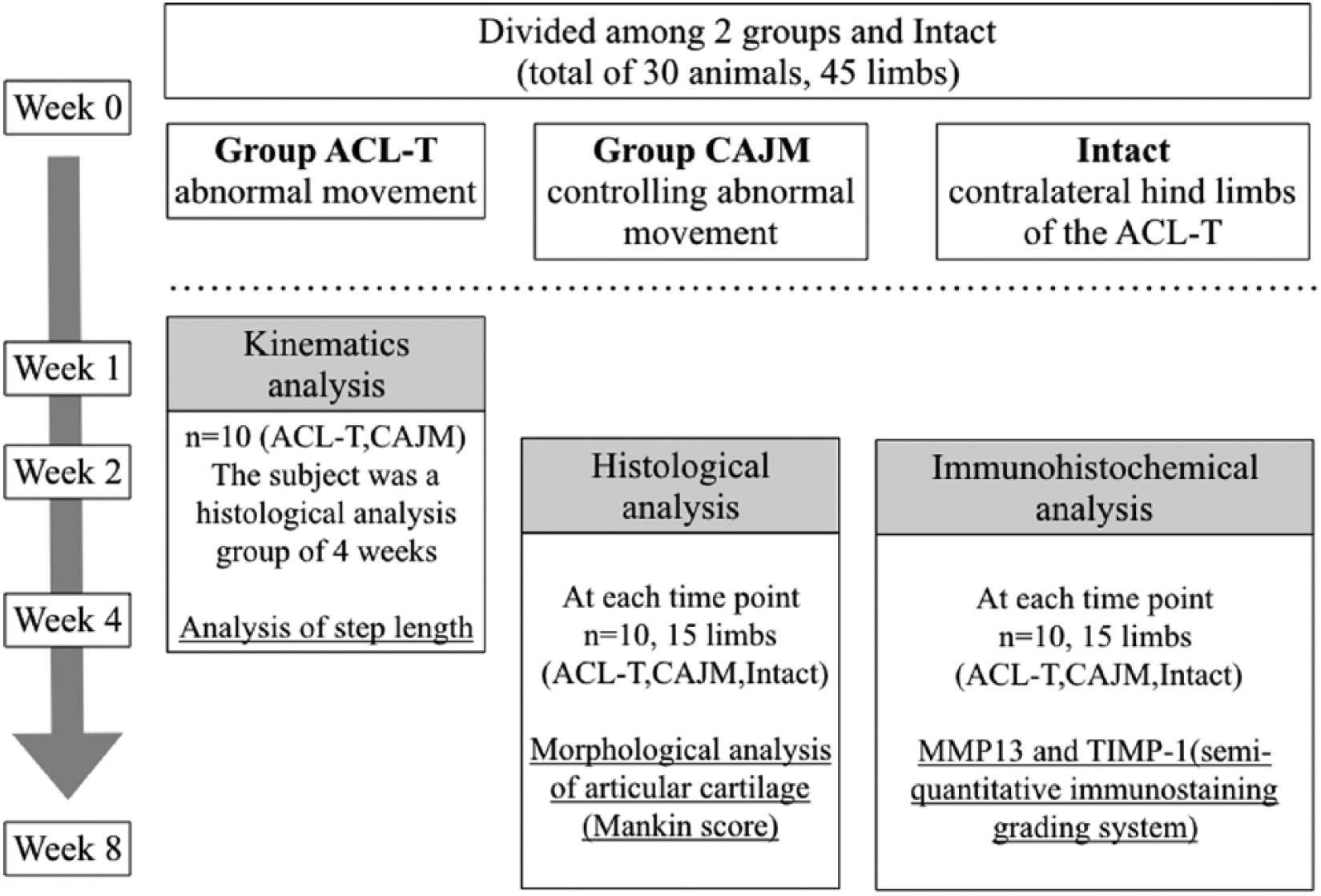
Experimental design. Histological and Immunohistochemical analyses were performed at 2 weeks, 4 weeks, and 8 weeks. These analyses involved the ACL-T group and CAJM group (for each group n = 5; total of 30 animals; 45 limbs including constralateral hind limbs of the ACL-T group). Kinematic analysis was performed at 1 week, 2 weeks, and 4 weeks. This analysis involved the ACL-T group and CAJM group (for each group n = 5; total of 10 animals).
All rats were housed in plastic cages (2 animals/cage) under room temperature (23°C) and a 12-hour light-dark cycle. Before and after surgery, rats were permitted unrestricted movement within the cage and had free access to solid feed and water.
Surgical Procedures
Surgical operations were performed on the right hind limbs in accordance with our previous study.19 -21 Animals were subjected to anesthesia using somnopentyl (0.9 mL/kg), and for analgesia, a reference amount of 1 mL/kg repetan was injected subcutaneously. In the ACL-T group, the right knee joint was incised longitudinally, the medial capsule of the right knee joint was exposed, and the ACL was completely transected at the medial intercondylar eminence of the tibia.
In the CAJM group, after the ACL transection, a bone hole was created at the tibial tuberosity in the mediolateral direction using a rotary drill. Furthermore, the anterior translation of the tibia was controlled by applying a 3-0 nylon thread through the hole and securing the tibia to the posterior aspect of the distal femur. The nylon thread, therefore, provided a posteriorly directed traction force on the tibia to resist anterior motion over the femoral condyles. In both groups, abnormal tibia anterior movement was manually assessed during and after surgery to confirm tibia movement. The joint capsule and the skin were sutured and closed. During the procedure, care was taken to avoid injury to the articular cartilage. Anterior joint instability of the tibia only differed between the ACL-T and CAJM groups.
Kinematics Analysis
Ten rats (ACL-T n = 5, CAJM n = 5) were used for histological analysis at 4 weeks. The ratio of the step length of the contralateral limb in the gait distance (sum of the step length on both sides) in a gait cycle was calculated as follows:
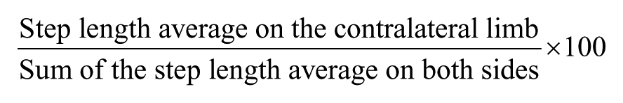
The effect of individual differences reported in previous studies was also considered in the present study,22,23 and the average number of stable steps of 2 to 3 gait cycles was analyzed.
At 1, 2, and 4 weeks after surgery, the rats were made to walk on a path consisting of a clear acrylic board ( Fig. 2 ), which was captured from the bottom, using an iPad Mini 2 tablet terminal (Apple Japan, Tokyo, Japan). The gait path was made to taper using styrene foam, considering that rats prefer dark and narrow places. The video recording was obtained as an output in the form of a frame-divided image every 0.3 seconds. Thereafter, using ImageJ software (Developer Wayne Rasband at the National Institutes of Health), the actual distance was corrected and the step length was calculated using the Pythagorean theorem on the basis of the distance in the x-axis (horizontal direction) and y-axis (longitudinal direction).
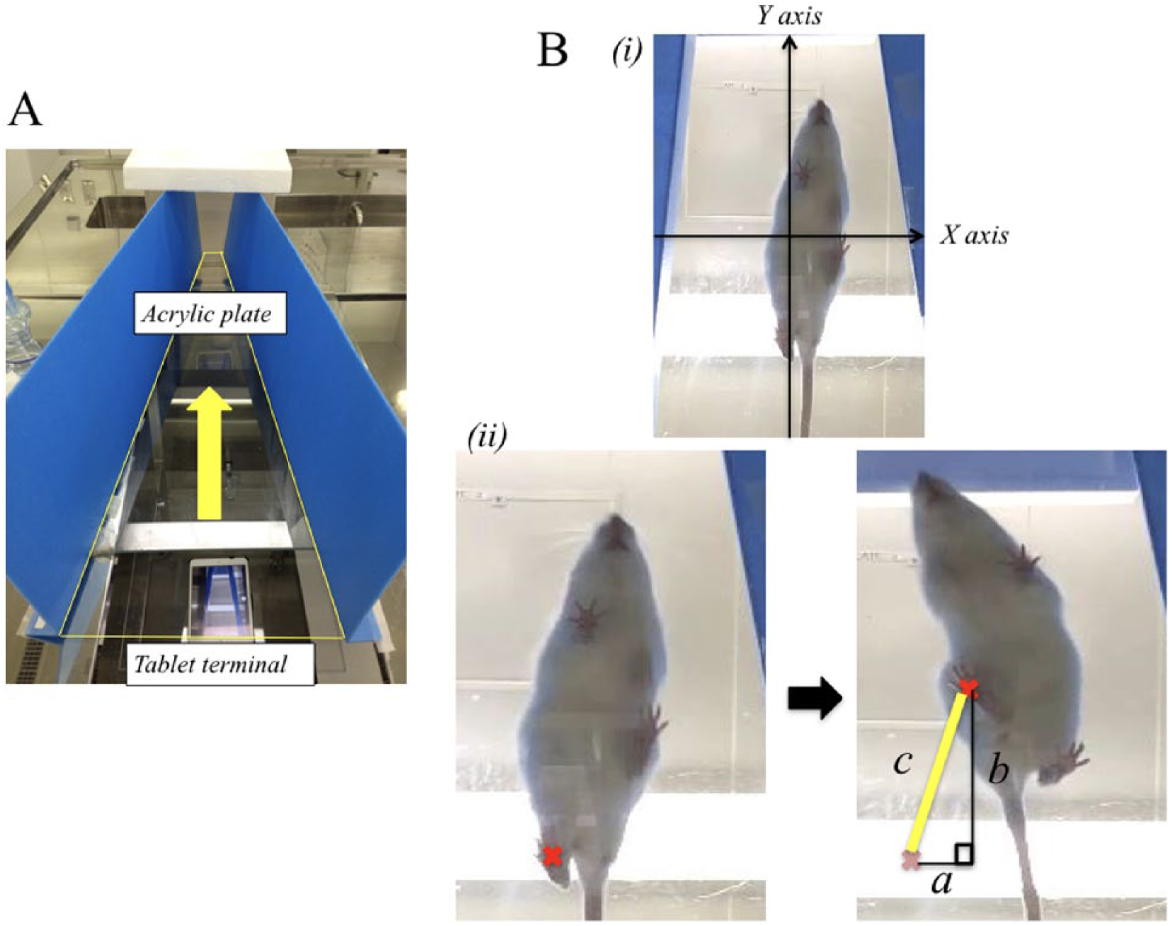
Kinematic analysis. (A) Picture of experimental enviroment. A tablet terminal was placed under a transparent acrylic plate, and as rats prefer narrow places, gait paths were made narrower. The gait of each rat was recorded from the bottom using the tablet terminal. Yellow arrows indicate the walking direction of the rats. (B) (i) Image of gait recorded from below. The horizontal direction corresponds to the x-axis and the longitudinal direction corresponds to the y-axis. (ii) Example of step length analysis. The distance of movement from the cross mark in the left panel to the cross mark in the right panel was considered to be the step length. Step length was calculated from the moving distance of the x-axis and the moving distance of the y-axis using the Pythagorean theorem.
Histological Analysis
Both groups of rats (n = 30, each group n = 5) were euthanized at 2, 4, and 8 weeks, and the knee joints (including contralateral hind limbs of the ACL-T group as the intact control) were collected. Knee joints were fixed in a 4% paraformaldehyde/phosphate-buffered saline (PBS) solution for 48 hours at 4°C and placed in a 10% EDTA-based solution at pH 7.4 for decalcification for approximately 90 days. After complete decalcification, the knee joint was immersed in sucrose solutions of different concentrations (10% for 4 hours, 15% for 4 hours, and 20% for 12 hours) and embedded in optimal cutting temperature compound (O.C.T. Sakura Finetek Japan, Tokyo, Japan). Sections of the knee articular cartilage were cut along the sagittal plane at 14-µm thickness, using a Leica CM 3050 S cryostat (Leica Microsystems AG, Wetzlar, Germany).
All slides were stained with Safranin O and Fast Green to evaluate the Mankin score. 27 The Mankin score was evaluated as the sum of points for structure (0-6 points), cells (0-3 points), Safranin O staining (0-4 points), and tidemark integrity (0-1 point), as a semiquantitative evaluation scale that grades normal cartilage (0 point) to severe degeneration (14 points). Tibia articular surfaces were assessed and sections with randomly assigned numbers were examined using a light microscope under 40× magnification. Sample scores were evaluated by 2 independent observers (KM and SF).
Immunohistochemical Analysis
Immunohistochemical localization of MMP13 and TIMP-1 was determined using sections of the tibia articular surfaces. Sections were air-dried for 30 minutes and washed 3 times for 5 minutes with PBS. Endogenous peroxidase was inactivated by incubating the sections in a 0.3% H2O2/ethanol solution for 30 minutes. After blocking with 0.1% bovine serum albumin/PBS, sections were incubated overnight at 4°C with a rabbit polyclonal anti-matrix metallopeptidase 13 (MMP13) antibody (dilution 1:250; Bioss, Woburn, MA) or anti-tissue inhibitor of metalloproteinase-1 (TIMP-1) antibody (dilution 1:250; Bioss, Woburn, MA). A streptavidin-biotin-peroxidase complex was then formed at room temperature (approximately 23°C), using an ABC kit (Vector Laboratories, Burlingame, CA). Sections for immunohistochemical analysis were stained using Dako Liquid DAB+ Substrate Chromogen System (Dako, Glostrup, Denmark). Furthermore, cell nuclei were stained using hematoxylin and observed with a Leica DM 250 optical microscope (Leica Microsystems AG, Wetzlar, Germany).
For the analysis, the percentage of positive cells and intensity of immunostaining for MMP13 and TIMP-1 were assessed using a semiquantitative immunostaining grading system. 28 The percentage of positive cells was evaluated as follows: 0, no visible staining; 1, <5% of cells and/or matrix positive; 2, 6% to 24% of cells and/or matrix positive; 3, 25% to 49% of cells and/or matrix positive; 4, 50% to 75% of cells and/or matrix positive; 5, >75% of cells and/or matrix positive. The intensity of immunostaining was evaluated as follows: 0, no visible staining; 1, minimal staining; 2, moderate staining; 3, marked staining. These scores were summed and evaluated using ranges from 0 to 8, with higher scores indicating more advanced cartilage degeneration. Sections with randomly assigned numbers were examined through light microscopy under 40× magnification. Sample scores were evaluated by 2 independent observers (KM and SF).
Statistical Analysis
All analyses were performed using JMP pro version 12.1 for Windows (SAS Institute Japan, Tokyo, Japan). The normality of the data distribution was assessed using the Shapiro-Wilk test of normality. Because all of the data were nonparametric, intergroup differences were evaluated using the Kruskal-Wallis test, with the Steel-Dwass test used for post hoc analysis. P < 0.05 was considered statistically significant.
Results
Kinematic Analysis
The ratio of the step length of the contralateral limb to the total step length in a gait cycle is shown in Figure 3 . The ratio is shown with the median and interquartile range. At 1 week, ACL-T group: 50.1 (47.6-50.2), CAJM group: 45.9 (43.8-49); at 2 weeks, ACL-T group: 49.3 (45.6-53.0), CAJM group: 45.2 (41.9-50.1); at 4 weeks, ACL-T group: 47.3 (46.8-49.1), CAJM group: 56.2 (47.5-56.7). Although the ACL-T group tended to show a temporally increased step length, there were no significant differences at any time point when both groups were compared.
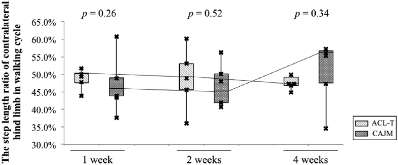
Analysis of the ratio of step length of the contralateral limb in one walking cycle. the median step length of the ACL-T group and CAJM group at each time point are shown. When the ratio was 50% or more, the ration of the step length of the contralateral limb was high, and when the ratio was 50% or less, the ratio of the step length of the operated limb was high. Crossmarks indicate individual data.
Histological Analysis
The results of Safranin O-Fast Green staining are described in
Figure 4A
. At all the time points, no obvious cartilage degeneration was observed at Intact compared with other groups, whereas in the ACL-T group, chondrocytes became enlarged and clustered. Loss of Safranin O staining and surface irregularities were observed at 4 weeks (
Fig. 4A
,
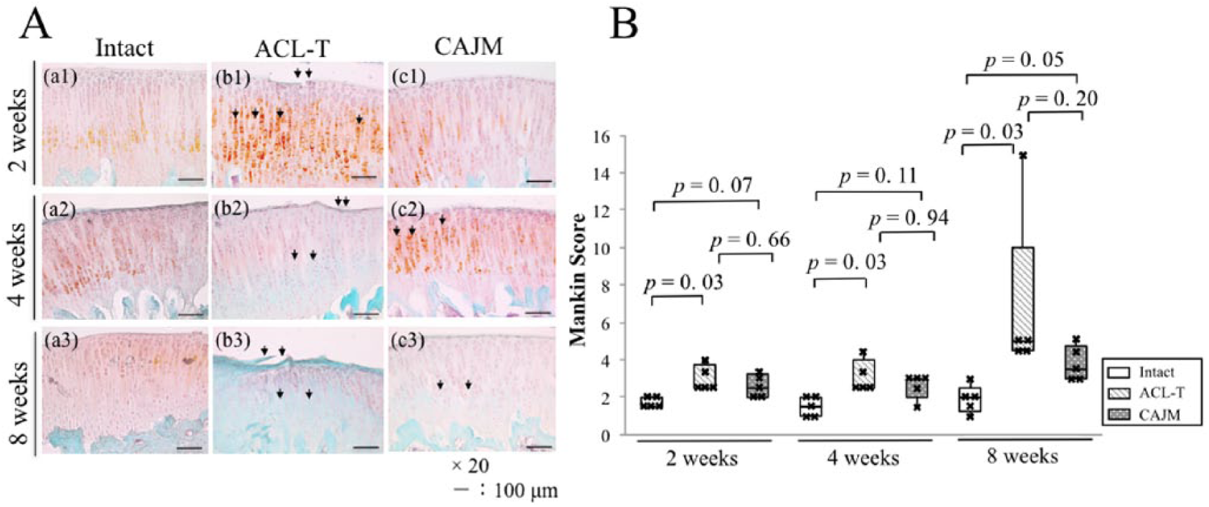
(A) Representative Safranin O–Fast Green staining images from each group (Intact, ACL-T group, CAJM group) at 2 weeks, 4 weeks, and 8 weeks. The front of the tibia was analyzed. Intact (a1–a3), ACL-T group (b1–b3), and CAJM group (c1–c3). Black arrow indicates the degenerated site of cartilage. (B) Mankin scores for each group at 2 weeks, 4 weeks, and 8 weeks (Intact, ACL-T group, CAJM group). Cross marks indicate individual data.
The results for the Mankin score ( Fig. 4B ) are described below; median and interquartile range are shown. At 2 weeks, the scores were as follows: intact group: 1.5 (1.5-2), ACL-T group: 2.5 (2.5-3.75), CAJM group: 2.5 (2-3.25); at 4 weeks: intact group: 1.5 (1-2), ACL-T group: 2.5 (2.5-4), CAJM group: 3 (2-3); at 8 weeks: intact group: 2 (1.25-2.5), ACL-T group: 5 (4.5-10), CAJM group: 3.5 (3-4.75). At all the time points, the ACL-T group showed a significant increase in Mankin score compared with the intact group (P < 0.05), whereas no significant difference was observed in the CAJM group compared with that in the intact group.
Immunohistochemical Analysis
The results for immunohistochemical staining of MMP13 and TIMP-1 are described in Figure 5 . Results of a semiquantitative immunostaining grading system are described below; median and interquartile range are shown ( Fig. 5B ). Both MMP13 and TIMP1 were expressed in chondrocytes from the surface to the deep layer of cartilage.
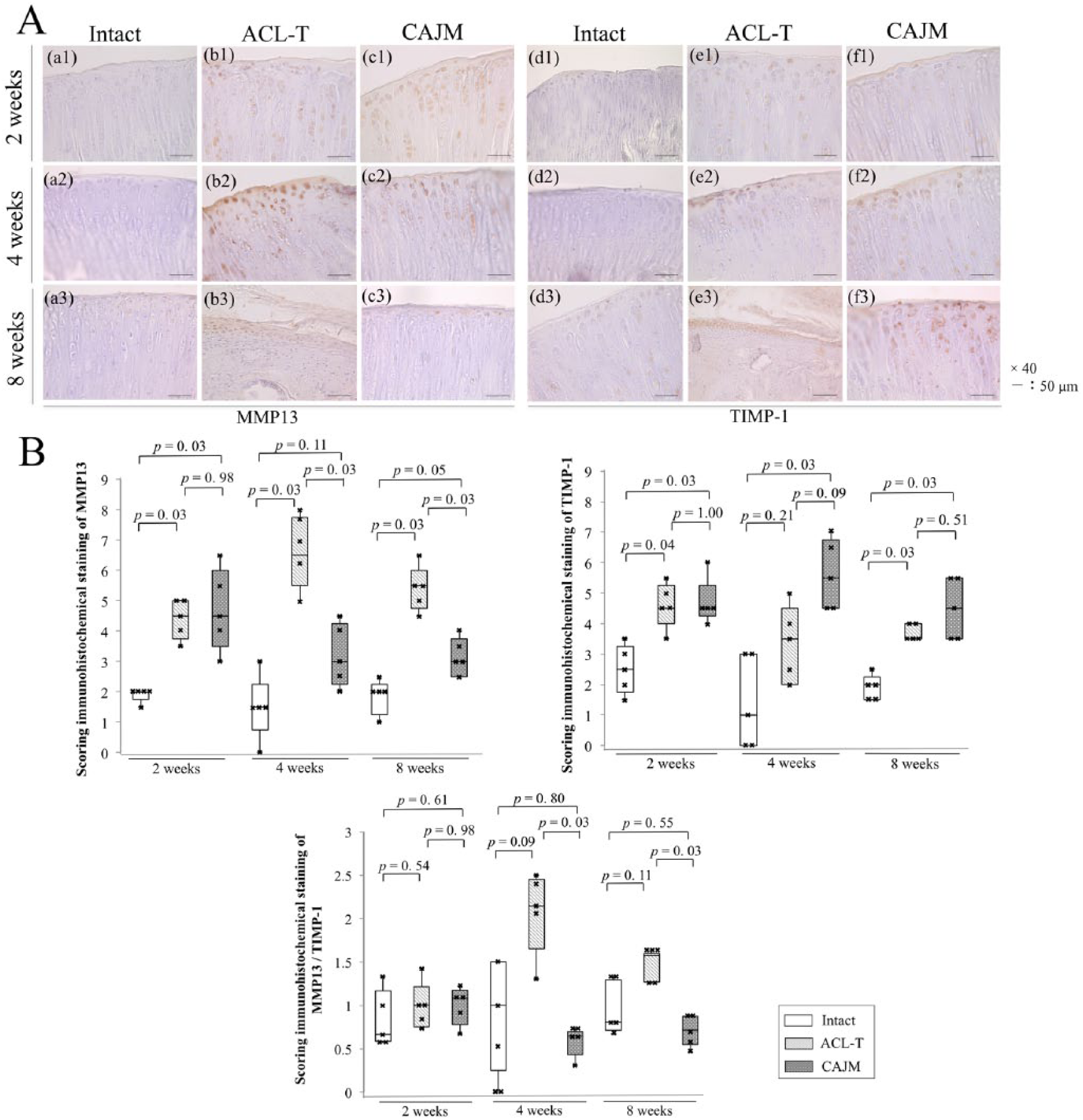
(A) Representative immunostaining images of MMP13 [Intact (a1–a3), ACL-T group (b1–b3), CAJM group (c1–c3)], and TIMP-1 [Intact (d1–d3), ACL-T group (e1–e3), CAJM group (f1–f3)] at 2 weeks, 4 weeks, and 8 weeks. The front of the tibia was analyzed. (B) Results of a semi-quantitative grading of immunostaining of each group at 2 weeks, 4 weeks, and 8 weeks (Intact, ACL-T group, and CAJM group). Scoring results for MMP13 (left panel), TIMP-1 (right panel), and the MMP13/TIMP-1 relationship (lower panel). Cross marks indicate individual data.
For MMP13, immuno-positive cells were observed at all the time points in the intact group (
Fig. 5A
, a1-a3). In the ACL-T group, an increasing percentage of positive cells and a higher intensity of immunostaining were observed at 2 weeks (
Fig. 5A
,
TIMP-1-immunopositive cells were observed at all the time points in the intact group (
Fig. 5A
,
The immunohistochemical staining results for the MMP13/TIMP-1 relationship ( Fig. 5B ), at 2 weeks, intact group: 0.67 (0.59-1.17), ACL-T group: 1.0 (0.75-1.21), CAJM group: 1.08 (0.78-1.17); at 4 weeks: intact group: 1.0 (0.25-1.50), ACL-T group: 2.14 (1.65-2.45), CAJM group: 0.64 (0.43-0.70); at 8 weeks, intact group: 0.80 (0.71-1.29), ACL-T group: 1.57 (1.27-1.60), CAJM group: 0.71 (0.55-0.87). There was no significant difference at 2 weeks in all groups; however, in the ACL-T group, MMP13 was significantly upregulated after 4 weeks (P < 0.05).
Discussion
The commonly used ACL-T model of OA exhibits high knee instability that causes anterior translation of the tibia. 29 However, the influence of the difference in mechanical stress due to abnormal articulation control on gait and cartilage degeneration has not been clarified. Our previous study20,21 reported that the CAJM model significantly controlled abnormal tibia anterior movement compared with the ACL-T model, and suggested the involvement of joint instability and cartilage degeneration. In addition, it was also reported that IL-1β expression was not different in the CAJM group and the ACL-T group at 4 weeks postoperatively; however, TNF-α was downregulated. 20 No studies have evaluated the effect of abnormal joint movement on MMP13 and TIMP-1 expression. Therefore, the present study investigated the effect of controlling abnormal knee instability on gait performance and the localization of MMP13 and TIMP-1 in chondrocytes through kinematic and histological analyses.
Here, kinematic analysis indicated that step length was not significantly different between the ACL-T and CAJM groups until 4 weeks. Previous studies reported that gait disorder develops after 12 weeks, suggesting that gait change occurs after severe cartilage degeneration. 22 Similarly, in previous studies involving humans, in comparison with healthy subjects, there was no difference in step length in mild OA patients 26 ; however, step length decreased in severe OA patients. 30 Generally, because the ACL-T model is a mild model among rodent surgical knee OA models that induce chronic OA, 8 change in step length was not considered to have occurred during this study period. In contrast, in the rat medial meniscus tear model, step length change was observed after 2 weeks, but cartilage degeneration was not observed for 6 weeks. 31 There is also a report that cartilage degeneration after ACL injury is most related to changes in load on cartilage caused by gait kinematic changes.5,6,10 Based on these findings, the reason for no difference in kinematic data in this study might be that early OA degeneration started before any change in a parameter, such as step length, that could be visualized. The early OA mechanism from the kinematic aspect cannot be elucidated unless a detailed and fine evaluation of abnormal movement can be achieved.
In the histological analysis, we observed that articular cartilage degeneration progressed over time in the ACL-T group, but specifically, that according to the Mankin score at 8 weeks, the ACL-T group underwent extensive degeneration than the CAJM group. Therefore, the CAJM model showed a tendency to delay degeneration as observed in previous studies,20,21 and may have suppressed secondary cartilage degeneration after ACL injury. These results suggest that the differences in intra-articular condition, such as joint instability, influenced both models. Despite the absence of kinematic changes in gait, the kinematic elements of small intra-articular capsular movement may induce mechanical stress, possibly resulting in cartilage degeneration.
Importantly, this intra-articular change in condition may have affected MMP13 and TIMP-1 localization in chondrocytes. Immunohistochemical analysis of MMP13 revealed increasing positive-cell number and staining intensity in the ACL-T group until 8 weeks, whereas in the CAJM group, there were no significant differences after 4 weeks, compared with the intact control. TIMP-1 showed a strong immunopositive reaction, with a high positive-cell percentage in the CAJM group at all the time points. During cartilage degeneration, TIMP-1 binds to MMP13 at a 1:1 molar ratio and inhibits the enzymatic activity of MMP molecules. 17 Consequently, the expression ratio of MMP13 was significantly increased in the ACL-T group compared to the CAJM group after 4 weeks, although no significant difference was observed in all groups at 2 weeks. In both groups, a positive reaction was observed for MMP13 in the surface and deep layer of cartilage, and the TIMP-1 expression sites were similar. Kamekura et al. 8 reported that MMP13 was upregulated in chondrocytes from the surface layer to the deep layer of cartilage in the ACL-T model, similar to the present study. MMP13 and TIMP-1 localized in chondrocytes of the cartilage surface layer would be most sensitive to mechanical stress caused by differences in joint movement. The strong expression of MMP13 in the ACL-T group, and TIMP-1 in the CAJM group, was also observed in the deep layer, indicating the possibility of the difference in mechanical stress affecting future cartilage degeneration. Chaudhari et al. 5 reported that changes in cartilage mechanical stress occur before biological changes and cause subsequent biological changes. The present results indicate that mechanical stress due to abnormal joint movement affected MMP13 and TIMP-1 localization from the surface to the deep layer of the cartilage, resulting in progression of articular cartilage degradation.
The present study had the following limitations. First, we could not quantify the extent of abnormal joint movement control in the CAJM model. However, previously, CAJM showed significant improvement in the abnormal tibia anterior translation compared with ACL-T,20,21 and there was a marked difference in joint movement between both models. We intend to further investigate the changes in joint movement in future studies. Second, the kinematic analysis was insufficiently sensitive to capture the difference in gait. Additionally, a kinetic analysis may also be required to fully assess gait. Moreover, the step length for the contralateral hind limbs, a parameter measured in this study, was affected by the supportability of the operated hind limbs and other factors. In a previous study using the OA model, the load on the operated hind limb and the vertical floor reaction force were decreased.31,32 Integrating and interpreting the results of kinematic analysis and kinetic analysis could reveal novel factors associated with gait changes. We intend to investigate gait changes after 4 weeks in a subsequent study. Third, the pain level and amount of weight bearing during gait were not quantified. The mechanism underlying cartilage degeneration and its effects on gait is the primary cause of pain, which results from cartilage degeneration and inflammation. Because CAJM involves a large surgical invasion, it is possible that cartilage degeneration was delayed owing to pain and the decrease in weight bearing compared with ACL-T. However, there was no difference in step length between the 2 groups, and cartilage degeneration before gait change progressed was more prominent in the ACL-T group. Hence, we considered that the effect of loading amount and pain levels on the present results was small. In future, we intend to investigate pain and weight levels during gait. Fourth, the surgical method difference between the ACL-T model and the CAJM model may have influenced the expression of inflammatory cytokines and the downstream cascade affecting cartilage metabolism. However, the expression ratio of MMP13 was significantly increased in the ACL-T group compared to the CAJM group after 4 weeks, although there was no significant difference in all groups at 2 weeks. In both the CAJM group and the ACL-T group, the ACL was transected. It has been reported that acute joint trauma reaches the peak of inflammation in approximately 2 days, and catabolism decreases in approximately 10 days. 9 In addition, when comparing the ACL-T model with the bone tunnel model and CAJM model in a previous study, the anterior translation of the tibia was similar. 21 It was therefore not possible to demonstrate the effect of the difference in surgery between the ACL-T and CAJM model on the expression of inflammatory cytokines, and there was a high possibility that the kinematic difference affected the present results. Hence, we wish to evaluate the effects of inflammation in future studies in models that only open the bone hole.
Fifth, control rats were not used, and intact, that is, the contralateral hindlimb of the ACL-T group was used. In future studies, it is necessary to verify these effects using control rats. However, the important point in this study was the change over time in the difference between ACL-T model and CAJM model. Finally, only the localization of 2 significant knee OA molecules, MMP13 and TIMP-1, in chondrocytes was examined; however, because many other catabolic molecules are also involved, further studies are required to determine metabolism. Furthermore, effects on anabolism were not evaluated. Therefore, we could not determine whether the difference in joint movement eventually affected catabolism or anabolism. The present results only indicate that greater levels of MMP13 localize to the tibia articular cartilage in the ACL-T group than in the CAJM group. Future studies with a larger sample size and presenting additional data is necessary to investigate the balance between catabolic and anabolic factors of articular cartilage.
Conclusions
To clarify the effects of controlling abnormal joint movement on cartilage, kinematic and histological analyses were performed using the ACL-T and CAJM models. Although the kinematic parameters observed in step length did not change, controlling abnormal joint movement decreased MMP13 and TIMP-1 localization in chondrocytes from the surface layer to the deep layer of cartilage. This study revealed that the difference in tibia anterior movement may acts as a difference in mechanical stress, and affect cartilage degeneration. Future studies are required to further investigate details through kinetic analysis and other analyses.
Footnotes
Acknowledgments and Funding
Part of this study was supported by the Japan Physical Therapy Association (Approval Number: H27-B30).
Author’s Note
Katsuya Onitsuka is also affiliated to Graduate Course of Health and Social Services, Graduate School of Saitama Prefectural University, Saitama, Japan.
Declaration of Conflicting Interests
The author(s) declared no potential conflicts of interest with respect to the research, authorship, and/or publication of this article.
Ethical Approval
The study was approved by the Animal Research Committee of Saitama Prefectural University (Approval Number: 27-7).
Animal Welfare
The present study followed international, national, and/or institutional guidelines for humane animal treatment and complied with relevant legislation.