
Abstract
Objective
Curcumin monoglucuronide (TBP1901) is highly water soluble and can convert to free form curcumin, which has pharmacological effects, on intravenous administration. This study aimed to investigate the effectiveness of TBP1901 intra-articular injections in an osteoarthritis (OA) rat model.
Methods
Sixty-four male Wistar rats (12 weeks old) who underwent destabilized medial meniscus (DMM) surgery were randomly separated into the TBP1901 injection or saline solution (control) injection group. They were sacrificed at 1, 2, 6, or 10 weeks postoperatively (weeks 1, 2, 6, and 10; n = 8 for each group). TBP1901 (30 mg/mL) or saline solution of 50 μL was injected into the knee joints twice a week during weeks 1 and 2 to investigate the effects in the acute phase of posttraumatic (PT) OA or once a week during weeks 6 and 10 to investigate it in the chronic phase of PTOA. Histology, immunohistochemistry, and micro-computed tomography were performed to evaluate the changes in OA.
Results
TBP1901 injections significantly reduced synovial inflammation at weeks 1 and 2, and tumor necrosis factor-α expression in the articular cartilage at week 6. The TBP1901 injections also significantly suppressed articular cartilage damage, subchondral bone (SB) plate thickening, SB plate perforation, and osteophyte formation at week 10.
Conclusions
TBP1901 intra-articular injections suppressed synovial inflammation in the acute phase of PTOA in DMM rats. In the chronic phase, TBP1901 suppresses articular cartilage damage and regulates SB plate changes.
Introduction
Osteoarthritis (OA) is a progressive joint disease characterized by articular cartilage loss, subchondral bone (SB) alterations, and osteophyte formation. OA progression affects an individual’s activities of daily living. Inflammation plays a central role in OA pathogenesis. It contributes to chondrocyte apoptosis, extracellular matrix disruption, and cartilage degeneration through pro-inflammatory cytokines such as interleukin (IL)-1β, IL-6, and tumor necrosis factor-α (TNF-α).1-4
Curcumin, a polyphenol compound present in turmeric (Curcuma longa, long used as a spice and herbal medicine), has many positive health benefits, including anti-inflammatory, 5 antioxidative, 6 anticancer, 7 and antidepressant 8 effects. Oral curcumin ingestion for OA was shown to reduce pain and improve function in clinical studies.9-11 Animal studies have shown that curcumin inhibits OA progression by decreasing inflammation by blocking the Toll-like receptor/nuclear factor-κB (TLR4/NF-κB) signaling pathway and inhibiting chondrocyte apoptosis through enhanced autophagy.12-16 Curcumin has few side effects; therefore, its use is expected to be a potential adjunct to pharmaceutical therapies. On the other hand, orally ingested curcumin has low bioavailability because of limited absorption with low hydrophilicity and conjugation in the intestinal tract.17-20 Only a few ingested curcumin molecules are absorbed and present in the conjugated form (mainly curcumin monoglucuronide [CMG]) in the blood.21,22 To improve its low absorption, bio-optimization by liposomal delivery systems, chemical modification of curcumin, nanoparticulation of curcumin, and use of polysorbate as an emulsifier have been performed.23-26 However, the low absorbability and hydrophilicity remain, although their efficacy is greater than that of pure curcumin. To overcome the disadvantages of oral curcumin ingestion, intra-articular administration, an ideal drug delivery method commonly used in OA therapies, has been used to deliver a concentrated therapeutic dose of curcumin into the knee joint and has been shown to suppress articular cartilage degeneration and synovial inflammation in an OA rat model.24,27 We hypothesized that the intra-articular administration of CMG could overcome the issue of low hydrophilicity due to its high water solubility, could be absorbed into the deep layer of the cartilage and maximize its pharmacological effects. CMG exhibits 10-fold lower antioxidative activity than unconjugated, free form curcumin. 28 Therefore, CMG must be metabolized to free form curcumin in vivo to execute its antioxidative activity. 29 β-Glucuronidase is an important hydrolase for the deconjugation of CMG to free form curcumin. It is released from neutrophils, injured cells at sites of inflammation, and cells in bone.30-33 In addition, β-glucuronidase secretion is enhanced in the articular cartilage, synovium, and synovial fluid with inflammation and OA.34-37 Therefore, administered CMG would be deconjugated to free form curcumin in OA joint. To confirm our hypothesis, we utilized the synthesized CMG (TBP1901), which has already been investigated and demonstrated the efficacy of intravenous administration in tumor-bearing mice with colorectal cancer. 29 TBP1901, which has high water solubility, may be deconjugated to free form curcumin in OA joint and suitable for intra-articular administration. Although the efficacy of intra-articular administration of TBP1901 is unknown, the pharmaceutical value of curcumin will be enhanced if it is shown to have an anti-osteoarthritic effect in intra-articular administration.
To verify the efficacy of TBP1901, we chose rats with destabilized medial meniscus (DMM) as an OA animal model.38,39 Although posttraumatic (PT) OA models include anterior cruciate ligament transection, meniscus resection, combination with ligament transection and meniscectomy, and DMM model, 40 the DMM model was chosen to be the slower progress model and closer to human OA progression. 38 PTOA has an acute phase and a chronic phase for each period from trauma. 41 Although curcumin is effective for treating chronic inflammation, 42 it was recently reported as effective for treating acute inflammation. 43 Therefore, to comprehensively confirm the efficacy of TBP1901 in DMM rats, we observed its effects from the acute phase to the chronic phase of PTOA.
This study aimed to investigate the effectiveness of intra-articular administration of TBP1901 on inflammation and OA progression in a rat model with DMM.
Methods
Animal Preparation and Surgical Procedures
This study was conducted in accordance with the ARRIVE guidelines. 44 Sixty-four male Wistar rats (12 weeks old), purchased from SHIMIZU Laboratory Supplies Co. Ltd. (Kyoto, Japan), were placed in a plastic cage with paper bedding on a 12-hour light/dark cycle at a constant temperature. The rats could move freely in cages and had free access to food and water. The rats were randomly separated into TBP1901 and saline solution (control) intra-articular injection groups (n = 32 for each group). All rats underwent DMM surgery for the OA model.38,39 Briefly, DMM surgery was performed with an anteromedial capsule incision and transection of the medial meniscotibial ligament in the right knee under anesthesia with 1.0 mL/kg pentobarbital sodium (Somnopentyl; Kyoritsu Seiyaku Corp., Tokyo, Japan). The DMM rat model rapidly progresses to moderate OA with severe inflammation by 4 weeks postoperatively and to severe OA by 8 weeks postoperatively, by which time inflammation is milder.39,45,46 Therefore, the rats were sacrificed at 1 and 2 weeks postoperatively to confirm the effect of TBP1901 as the acute phase of PTOA and at 6 and 10 weeks postoperatively to confirm the effect as the chronic phase of PTOA (n = 8 for each).
The synthesis of TBP1901 was performed as described previously. 29 TBP1901 and saline intra-articular injections of 50 μL were administered to the right knee joints through the patellar tendon. The concentration of TBP1901 was chosen to be 30 mg/mL (1.5 mg/50 μL) based on a previous study, 29 which showed no abnormalities in mice administered with 125 mg/kg of TBP1901, and a preliminary study (Supplementary Fig. S1). The preliminary study showed no significant effects on TBP1901 injections at 10 mg/mL for 6 weeks in DMM rats. The rats sacrificed at 6 and 10 weeks postoperatively were injected once a week from 1 week postoperatively. All rats were injected an hour before sacrifice to confirm intra-articular curcumin by fluorescence observation. The rats sacrificed at 1 and 2 weeks postoperatively were injected twice a week since the observation periods were short. The rats sacrificed at 1 week were injected on days 3 and 7, and the rats sacrificed at 2 weeks were injected on days 3, 7, 10, and 14. One hour after the final intra-articular injection in each observation period, the rats were sacrificed by a lethal dose of pentobarbital sodium, and their right knee joints were harvested for micro-computed tomography (micro-CT), histochemical, and immunohistochemical analyses. Body weight was measured weekly for all rats.
Micro-CT Analysis
After fixation of the knees with 4% paraformaldehyde overnight, the tibiofemoral (TF) joints were scanned using a micro-CT system (SMX-100CT, Shimadzu, Kyoto, Japan) at 43 kV and 43 μA with a scan time of approximately 10 minutes. After scanning, the 3-dimensional (3D) reconstruction and assessment of the TF joint were performed using ImageJ (National Institutes of Health, Bethesda, MD, USA) and Amira (version 5.5, EFI Visualization Science Group, Burlington, MA, USA) software. The SB plate thickness, number of SB plate perforations (holes), diameter of SB plate perforations, bone volume (BV), total volume (TV), and osteophyte volume in the medial tibia SB (above epiphyseal plate) were calculated according to protocols described in previous studies.39,47 SB plate thickness was measured at the thickest region around the center of the medial articular surface. The diameter of the SB plate perforation was defined as the largest diameter of the largest perforation in each sample. To assess the bone density of the SB, bone volume was divided by the total volume (BV/TV).
Histological Analysis
After micro-CT, the knee joint samples were decalcified in 10% ethylenediaminetetraacetic acid (EDTA) for 3 to 4 weeks and cut along the mid-sagittal plane at the halfway point. The DMM model causes rapid OA progression in the TF joint and also affects the patellofemoral (PF) joint. 48 Therefore, the histological evaluation was performed on the TF and PF joints. After decalcification, the samples were paraffin-embedded and cut into 6-μm sections at 50-μm intervals. Fluorescence histochemistry was performed using sections with an epifluorescence microscope (MVX10, Olympus Corporation, Tokyo, Japan) to confirm the presence of curcumin in the articular cartilage and synovium. Curcumin is a fluorescent substance with an excitation wavelength of 300 to 550 nm (maximum excitation wavelength: 467 nm) and an emission wavelength of 548 to 600 nm (maximum emission wavelength: 571 nm). 49 Fluorescence detection is used for curcumin quantification. 50 Based on these reports, curcumin fluorescence was observed with the mirror unit of U-MYFPHQ/XL (Olympus Corporation, Tokyo, Japan). In addition, other paraffin sections were stained with hematoxylin and eosin (H&E) to evaluate inflammation of the synovium in knee joints and safranin O/fast green to evaluate the severity of cartilage degeneration. Inflammation was assessed using an inflammation scoring system described in a previous study. 51 Three membrane features (synovial lining cell layer, stroma cell density, and inflammatory infiltrate) were assessed in the whole knee joint as 0 (none), 1 (slight), 2 (moderate), or 3 (strong). The inflammation score was determined as the summed score for all parameters. The values of the parameters were summarized and interpreted as follows: 0-1, no synovitis; 2-4, low-grade synovitis; and 5-9, high-grade synovitis. Cartilage degeneration was assessed using the Osteoarthritis Research Society International (OARSI) score 52 and the modified Mankin (MM) score. 53 The OARSI score consists of 6 grades and 4 stages on a scale from 0 (intact) to 24 (severe damage). The MM score consists of 3 features (pericellular matrix staining, spatial arrangement of chondrocytes, and interterritorial matrix staining) on a scale from 0 (intact) to 8 (severe). Cartilage degeneration was evaluated in the medial TF and PF joints. The maximum score was used for all scoring systems and samples.
Immunohistochemical Analysis
Immunohistochemistry of type I collagen, type II collagen, IL-1β, IL-6, and TNF-α was performed to determine the principal collagen expression and inflammation in the articular cartilage. Antigen retrieval was performed for 20 minutes by heating with HistoVT One (Nacalai Tesque, Inc., Kyoto, Japan). Blocking was performed using 0.3% H2O2 for 15 minutes and 5% goat serum for 20 minutes at room temperature. The sections were then incubated at 4 °C overnight with primary antibodies against type I collagen (AB755P, diluted 1:1000; Merck KGaA, Darmstadt, Germany), type II collagen (diluted 1:100; Kyowa Pharma Chemical Co., Ltd., Toyama, Japan), IL-1β (ab9722, diluted 1:100; Abcam Co., Cambridge, UK), IL-6 (ab9324, diluted 1:250; Abcam Co., Cambridge, UK), and TNF-α (ab6671, diluted 1:100; Abcam Co., Cambridge, UK). Detection was performed using the streptavidin-biotin-peroxidase complex technique with an Elite ABC kit (Vector Laboratories, Burlingame, CA, USA). Immunoreactivity was visualized by incubation with a diaminobenzidine solution (Vector Laboratories, Burlingame, CA, USA) followed by counterstaining with hematoxylin. The primary antibody was omitted from negative control slides. The expression of type I and type II collagen in the medial tibia and patella cartilage was analyzed by measuring the minimum and mean pixel intensity values using TIFF images (magnification ×40) taken by microscopy and ImageJ, respectively, on a scale of 0 (minimum; dark) to 255 (maximum; no staining), according to a previous study. 54 The expression of IL-1β, IL-6, and TNF-α in the medial tibia and patella cartilage was analyzed by measuring the percentage of positive chondrocytes to detect the severity of cartilage inflammation within the middle region of the medial tibia with an anteroposterior width of 0.5 mm or the central region of the patella with a superoinferior width of 0.5 mm.
Statistical Analyses
The median and interquartile range (IQR) of the inflammation score, OARSI score, and MM score (nonparametric data) were calculated for each group. The Wilcoxon test was used to compare differences between the 2 groups. In addition, the mean and 95% confidence intervals (CIs) of body weight, immunohistochemical, and micro-CT analysis data (parametric data) were calculated, and Welch’s t test was used to compare differences between the groups. The normality of the data was assessed using normal quantile–quantile plots and the Shapiro-Wilk test. Statistical significance was set at P < 0.05. Statistical analyses were performed using JMP Pro 14 software (SAS Institute Inc., Cary, NC, USA).
Results
Body weight is shown in Supplementary Figure S2. Body weight in the TBP1901 group was significantly lower than that in the saline control group at week 1 (mean [95% CIs]: 276.59 g [272.65–280.54]; TBP1901, 282.31 g [278.40–286.23]; control, P = 0.040), but the difference was small. There were no significant differences between the groups at weeks 2, 6, and 10. No other adverse events were observed.
Histology
The fluorescence expression of curcumin was confirmed on the tibial articular cartilage and was found on the synovium in the TBP1901 group but not in the saline control group (
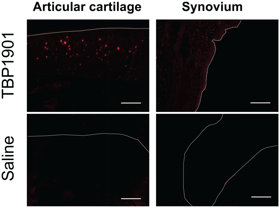
Fluorescence microscopy images of the articular cartilage and synovium of knee joints. Representative histological images. Curcumin fluorescence (red) was confirmed in the articular cartilage and slightly in the synovium of rats injected with TBP1901 intra-articularly. The dotted line indicates the surface line of the articular cartilage and synovium. Magnification: ×125. Scale bar: 100 μm.
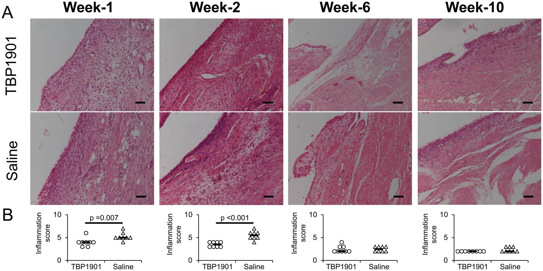
Histological images stained with hematoxylin and eosin and synovial inflammation scores in knee joints. (
Articular cartilage damage of the medial tibia was found as fissures and matrix staining depletion at week 1 in both groups. Cartilage damage did not change much until week 6 in both groups. In week 10, the cartilage damage in most of the TBP1901 did not progress, but it progressed and eroded in most of the saline control (
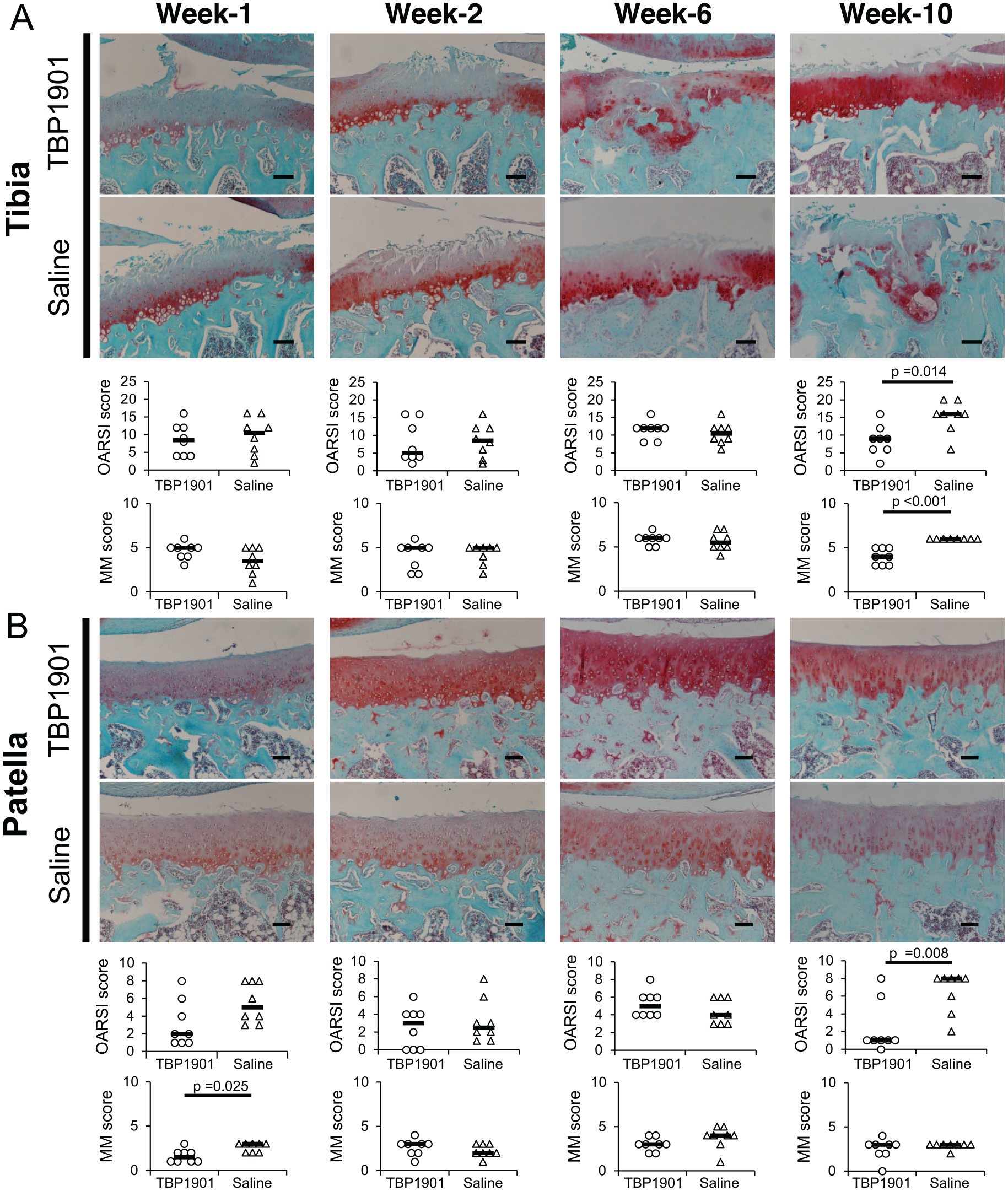
Histological images of safranin O/fast green–stained specimens and osteoarthritis (OA) scores. (
Immunohistochemistry
Tibiofemoral Joint
The expression of type I collagen was confirmed on the surface of the articular cartilage in the medial tibia during all observation periods. There were no significant differences between the groups (
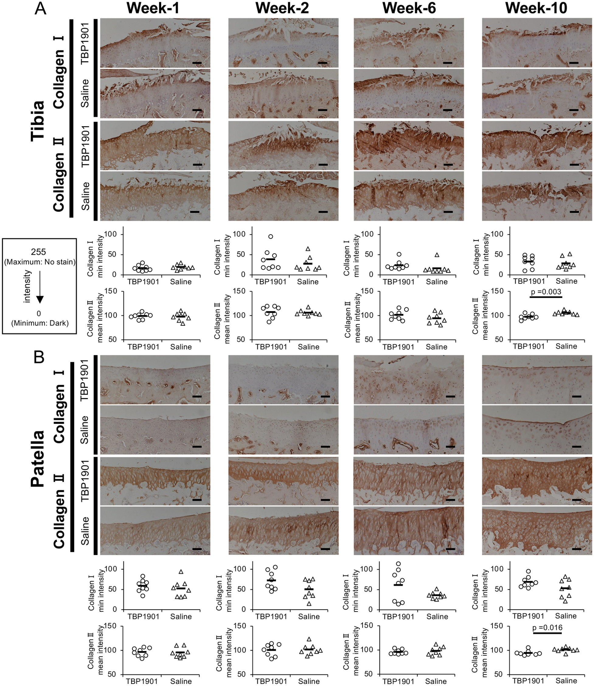
TBP1901 intra-articular injections increased type II collagen expression at 10 weeks. (
The expression of pro-inflammatory cytokines IL-1β, IL-6, and TNF-α was confirmed in the articular cartilage (
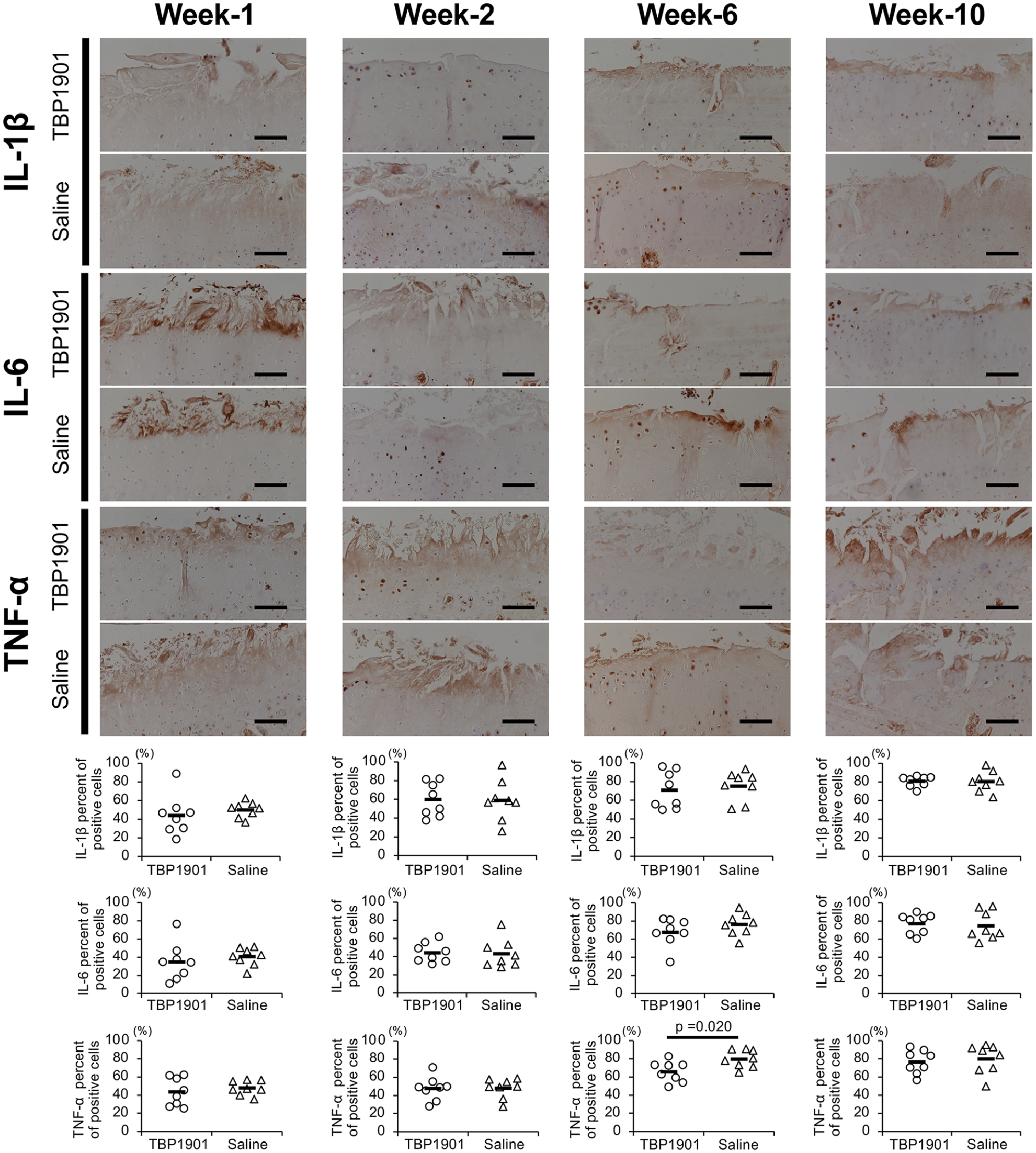
TBP1901 intra-articular injections suppressed the expression of pro-inflammatory cytokines in the articular cartilage of the tibiofemoral joint. Representative immunohistochemical images of interleukin (IL)-1β, IL-6, and tumor necrosis factor-α (TNF)-α in the articular cartilage of the tibia are shown in the upper row. The percentages of positive cells are shown in the lower row. The percentage of TNF-α positive cells in the TBP1901 group was significantly reduced at week 6 compared with that in the saline control group. Values are the means of the TBP1901 and saline control groups at several time points (n = 8 each). P values were calculated using Welch’s t test. Magnification: ×200. Scale bar: 100 μm.
Patellofemoral Joint
The expression of type I collagen was almost undetectable in the patellar articular cartilage at weeks 1 and 2. At week 6, it was confirmed in the peri-chondrocytes and the middle layer of the cartilage at week 6 in the TBP1901 group. At week 10, it was confirmed on the cartilage surface (
The pro-inflammatory cytokines IL-1β, IL-6, and TNF-α were more highly expressed in the articular cartilage of the patella than in the tibia (
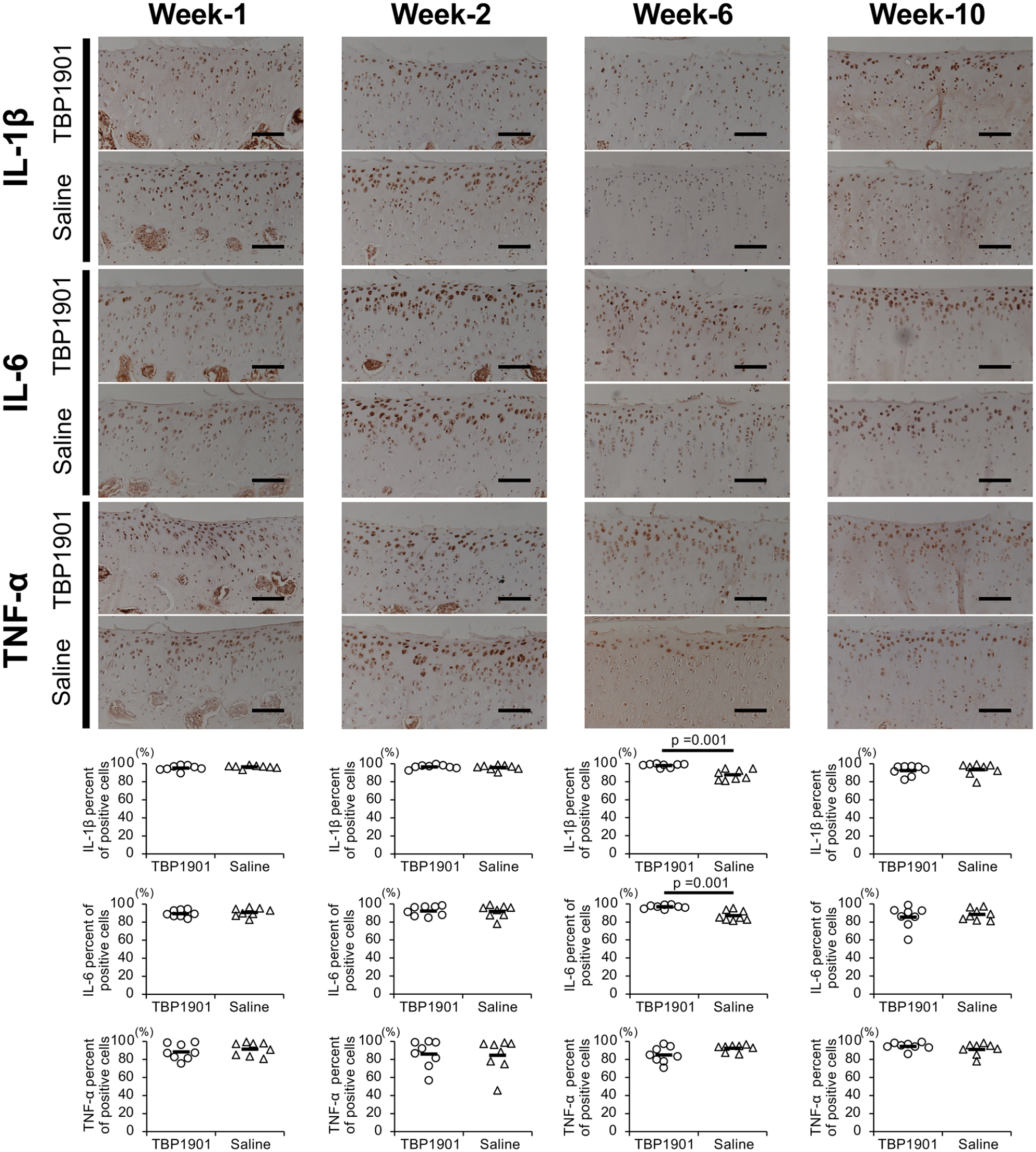
TBP1901 intra-articular injections did not suppress the expression of pro-inflammatory cytokines in the articular cartilage of the patellofemoral joint. Representative immunohistochemical images of interleukin (IL)-1β, IL-6, and tumor necrosis factor-α (TNF)-α in the articular cartilage of the patella are shown in the upper row. The percentages of positive cells are shown in the lower row. The percentages of IL-1β and IL-6 positive cells were significantly higher at week 6 in the TBP1901 group than in the saline control group. Values are the means of the TBP1901 and saline control groups at several time points (n = 8 each). P values were calculated using Welch’s t test. Magnification: ×200. Scale bar: 100 μm.
Micro-CT Analysis
DMM surgery with saline or TBP1901 injections induced SB changes and osteophyte formation (
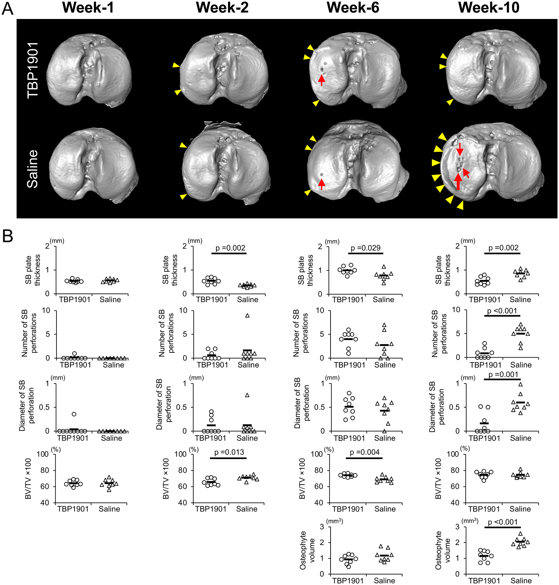
TBP1901 intra-articular injections regulated subchondral bone (SB) plate formation and prevented excessive bone formation. (
The BV/TV tended to increase from week 1 to week 10 in both groups (
The osteophytes had formed from week 2 (quantitative data not shown). In the TBP1901 group, the osteophyte volume tended to be lower than that in the control group at week 6. In week 10, that in the TBP1901 group was significantly lower than that in the control group (P < 0.001).
Discussion
In this study, we performed an intra-articular injection of TBP1901 for 1, 2, 6, or 10 weeks in DMM rats to investigate its effects on inflammation and OA progression. First, the fluorescence of curcumin was confirmed in the articular cartilage and synovium of the TBP1901 group. This indicates that TBP1901 is incorporated into the cartilage and synovium and might induce biological effects. In addition, we found that TBP1901 intra-articular injections suppressed synovial inflammation in the acute phase and degeneration of cartilage and SB in the chronic phase (
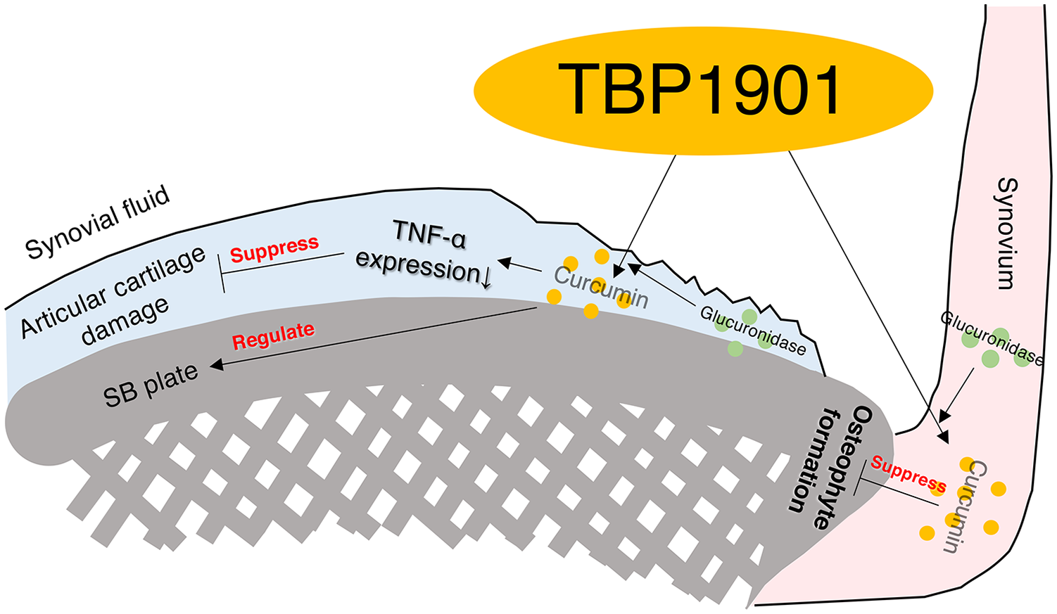
Graphical abstract of the effects of the intra-articular injection of TBP1901 on the suppression of osteoarthritis (OA) development in an OA rat model (destabilized medial meniscus). Curcumin was absorbed in the articular cartilage and synovium by the TBP1901 injection. Curcumin suppressed inflammation, osteophyte formation, articular cartilage, and subchondral bone (SB) pathology.
Suppression of Inflammation and Articular Cartilage Degeneration by TBP1901 Intra-Articular Injections
This study showed the suppression of synovial inflammation in the acute phase of PTOA by TBP1901 intra-articular injections in an OA rat model. The mammalian target of the rapamycin (mTOR) pathway plays an important role in regulating cell growth, proliferation, differentiation, and apoptosis. It has been reported that curcumin inhibits synovial inflammation and hyperplasia via the mTOR pathway in rats with collagen-induced arthritis. 55 The present study showed the suppression of synovial inflammation in a rat model of PTOA. In contrast, TBP1901 did not suppress cartilage damage during the acute phase. In a previous report, oral administration of curcumin or curcumin-loaded poly lactic-co-glycolic acid nanoparticles for 14 days showed a preventative effect on articular cartilage in a monoiodoacetate (MIA) model of osteoarthritis in rats. 56 MIA inhibits glyceraldehyde-3-phosphate dehydrogenase of the Krebs cycle, leading to the death of chondrocytes, leading to unique pathophysiology that does not correlate with PTOA. 57 In the present study, the progression of cartilage damage by DMM surgery may have been too rapid for TBP1901 to be sufficiently effective in the acute phase.
In the chronic phase, articular cartilage degeneration is suppressed by TBP1901. Curcumin suppresses inflammatory cytokines, such as TNF-α, IL-1β, and IL-6. It inhibits the production of pro-inflammatory mediators and matrix-degrading enzymes, metalloproteinase (MMP)-3 and MMP-13.58,59 In the present study, TNF-α expression in the cartilage was also reduced by TBP1901 injections. In addition, type II collagen deposition was higher in the TBP1901 group at week-10. It has been reported that curcumin promotes chondrogenic differentiation, proliferation, and migration of mesenchymal stem cells (MSCs).60,61 These effects may improve articular cartilage conditions. Furthermore, SB and articular cartilage interact with each other and work as a unit. 62 The protection of the SB damage by TBP1901 injections may have suppressed cartilage degeneration.
SB Improvement by TBP1901 Intra-Articular Injections
There are few reports on the effects of curcumin on SB. The present study evaluated the effect of TBP1901 on SB in detail using μ-CT. Some reports have shown that curcumin attenuates bone resorption by suppressing osteoclast proliferation 63 and osteoblast apoptosis. 64 The SB plate thins as bone remodeling increases in early OA and thickens as the remodeling balance changes as OA progresses.65,66 The attenuation of bone resorption may have suppressed the SB plate thinning in the acute phase and the SB plate thickening and perforating in the chronic phase in the present study. A previous study demonstrated histologically that the oral administration of curcumin reduced SB plate thickness in the chronic phase, 12 similar to our results.
In addition, TBP1901 injection suppressed osteophyte formation. Osteophytes are formed by synovial cells in a process similar to endochondral ossification and limit range of motion. 67 Transforming growth factor-β (TGF-β) and macrophages play an important role in osteophyte formation.68,69 In a previous report, the administration of oral curcumin suppressed TGF-β activation in the synovium. 70 TBP1901 injections may have suppressed the osteophyte formation by the inactivation of TGFβ on synovium. The remarkable effects on SB in our study may have been due to the presence of rich β-glucuronidase, which hydrolyzes TBP1901 to free form curcumin in the bone and synovium.29,32
Limitations and Conclusion
The present study has some limitations. First, it is still unclear when and how much TBP1901 should be administered, although it was administered at a dose of 30 mg/mL by intra-articular injection from postoperative day 3 or 7 in this study. Additional investigations are necessary to determine the optimal postoperative timing and dose of TBP1901. Second, the working mechanism of TBP1901 in SB and osteophytes is unclear. Additional comprehensive studies focusing on TGF-β and macrophages are required. Third, although TBP1901 suppressed articular cartilage in a rat OA model, its effects on human OA are unclear. Fourth, it has not been determined whether TBP1901 is more effective than traditional curcumin or other curcumin-related products. We plan to conduct comparative studies to demonstrate the efficacy of TBP1901 in the future.
In conclusion, this study showed that intra-articular injections of TBP1901 reduced synovial inflammation at weeks 1 and 2 in the acute phase of PTOA. In the chronic phase of PTOA, TBP1901 reduced TNF-α expression at week 6 and suppressed articular cartilage degeneration, SB plate thickening, SB plate perforation, and osteophyte formation at week 10.
Supplemental Material
sj-pdf-1-car-10.1177_19476035211043202 – Supplemental material for Intra-Articular Injections of Curcumin Monoglucuronide TBP1901 Suppresses Articular Cartilage Damage and Regulates Subchondral Bone Alteration in an Osteoarthritis Rat Model
Supplemental material, sj-pdf-1-car-10.1177_19476035211043202 for Intra-Articular Injections of Curcumin Monoglucuronide TBP1901 Suppresses Articular Cartilage Damage and Regulates Subchondral Bone Alteration in an Osteoarthritis Rat Model by Akihiro Nakahata, Akira Ito, Ryo Nakahara, Atsuhiro Kishimoto, Atsushi Imaizumi, Tadashi Hashimoto, Shogo Mukai, Yasuaki Nakagawa and Hiroshi Kuroki in CARTILAGE
Supplemental Material
sj-pdf-2-car-10.1177_19476035211043202 – Supplemental material for Intra-Articular Injections of Curcumin Monoglucuronide TBP1901 Suppresses Articular Cartilage Damage and Regulates Subchondral Bone Alteration in an Osteoarthritis Rat Model
Supplemental material, sj-pdf-2-car-10.1177_19476035211043202 for Intra-Articular Injections of Curcumin Monoglucuronide TBP1901 Suppresses Articular Cartilage Damage and Regulates Subchondral Bone Alteration in an Osteoarthritis Rat Model by Akihiro Nakahata, Akira Ito, Ryo Nakahara, Atsuhiro Kishimoto, Atsushi Imaizumi, Tadashi Hashimoto, Shogo Mukai, Yasuaki Nakagawa and Hiroshi Kuroki in CARTILAGE
Supplemental Material
sj-pdf-3-car-10.1177_19476035211043202 – Supplemental material for Intra-Articular Injections of Curcumin Monoglucuronide TBP1901 Suppresses Articular Cartilage Damage and Regulates Subchondral Bone Alteration in an Osteoarthritis Rat Model
Supplemental material, sj-pdf-3-car-10.1177_19476035211043202 for Intra-Articular Injections of Curcumin Monoglucuronide TBP1901 Suppresses Articular Cartilage Damage and Regulates Subchondral Bone Alteration in an Osteoarthritis Rat Model by Akihiro Nakahata, Akira Ito, Ryo Nakahara, Atsuhiro Kishimoto, Atsushi Imaizumi, Tadashi Hashimoto, Shogo Mukai, Yasuaki Nakagawa and Hiroshi Kuroki in CARTILAGE
Research Data
sj-pptx-1-car-10.1177_19476035211043202 – Supplemental material for Intra-Articular Injections of Curcumin Monoglucuronide TBP1901 Suppresses Articular Cartilage Damage and Regulates Subchondral Bone Alteration in an Osteoarthritis Rat Model
sj-pptx-1-car-10.1177_19476035211043202 for Intra-Articular Injections of Curcumin Monoglucuronide TBP1901 Suppresses Articular Cartilage Damage and Regulates Subchondral Bone Alteration in an Osteoarthritis Rat Model by Akihiro Nakahata, Akira Ito, Ryo Nakahara, Atsuhiro Kishimoto, Atsushi Imaizumi, Tadashi Hashimoto, Shogo Mukai, Yasuaki Nakagawa and Hiroshi Kuroki in CARTILAGE
Footnotes
Acknowledgments and Funding
The author(s) disclosed receipt of the following financial support for research, authorhship and/or publication of this article: This study was supported in part by JSPS KAKENHI Grant Numbers JP18H03129 and JP18K19739, and a sponsored research grant from Therabiopharma Inc. to HK. We would like to thank Editage (![]() ) for English language editing.
) for English language editing.
Declaration of Conflicting Interests
The author(s) declared the following potential conflicts of interest with respect to the research, authorship, and/or publication of this article: This study was supported in part by a sponsored research grant from Therabiopharma Inc. to HK. AIm and TH have equity in Therabiopharma Inc. and serve on the board of directors of Therabiopharma Inc. AK is an employee of Therabiopharma Inc.
Ethical Approval
This study was approved by the animal research committee of Kyoto University (approval number: Med Kyo 19016).
References
Supplementary Material
Please find the following supplemental material available below.
For Open Access articles published under a Creative Commons License, all supplemental material carries the same license as the article it is associated with.
For non-Open Access articles published, all supplemental material carries a non-exclusive license, and permission requests for re-use of supplemental material or any part of supplemental material shall be sent directly to the copyright owner as specified in the copyright notice associated with the article.