
Abstract
The sirtuin family has emerged as important regulators of diverse physiological and pathological events, including life-span extension, neurodegeneration, age-related disorders, obesity, heart disease, inflammation, and cancer. In mammals, there are 7 members (SIRT1-SIRT7) in the sirtuin family, with the function of SIRT1 being extensively studied in the past decade. SIRT1 can deacetylate histones and a number of nonhistone substrates, which are involved in multiple signaling pathways. Numerous studies have suggested that SIRT1 could act as either a tumor suppressor or tumor promoter depending on its targets in specific signaling pathways or in specific cancers. This review highlights the major pathways regulated by SIRT1 involved in tumorigenesis.
Keywords
Introduction
Sirtuins have drawn emerging attention due to their diverse roles in various physiological and pathological events, including life-span extension, neurodegeneration, age-related disorders, obesity, heart disease, inflammation, and cancer.1-7 In mammals, there are 7 members (SIRT1-SIRT7) in the sirtuin family, which belong to class III histone deacetylases (HDACs) and show different functions, structure, and localization. 8 SIRT1 is a mammalian homolog of yeast silent information regulator 2 (sir2) and is the most extensively studied sirtuin member.9,10 SIRT1 can deacetylate histones11,12 and a number of nonhistone substrates. p53 is a well-known substrate of SIRT113,14 as well as other substrates with diverse functions involved in multiple signaling pathways.
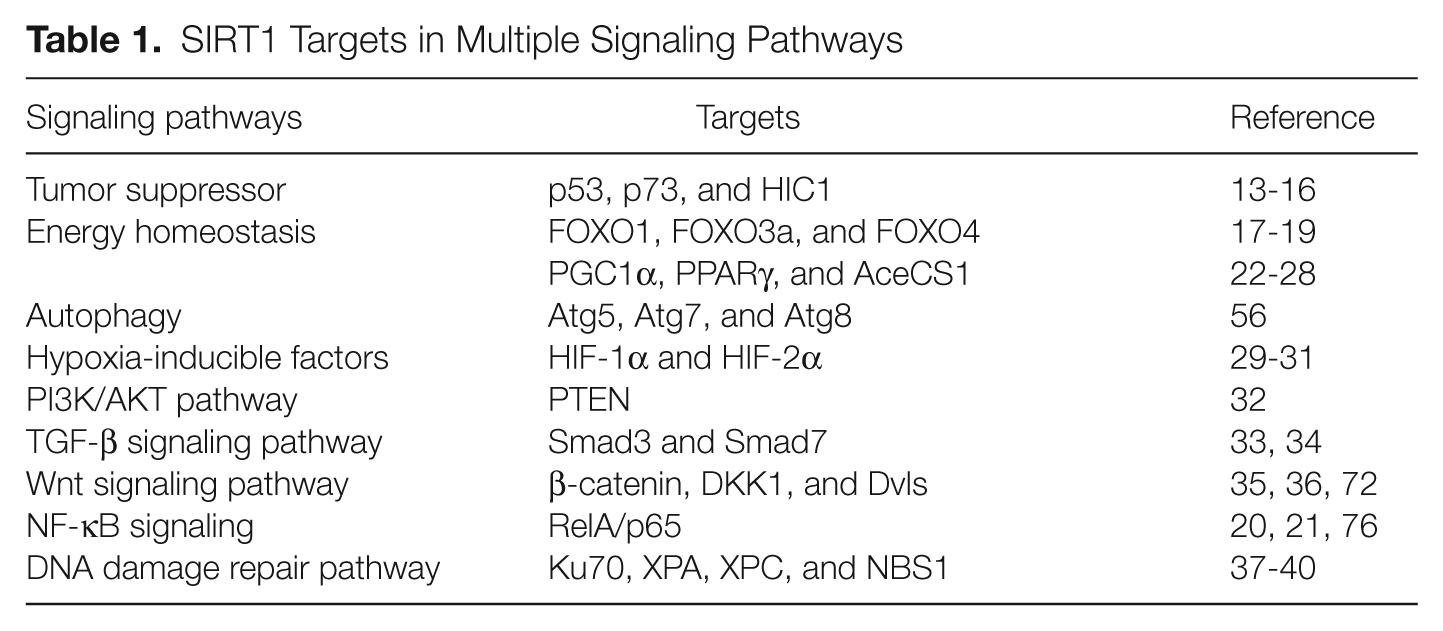
The role of SIRT1 in cancers has been extensively studied in the past decade. However, controversy regarding cancer and sirtuins exists and is still under debate since it could act as either a tumor suppressor or tumor promoter depending on the cellular context or its targets in specific signaling pathways or specific cancers. For example, since SIRT1-mediated deacetylation suppresses the functions of several tumor suppressors including p53, p73, and HIC1, it has been suggested that SIRT1 has a promoting function in tumor development and progression.13-16 In contrast, SIRT1 may have a suppressive activity in tumor cell growth by suppressing NF-κB,17-19 a transcription factor playing a central role in the regulation of the innate and adaptive immune responses and carcinogenesis, the dysregulation of which leads to the onset of tumorigenesis and tumor malignancy.20,21 In addition to the above 2 pathways, this review will highlight the main SIRT1 substrates involved in energy homeostasis,22-28 hypoxia response,29-31 the PI3K/AKT pathway, 32 the transforming growth factor-β (TGF-β) signaling pathway,33,34 the Wnt signaling pathway,35,36 and the DNA damage repair pathway37-40 in cancer cells (Table 1).
SIRT1 Targets in Multiple Signaling Pathways
Specific Signaling Pathways Regulated by SIRT1
SIRT1-p53 axis
p53, the guardian of the genome, 41 is one of the most important tumor suppressors and is frequently mutated in human solid tumors. 42 p53 can be both transcriptionally and posttranslationally regulated. One of the posttranslational modifications is acetylation. SIRT1 has been found to target p53 for deacetylation and to attenuate p53-mediated transcriptional activity.13,14 Overexpression of wild-type SIRT1 removed p53 acetylation, but overexpression of catalytically inactive SIRT1 failed to do so, suggesting that SIRT1 regulation of p53 transcriptional activity depends on its deacetylase activity. 13 SIRT1 deacetylase activity is NAD dependent, and inhibition of SIRT1 by NAM (nicotinamide) enhanced p53 acetylation. 14 More intriguingly, SIRT1 can exert a second layer of regulation on p53 activity by deacetylating p300, which is a histone acetyltransferase of p53. 43 Since the acetylation of p300 promotes its binding to p53, inhibition of SIRT1 would lead to the elevation of p300-mediated p53 activation and its target gene transcription. 44
The most important function of activated p53 is to induce cell cycle arrest, apoptosis, and DNA repair. SIRT1 has been demonstrated to reduce p53-mediated apoptosis2,14 and negatively regulate p53-induced cellular senescence. 45 Therefore, inhibition of SIRT1 activity, which leads to elevated p53 acetylation and transactivation and results in enhanced apoptosis and cytostasis, would be beneficial for cancer treatment. However, this must be carefully evaluated since the activation of p53 has dual functions. On one hand, activated p53 contributes to tumor cell death; on the other hand, although occurring in a small population, p53 induces cancer cell cycle arrest, which can be reversed by treatment itself or other stimuli, 46 and this could accumulate further mutations and compromise the killing effect of the therapy. Future studies need to be explored to investigate the differential regulation of p53 activity on cell cycle arrest and apoptosis by SIRT1 inhibitors or p53 activators.
Interestingly, SIRT1 interacts with hypermethylated in cancer 1 (HIC1), a tumor suppressor and transcriptional repressor that is epigenetically inactivated but not mutated in human cancers 47 to form a transcriptional repression complex, which directly binds the SIRT1 promoter and represses SIRT1’s transcription. Depletion of HIC1 expression results in an upregulated SIRT1 level, which deacetylates p53 and attenuates p53 function, thus allowing cells to bypass apoptosis, survive DNA damage, and promote tumorigenesis. Inhibition of SIRT1 expression in HIC1−/− cells abolishes the resistance to apoptosis. Since the HIC1 promoter is hypermethylated and epigenetically silenced by age, the resultant upregulation of SIRT1 may promote the survival of aging cells and increase cancer risk in mammals. 16
SIRT1 and FOXO
The first evidence of SIRT1 as a regulator of the forkhead O family (FOXO) transcription factors was the observation that Sir2 extends the life span of Caenorhabditis elegans through DAF-16, a homolog of mammalian FOXO3a. 48 Subsequent studies indicate that SIRT1 regulates a variety of cellular processes through deacetylating FOXO family transcription factors including FOXO3a, FOXO1, and FOXO4.23,24,49,50 FOXO proteins are phylogenetically conserved and regulate key physiological functions, including cell proliferation, cell differentiation, and survival, and their dysregulation is associated with tumorigenesis. While the molecular mechanisms for such an effect remain to be studied, SIRT1-mediated deacetylation of FOXO proteins can either activate or inhibit their transcriptional activity. For example, SIRT1-mediated activation of FOXO3a induces cell cycle arrest and oxidative stress resistance but inhibits FOXO3a-induced cell death. 22 Recently, Wang et al. 51 reported that deacetylation of FOXO3 by SIRT1 leads to Skp2-mediated FOXO3 ubiquitination and degradation, implying that SIRT1 regulates the transcriptional activities of FOXO family proteins through multiple mechanisms.
FOXO3a also regulates SIRT1 expression.52,53 Acute nutrient withdrawal augments both SIRT1 and FOXO3a expression, 52 but knockdown of FOXO3a inhibited starvation-induced SIRT1 expression. Under normal nutrient conditions, SIRT1 expression is repressed by p53. 52 In contrast, p53 interacts with FOXO3a under starvation conditions and stimulates SIRT1 transcription through 2 p53 binding sites in the SIRT1 promoter. p53 is required for starvation-induced FOXO3a to stimulate SIRT1 expression since SIRT1 expression was not induced in starved p53-deficient mice. Thus, SIRT1, p53, and FOXO3a, 3 proteins implicated in aging, constitute a nutrient-sensing pathway in mammalian cells. 52 Since cancer is an age-related disease, these studies may provide a clue to understand the complex role of SIRT1 in certain kinds of tumors.
SIRT1 and autophagy
There are 3 major protein degradation pathways in mammalian cells: the proteasome, the lysosome, and the autophagosome pathways. All of them use ubiquitin as a common denominator to target substrates for degradation. 54 Autophagosome-mediated degradation is an evolutionarily conserved, cellular housekeeping process for the clearance of long-lived proteins, malfunction molecules, and organelles that harbor mutations beneficial for cancer development55,56 and is required for the development of glands such as in the breast and prostate. SIRT1 has been reported to repress androgen-responsive gene expression and induced autophagy as revealed by genome-wide gene expression analysis in SIRT1−/− prostates. 57 SIRT1 induced autophagy by promoting autophagosome maturation and completion in cultured prostate cancer cells. Depletion of SIRT1 expression resulted in prostatic intraepithelial neoplasia (PIN) associated with reduced autophagy, thus providing a checkpoint function and a tumor suppressor role of SIRT1 in the development of PIN. 57
Mechanically, SIRT1 interacts with several autophagy (ATG) proteins, such as Atg5, Atg7, and Atg8, to regulate autophagy. 58 Atg5 and Atg7 play central roles in the formation and targeting of the Atg12 conjugation system, whereas Atg8, also called LC3 in mammals, represents the second conjugation system in autophagosome formation.56,59 Moreover, overexpression of SIRT1 significantly stimulates the basal level of autophagy, but depletion of SIRT1 in mice attenuates the ability to undergo autophagy during starvation. Consistent with the above observations, SIRT1−/− mice and Atg5−/− mice exhibit similar phenotypes, such as the accumulation of damaged mitochondria and deficiencies in energy homeostasis. The regulation of autophagy by SIRT1 is deacetylase dependent since depletion of SIRT1 expression leads to an increase in the acetylation level of these autophagy proteins both in cultured cells and in embryonic and neonatal tissues, and reconstitution with wild-type but not a deacetylase-inactive mutant of SIRT1 restores autophagy in these cells. 58 These studies suggest that SIRT1-mediated deacetylation of ATG proteins plays an important role in regulating both the autophagic uptake of cellular proteins during starvation and the degradation of damaged organelles as a means of housekeeping during aging.
SIRT1 in TGF-β signaling
TGF-β and related growth factors are involved in the regulation of a variety of cellular events, including cell growth, differentiation, migration, and apoptosis, and play key roles in many physiological and pathological processes.60,61 Inhibitory Smads, including Smad6 and Smad7, are key modulators of TGF-β family signaling through a negative feedback circuit.62,63 SIRT1 has been demonstrated to participate in the regulation of TGF-β signaling by interacting with and deacetylating Smad7 33 and Smad3. 34
SIRT1 directly interacts with the N-terminus of Smad7 to reverse p300-mediated acetylation of 2 lysine residues (Lys-64 and Lys-70) on Smad7. Overexpression of SIRT1 led to a reduction of Smad7 levels in mesangial cells and attenuated both Smad7- and TGF-β–induced mesangial cell apoptosis, whereas SIRT1 knockdown enhanced apoptosis. Further biochemical studies reveal that the deacetylation of Smad7 by SIRT1 promotes its ubiquitination, enhances Smad ubiquitination regulatory factor 1 (Smurf1)–mediated degradation, and thereby inhibits TGF-β–dependent apoptosis. 33
Not only Smad7 but Smad3 also can be deacetylated by SIRT1. SIRT1 reversed TGF-β–induced acetylation but not phosphorylation of Smad3 and inhibited TGF-β1–induced upregulation of collagen IV and fibronectin mRNA levels. SIRT1 knockdown abolished the inhibitory effect of resveratrol, an activator of SIRT1, on Smad3 acetylation. Resveratrol inhibited the acetylation but not phosphorylation of Smad3 at day 2 of unilateral ureteral obstruction (UUO), and resveratrol treatment inhibited interstitial fibrosis at day 7 of UUO. These studies suggest a pathological role for Smad3 acetylation in renal fibrosis, and deacetylation of Smad3 by SIRT1 or SIRT1 activators may be a novel therapeutic strategy for fibrotic disease. 34 Interestingly, SIRT1 expression is under the control of TGF-β signaling. SIRT1 expression can be induced by TGF-β1 in mouse lung epithelial cells, human lung fibroblast cells, and mouse monocyte/macrophage cells, and its expression is significantly decreased in both macrophages and nonmacrophages (lung parenchymal cells, including epithelial and mesenchymal cells) of Smad3-null lungs, suggesting a TGF-β signaling–mediated regulatory mechanism of SIRT1 expression through Smad3. 64
SIRT1, Wnt signaling, and cancer
The Wnt/β-catenin signaling pathway plays an important role in controlling stem cell differentiation, embryo development, as well as tumorigenesis.65,66 Constitutive activation of the Wnt/β-catenin signaling pathway has been found in 90% of colorectal cancers67,68 and in many other cancers such as prostate, breast, and ovary cancer and melanoma. β-catenin is tightly regulated by a variety of posttranslational modifications, including acetylation. One previous study suggests that CBP acetylates β-catenin at K49, which negatively regulates its transcriptional activity. 69 β-catenin can also be acetylated by p300 at K345, which promotes the binding of β-catenin to TCF4 but represses the β-catenin–androgen receptor interaction. 70 SIRT1 removes the acetylation of β-catenin at K345, 71 and deacetylation of β-catenin by SIRT1 suppresses its ability to activate transcription and drive cell proliferation. Moreover, SIRT1 promotes the cytoplasmic retention of β-catenin and thus suppresses intestinal tumor formation. 71 SIRT1 also suppresses β-catenin through pancreatic adenocarcinoma upregulated factor (PAUF) in pancreatic cancer cells. The overexpression of SIRT1 or resveratrol treatment suppressed β-catenin protein levels and its transcriptional activity in Panc-PAUF cells. Conversely, SIRT1 knockdown by siRNA enhanced β-catenin expression and transcriptional activity. 72 Interestingly, SIRT1 expression is under the control of β-catenin since the absence of β-catenin leads to a significant decrease in SIRT1 mRNA and protein levels in sham-treated knockout mice compared to nontreated controls. 73
SIRT1 seems to regulate the expression of DKK1, a Wnt signaling inhibitor. Tobacco smoke condensate (TSC) exposure decreases H4K16 acetylation and increases H3K27 trimethylation within the DKK1 promoter, and siRNA- mediated knockdown of SIRT1 partially abrogated TSC-mediated inhibition of DKK1 expression. Knockdown of SIRT1 also increased DKK1 expression in untreated Calu-6 cells. 74
Dishevelled (Dvl) is a key component in the canonical Wnt signaling pathway and is required for LRP6 phosphorylation, which in turn is essential for subsequent steps of signal transduction, such as Axin recruitment and cytosolic β-catenin stabilization. 75 SIRT1 serves as a regulator of Wnt signaling by targeting Dvl. 36 SIRT1 and Dvl proteins form a complex in vivo, and loss of SIRT1 function leads to a significant decrease in the levels of all 3 Dvl proteins and Wnt target gene expression. In addition, Wnt-stimulated cell migration is inhibited by the SIRT1 inhibitor, suggesting a role of SIRT1 in the regulation of the Wnt signaling pathway. 36
SIRT1 in NF-κB signaling
NF-κB plays a central role in the regulation of the innate and adaptive immune responses and carcinogenesis, the dysregulation of which leads to the onset of tumorigenesis and tumor malignancy.20,21 The activity of the prototypical NF-κB complex, a heterodimer of p50 and p65 subunits, is inhibited by IκB molecules, which associates with NF-κB and maintains the cytosolic localization of inactive NF-κB. Upon cytokine stimulation, IκB molecules are rapidly phosphorylated, polyubiquitylated, and degraded, and the released NF-κB dimmer translocates to the nucleus, where the p65 subunit undergoes posttranslational modifications, including reversible acetylation by p300, thus initiating target gene transcription.20,21 There are 3 acetylation sites, K218, K221, and K310, in the NF-κB amino acid sequence. The former 2 sites mediate its DNA binding activity, while the latter enhances the transcriptional activity of NF-κB. The acetylation of NF-κB can be reversed by 2 deacetylases, HDAC3 and SIRT1, which deacetylate NF-κB and suppress its transcriptional activity.18,76 SIRT1 specifically removes the acetylation of p65 at K310 and inhibits the prosurvival function of NF-κB. 18 SIRT1 not only inhibited p300-mediated NF-κB acetylation but also suppressed tumor necrosis factor α (TNFα)–induced NF-κB acetylation, thus leading to the inhibition of NF-κB–mediated CD40 expression in CRL-1730 endothelial cells. The inhibition of SIRT1 on NF-κB acetylation seems to be regulated by TNFα since SIRT1 expression is reduced following TNFα stimulation. 77
Posttranslational modifications of the RelA/p65 subunit of NF-κB, including acetylation and methylation, determine the strength and duration of its transcriptional activity. Not only acetylation but also methylation of NF-κB can be regulated by SIRT1. Set9-mediated methylation of K314 and K315, 78 which is important for the ubiquitination and degradation of chromatin-associated p65, is impaired by the acetylation of K310, as acetylation on K310 interferes with its interaction with Set9. Conversely, the interaction between RelA/p65 and Set9 is enhanced by SIRT1 since SIRT1 removes K310 acetylation of p65. 18 Mutation of lysine 310 to arginine (K310R) or overexpression of SIRT1 enhances p65 methylation. Conversely, depletion of SIRT1 by siRNA inhibits the methylation of RelA/p65, decreases its ubiquitination, prolongs the stability of chromatin-associated p65, and enhances the transcriptional activity of NF-κB. 78 Since the acetylation (K310) and methylation sites (K314 and K315) of p65 are in close proximity, there is competition between p300-mediated acetylation and Set9-mediated methylation. Thus, removal of the acetylation by SIRT1 prepares the site for incoming Set9 to methylate p65. These findings suggest cooperation between SIRT1-mediated deacetylation and Set9-induced methylation on RelA/p65 transcriptional repression.
SIRT1 seems to regulate cigarette smoke–induced proinflammatory mediator release via RelA/p65 NF-κB both in vitro and in vivo. 79 Cigarette smoke extract (CSE) exposure to MonoMac6 cells or inflammatory cells in the lungs of rats caused dose- and time-dependent decreases in SIRT1 activity and an increase of NF-κB–dependent proinflammatory mediator release. Sirtinol, an inhibitor of SIRT1, augmented CSE-mediated proinflammatory cytokine release, 79 whereas resveratrol, an activator of SIRT1, functions in an opposite manner to that of sirtinol.79,80 CSE-mediated inhibition of SIRT1 was associated with increased NF-κB levels. Moreover, the interaction between SIRT1 and NF-κB was disrupted by cigarette smoke exposure, leading to elevated RelA/p65 acetylation in MonoMac6 cells, suggesting that SIRT1 regulation of NF-κB acetylation may be important for proinflammatory mediator release when exposed to cigarette smoke in the lungs, 79 which is the leading cause of non–small cell lung carcinoma. 81
Breast cancer–associated protein 3 (BCA3) has been reported to suppress NF-κB–dependent transcription by binding to p65 in a neddylation-dependent manner. 82 Overexpression of wild-type BCA3 and SENP8, a NEDD8-specific protease, but not by neddylation-deficient BCA3 or a SENP8 mutant in DU145 and MCF7 cells suppressed NF-κB transcription and elevated TNFα-induced apoptosis by recruiting SIRT1. Conversely, silencing of endogenous BCA3 by siRNA reversed its suppressive effect on NF-κB transcription. These results suggest that there is crosstalk between neddylation and SIRT1 regulation of NF-κB transcription. 82 Since BCA3 binds to SIRT1 and the effect of BCA3 on TNFα-induced apoptosis is SIRT1 dependent, whether BCA3 is a SIRT1 substrate remains to be elucidated.
The above studies together suggested that the acetylation of NF-κB is very important for its transcriptional activation, and this can be inhibited by SIRT1 through its deacetylase activity. In contrast to this conclusion, the deacetylase activity of SIRT1 is not crucial for its effect on transducin-like enhancer of split 1 (TLE1)–mediated NF-κB repression since the catalytic mutant of SIRT1, SIRT1-H363Y, and its N-terminal fragment, which lacks the deacetylase catalytic domain, also repressed NF-κB– mediated transcription. 83 Therefore, SIRT1 may inhibit NF-κB–mediated transcription through both deacetylase-dependent and -independent manners, depending on its specific interaction partner and cellular context.
SIRT1 and AP-1 transcriptional activation
Recent studies have identified that SIRT1 suppresses the transcriptional activities of AP-1 in immune cells and cancers.1,84,85 A direct interaction between SIRT1 and the AP-1 transcription factor c-Jun appears to be responsible for this suppressive activity.1,84 Since acetylation of c-Jun by the acetyltransferase p300 at lysine residue 271 is essential for AP-1 transcriptional activity, 86 it is not surprising that SIRT1 deacetylates c-Jun to suppress AP-1 transcriptional activity. Further studies are needed to illuminate whether and, if yes, how SIRT1-mediated suppression of AP-1 is involved in tumorigenesis.
SIRT1 and the DNA damage pathway
Mammals are evolved to explore a number of pathways to sense specific DNA-damaging agents, repair specific types of DNA lesions, and ensure genomic stability. Mice defective in individual DNA repair pathways showed hypersensitivity to specific genotoxic stress and increased chromosomal instability, which are hallmarks of eukaryotic aging and cancer. Double-stranded breaks (DSBs) induced by ionizing radiation (IR) or other DNA-damaging agents are repaired by homologous recombination (HR) or nonhomologous end joining (NHEJ). Single-stranded DNA lesions are repaired via base excision repair or nucleotide excision repair (NER), which depends on the type of lesions.87,88 SIRT1 has been implicated in both DSB repair 89 and single-stranded DNA lesion repair. 39
SIRT1 plays a role in both HR and NHEJ. Inhibition of SIRT1 activity with NAM, the specific SIRT1 inhibitor S91211, 90 or stable knockdown of SIRT1 led to a reduction of both HR- and NHEJ-mediated repair. SIRT1 is also necessary for maintaining genomic stability since chromosomal aberrations significantly increased in SIRT1- deficient cells upon H2O2 treatment. The numbers of chromosomal fusions were significantly higher in the absence of SIRT1, whereas the frequency of chromatic breaks was comparable between knockdown and control embryonic stem cells. Chromosome fusions are due to a failure of DNA repair, thus supporting a role for SIRT1 in DSB repair. 89
Mutation-caused disruption of the NER pathway leads to xeroderma pigmentosum, a syndrome predisposed to develop skin cancer. The xeroderma pigmentosum C (XPC) protein, essential for initiating global genome NER by recognizing DNA damage and recruiting downstream factors, is a substrate of SIRT1. Ectopic expression of SIRT1 enhances XPC expression, whereas depletion of SIRT1 expression by siRNA or knockout suppressed XPC transcription and impaired global genome NER in a SIRT1 deacetylase–dependent manner. Moreover, SIRT1 levels are reversely correlated with human skin tumor progression in white patients, suggesting a possible role of SIRT1 in DNA repair 39 and tumor suppression.
The ATM protein kinase, a master sensor of DNA damage, is mutated in individuals with the radiosensitivity disorder ataxia telangiectasia. ATM is rapidly autophosphorylated at Ser1981, which leads to the conversion of the inactive dimmer form to active monomers following cellular exposure to IR. 91 Activated ATM phosphorylates multiple cellular substrates including NBS192,93 and deleted in breast cancer 1 (DBC1), a negative regulator of SIRT1. 94 NBS1, which is mutated in the human genetic disease Nijmegen breakage syndrome, serves as the regulatory subunit of the MRE11-RAD50-NBS1 (MRN) complex involved in DNA damage repair signaling. 40 Interestingly, NBS1 is a substrate of SIRT1, which associates with the MRN complex and maintains NBS1 in a hypoacetylated state. The deacetylation of NBS1 by SIRT1 is required for ATM-mediated NBS1 phosphorylation. 40 Therefore, SIRT1 plays a vital role in regulating the DNA damage response and maintaining genomic stability.
Not only NBS1 but ATM also targets DBC1 for phosphorylation to regulate SIRT1 activity. ATM-dependent phosphorylation of DBC1 at Thr454, which creates a second binding site for SIRT1, is required for the stress-induced DBC1-SIRT1 interaction, suggesting an important role of ATM-mediated enhancement of DBC1-SIRT1 interaction in cell fate determination following genotoxic stress. 94 Since 3 proteins, USP22, 2 DBC1, 94 and NBS1, 40 all bind to SIRT1 in response to DNA damage, and SIRT1 mRNA and protein levels are regulated by HuR through CHK2, 95 it is worth investigating whether USP22 is phosphorylated by ATM to regulate SIRT1 stability, whether USP22 interacts with DBC1 and NBS1 to involve SIRT1-mediated DNA damage response, whether USP22 and DBC1 are substrates of SIRT1, whether ATM-mediated DBC1 phosphorylation is through CHK2, and whether DBC1 can regulate NBS1 in response to genotoxic stress.
SIRT1 activity can also be regulated by posttranslational modification such as sumoylation in response to genotoxic stress. Sumoylation of SIRT1 at Lys 734 increased its deacetylase activity. Conversely, mutation of SIRT1 at Lys 734 or desumoylation by SENP1 reduced its deacetylase activity. Stress-inducing agents enhanced the binding of SIRT1 to SENP1, and cells depleted of SENP1 (but not of SENP1 and SIRT1) were more resistant to stress-induced apoptosis than control cells. Thus, stress-inducing agents recruit SENP1 to SIRT1, leading to both the desumoylation and inactivation of SIRT1 and the acetylation and activation of apoptotic proteins, to counteract the antiapoptotic activity of SIRT1. 96
The above studies together suggest that SIRT1 plays an important role in maintaining genomic stability and repairing DNA damage. In this regard, SIRT1 may act as a tumor suppressor through its role in DNA damage response and genome integrity. Indeed, Wang et al. 6 demonstrate that SIRT1+/– p53+/– mice develop tumors in multiple tissues, whereas activation of SIRT1 by resveratrol treatment shows reduced tumorigenesis. Consistent with their results, a metabolic syndrome–associated liver cancer model implied a role of SIRT1 in tumor suppression. In that model, wild-type mice developed multiple carcinomas, but SIRT1-tg mice had reduced susceptibility to liver cancer and exhibited hepatic protection from both DNA damage and metabolic damage. 97
Therapeutic Implications of SIRT1 Targeting in Cancer Chemotherapy
SIRT1 activators
Encouraged by these findings, many scientists have spent effort in exploring the therapeutic potential of SIRT1 activators or inhibitors, including natural and nonnatural small molecules, and were considering applying them to clinical practice. Resveratrol, a polyphenolic flavonoid with potent antioxidant properties, is the first reported SIRT1 activator. 98 A number of studies have demonstrated diverse biological effects of resveratrol including anti-inflammatory, antiangiogenic, antioxidant, proapoptotic, antiaging, anticancer, and metabolic modulating properties in mammalian cells. 99 Subsequently, much more efficacious and potent SIRT1 activators were suggested as potential therapeutics for the treatment of type 2 diabetes (e.g., SRT1720, SRT2183, and SRT1460), using a fluorescently labeled substrate in a fluorescence polarization assay. 100 However, it was later found that these effects mediated by resveratrol could only be demonstrated when the substrate was fluorescently tagged, 101 and these compounds (resveratrol, SRT1720, SRT2183, and SRT1460) did not enhance SIRT1 activity when using native peptide or protein substrates.101,102 Therefore, much more specific SIRT1 activators are still needed to be explored for the understanding of sirtuin biology and the treatment of SIRT1-related diseases.
SIRT1 inhibitors
Based on cell culture systems, many studies show that SIRT1 can inhibit apoptosis and senescence,2,22,103-105 suggesting that SIRT1 inhibition may be beneficial for treating certain types of cancer.104,106-108 EX527 was initially reported as a SIRT1 inhibitor, which was shown to increase p53 acetylation in the presence of etoposide. However, it had no effect on the expression of p53 target genes, cell viability, and proliferation in various tumor lines. 90 Followed by this, many other SIRT inhibitors such as cambinol, 109 sirtinol, 110 salermide, 107 JGB1741, 111 suramin analogs, 112 and the tenovins 113 are found and are effective in inducing cancer cell apoptosis. Inhibition of both SIRT1 and SIRT2 induced apoptosis in MCF-7 breast cancer cells, whereas knockdown of the individual proteins was ineffective, 114 suggesting a possible functional redundancy between SIRT1 and SIRT2 in certain types of cancer. Consistent with this finding, both sirtinol and salermide, dual SIRT1/2 inhibitors, but not EX527, enhanced p53 acetylation and stabilization in MCF-7 cells. 114 These observations remind us that dual SIRT1/2 inhibition or broad-spectrum sirtuin inhibitors may provide therapeutic benefits in cancer. However, the efficacy and safety of these inhibitors need to be fully assessed. There is still a long way to go for combating SIRT1-related diseases such as cancer.
Footnotes
Declaration of Conflicting Interests
The author(s) declared no potential conflicts of interest with respect to the research, authorship, and/or publication of this article.
Funding
The author(s) received the following financial support for the research, authorship, and/or publication of this article: This work was supported by research grants R01AI079056 and R56AI079056, the “The Type I Diabetes Pathfinder Award” (DP2DK083050), and their supplemental grants from the National Institutes of Health to D.F.