
Abstract
Objectives/Background
Prostate cancer is the second most diagnosed malignancy among men globally. In Ghana, the age-standardized mortality rate aligns with global trends, reflecting low survival rates largely due to limited access to cancer care and the high cost of therapies. This has increased reliance on traditional medicine, much of which remains scientifically unverified. This study aimed to evaluate the cytotoxic and pharmacological potential of Entandrophragma angolense stem bark extract and its bioactive metabolite, methyl angolensate, in prostate cancer models.
Methods
Cytotoxic activity was evaluated using the Alamar Blue assay across five prostate cancer cell lines (LNCaP, PC-3, DU-145, 22Rv1, VCaP) and one normal prostate epithelial line (RWPE-1). Molecular docking assessed the interaction of methyl angolensate with the androgen receptor (AR), using metribolone as a reference. Molecular dynamics (MD) simulations totaling 100 ns were performed to evaluate the stability of ligand–AR complexes. Pharmacokinetic and absorption, distribution, metabolism, excretion, and toxicity (ADMET) properties were assessed in silico using ADMETLab 2.0.
Results
The extract and methyl angolensate reduced viability by over 40% in AR-positive cell lines (LNCaP, VCaP, 22Rv1) while showing <10% effect on RWPE-1 cells. Methyl angolensate demonstrated a strong binding affinity to AR (ΔG = −16.5 kcal/mol), exceeding that of metribolone (ΔG = −7.6 kcal/mol). MD simulations confirmed stable AR–ligand binding over 100 ns. ADMET predictions showed acceptable solubility (logS = −3.899), lipophilicity (logP = 1.977), and minimal interaction with P-glycoprotein, though tissue distribution may be limited.
Conclusion
Methyl angolensate exhibits selective cytotoxicity, strong AR binding, stable molecular interaction, and promising pharmacokinetic properties. These findings support its potential as a novel antiandrogenic agent and validate the traditional use of E. angolense in prostate cancer treatment.
Introduction
Cancer remains one of the leading causes of death before the age of 70, posing a significant barrier to increasing life expectancy worldwide.1–3 In 2022, it was estimated that there were 20 million new cancer cases and 9.7 million cancer-related deaths globally. Prostate cancer accounted for 7.2% of all new cancer cases, ranking as the second most prevalent cancer after breast cancer, and was the eighth leading cause of cancer death worldwide. 4 Africa accounted for 14% of the global cancer mortality, trailing behind Asia, Europe, and Latin America & the Caribbean. 5 The continent also ranked sixth in both global incidence and prevalence. However, the relatively low reported values for Africa do not accurately reflect the true burden of prostate cancer in the region. This discrepancy is largely attributed to weak health management information systems and poor routine record-keeping. 6 Moreover, only a few African countries have population-based cancer registries, and those that do are often inadequately equipped and not regularly updated. 7
Prostate cancer incidence and mortality rates are generally higher in predominantly Black African populations compared to other racial groups. 6 For instance, prostate cancer incidence and mortality have been observed to be lower in Northern Africa compared to sub-Saharan Africa.8,9 In Ghana, prostate cancer is the second leading malignancy among men, accounting for approximately 21% of all male cancer cases. Current statistics in Ghana indicate an age-standardized mortality rate of 6.9 per 100 000, which is notably comparable to the global average of 7.8, reflecting a relatively low survival rate for prostate cancer patients. 10 The high mortality rate is primarily attributed to limited access to cancer treatment facilities, high cost of cancer therapies, and the exclusion of cancer treatment from the National Health Insurance Scheme (NHIS) in Ghana. These factors hinder access to sustained cancer care, particularly for underserved populations, resulting in high rates of treatment abandonment. 11
Due to the prohibitive costs of conventional cancer treatments, a significant portion of the Ghanaian population turns to traditional medicine. Approximately 75% of the population is said to rely on traditional herbal medicine for healthcare.12,13 The World Health Organization (WHO) encourages the use of traditional herbal remedies in developing countries, provided that these remedies are proven to be non toxic and effective. 14 The validation of several traditional medicinal plants has led to the development of well-known anticancer drugs of clinical significance, such as paclitaxel (derived from Taxus brevifolia), vincristine and vinblastine (derived from Vinca rosea). 15
Ghana is home to numerous medicinal plants and products that are used in traditional medicine for the management of various cancers, although many of these remedies have yet to be scientifically validated. In Ghanaian traditional medicine, a decoction of the stem bark of Entandrophragma angolense is used for treating breast, prostate, throat, and stomach cancers. 16 However, despite the promising traditional use, the cytotoxic effects and in silico elucidation of the antiandrogenic effect and pharmacokinetic behavior of its main metabolite methyl angolensate remain underexplored. Previous studies have demonstrated that Entandrophragma angolense extracts inhibit the growth and proliferation of prostate carcinoma cells, including LNCaP, DU-145, and PC-3 17 but could not track down any metabolite which could be responsible.The current study aims to update the cytotoxic activity of a 70% ethanol extract of Entandrophragma angolense and its primary metabolite, methyl angolensate, against a panel of prostate cancer cell lines: LNCaP, PC-3, DU-145, 22Rv1, VCaP, and the normal prostate epithelial cell line RWPE-1. This computational approach would provide insights into the absorption, distribution, metabolism, excretion, and toxicity (ADMET) profile of the compound, which is crucial for understanding its drug-like properties. Furthermore, molecular docking studies were conducted to explore the interaction between methyl angolensate and the androgen receptor (AR), a key player in the progression of prostate cancer. Due to the structure and molecular weight of methyl angolensate, it was deemed more suitable for docking with AR rather than cytochrome P450 17A1 (CYP17A1). Taken together, this study aims to evaluate both the biological and pharmacological potential of Entandrophragma angolense stem extract and methyl angolensate as a novel therapeutic agent for prostate cancer, with a particular focus on the interaction of the latter with the androgen receptor and its pharmacokinetic properties.
Materials and Methods
Plant Material Collection and Authentication
Entadrophragma angolense (Welw.) C.DC. stem bark was harvested from Kwahu–Asakraka (06°37.356'N/000°41.393'W) in the Eastern Region of Ghana. The plant was authenticated by Mr Clifford Asare, of the herbarium section of the Department of Herbal Medicine, Faculty of Pharmacy and Pharmaceutical Sciences, Kwame Nkrumah University of Science and Technology (KNUST), Kumasi, where a voucher specimen (KNUST/HM1/2020/SB029) has been deposited.
Reagents, Media, Cell Lines and Equipment
Prostate cancer cell lines LNCaP FGC CRL-1740™ were obtained from the American Type Culture Collection (ATCC) and cultured in RPMI-1640 medium supplemented with 2 mM L-glutamine, 10 mM HEPES, 1 mM sodium pyruvate, 10% fetal bovine serum (FBS), and 1% penicillin-streptomycin (Gibco™, Thermo Fisher Scientific, Waltham, MA, USA). Additional prostate cancer cell lines, including VCaP CRL-2876, PC-3 CRL-1435™, DU-145 HTB-81™, 22Rv1 CRL-2505™, and the non-tumorigenic RWPE-1 CRL-3607™, were generously provided by Prof. Mark Rubin from the Department of Biomedical Research (DBMR), University of Bern, Switzerland. The PC-3 CRL-1435™ and 22Rv1 CRL-2505™ cell lines were cultured in the same medium as LNCaP cells. VCaP CRL-2876™ and DU-145 HTB-81™ cells were cultured in Dulbecco's Modified Eagle Medium (DMEM), supplemented with 10% FBS, 1% antibiotic mix (100×), and 1 mM sodium pyruvate. RWPE-1 CRL-3607™ cells, a non-tumorigenic prostate epithelial cell line, were maintained in keratinocyte serum-free medium containing 0.05 mg/mL bovine pituitary extract, 5 ng/mL human recombinant epidermal growth factor, 1% penicillin (100 U/mL), and streptomycin (100 μg/mL; GIBCO). All cell lines were maintained at a passage number below 30 throughout the experiments, in accordance with standard cell culture protocols.18–21 Fluorescence-Machine
Plant Material Extraction
The harvested stem bark was chopped into smaller pieces, washed under running water, and allowed to dry in the shade for 14 days, turning them occasionally. Afterwards, the materials were milled into coarse powder using a mechanical grinder (Nulux, China) and kept sealed in paper bags until needed for use. Three successive weights of 700 g each of the powdered stem bark of Entadrophragma angolense were separately cold macerated with 5 L of 70%v/v ethanol for 72 h and successively filtered with cotton wool and Whatman filter paper No. 1. The mac was re-macerated for 24 h and the filtration process was repeated until it was exhausted. The filtrates were pooled together and concentrated under reduced pressure at 50 °C using a rotary evaporator (Buchi Rotavapor, Switzerland). The extract was finally dried in an oven (60 °C) and kept in a desiccator until required for use. The reddish-brown extract, yield = 19.09%w/w, was designated as extract (EA).
Purification of Entandrophragma angolense Stem Bark Extract
The weight of the extract (100 g) was taken and reconstituted in methanol. This was adsorbed onto 350 g of silica gel and loaded on the top of a pre-packed silica gel column. The surface of the silica was covered with a wad of cottonwool and the column eluted with gradient mixtures of petroleum ether, ethyl acetate and methanol. Seventy eluates of 100 mL aliquot were collected and their content analysed using thin layer chromatography with mobile phase composition of pet-ether/ethyl acetate 7:3; 1:1, 3:2. They were bulked according to their TLC profiles into six fractions designated B1 to B6, such that B1 (fractions eluted with 100% pet-ether to 70:30 pet-ether/ethyl acetate), B2 (60% pet-ether to 40:60 pet-ether/ethyl acetate, B3 (30% pet-ether to 10:90 pet-ether/ethyl acetate), B4 (100% ethyl acetate), B5 (90% ethyl acetate to 70:30 ethyl acetate/methanol and B6 (100% methanol). Fraction B2 revealed one major compound on TLC plate with some impurities. It was loaded (18 g) onto the silica gel column and eluted isocratically with pet-ether/ethyl acetate 1:1. The eluates corresponding to the compound profiled in B2 were bulked together and eluted again isocratically with the same mobile phase to afford a very pure form of the compound designated EA1 (13.1 g).
Spectroscopic Analysis of Compound EA1
A Bruker Fourier transform infrared (FT-IR) spectrometer was used for the identification of functional groups in compound EA1 by scanning between 400 and 4000 cm−1, with 4 cm−1 resolving power and a cumulative scan of 24 times. Nuclear magnetic resonance (NMR) spectra were acquired at 25 °C using a Bruker Adven 500 TM NMR spectrophotometer (Germany). Chemical shifts (δ) were measured in parts per million (ppm) using tetramethylsilane (TMS) as the internal standard. The coupling constants (J) were recorded in Hertz (Hz). Both one-dimensional and two-dimensional NMR analysis were conducted on the compound. Deuterated chloroform was used to dissolve the compound.
In Vitro Cytotoxicity Assay
LNCaP, PC-3, DU-145, 22rv1, VCaP, and RWPE-1 prostate cancer cell lines were seeded into 96-well culture plates at a density of 10 × 10³ cells per well, with VCaP cells seeded at 12 × 10³ cells per well and incubated overnight at 37 °C in a 5% CO2 and 90% humidity environment. Following the initial incubation period, the medium was replaced with fresh medium containing DMSO (control), the extract EA (10 µg/mL), compound EA1 and Abiraterone, each at a final concentration of 10 µM. The cells were then incubated for an additional 24 and 48 h under the same conditions. Cell viability was assessed using the Alamar Blue assay. Specifically, 0.05 mg/mL of Alamar Blue in phosphate-buffered saline (PBS) was added to each well post-incubation. The plates were incubated in the dark for 4 h at 37 °C, after which fluorescence was measured at an excitation wavelength of 550 nm and an emission wavelength of 590 nm. Cell viability was determined by comparing the fluorescence readings of the treated samples to those of the control samples (treated with DMSO), and percent viability was calculated relative to the mean viability of control samples. 22
In Silico Studies on Compound EA1
Molecular docking studies was conducted to explore the interactions of compound EA1 with the AR to assess its potential antiandrogenic effects in prostate cancer. Metribolone (R1881, Figure 1), a known androgen receptor antagonist, was used as a reference compound for comparison. The 3D structure of compound EA1 was obtained from the ZINC database 23 (Figure 1b). The compound was then prepared for docking using OpenBabel, an open-source chemical toolbox, to convert them into the appropriate file format (vina file) suitable for docking studies. For the docking simulations, the AR structure with Protein Data Bank (PDB) code 1E3G was retrieved from the PDB. 24 The 1E3G protein is a crystal structure of the ligand-binding domain of the human AR, which is highly relevant to prostate cancer progression, particularly in AR-positive lines (eg, LNCaP, VCaP, 22Rv1). It serves as a validated docking model for AR antagonists. 25 This structure represents the AR-Ligand complex and is widely used in molecular docking studies. The 3D structure of 1E3G was pre-processed by removing all water molecules, and other heteroatoms to avoid interference during docking (Figure 1). Hydrogens were added to the receptor using Discovery Studio Visualizer, a molecular modeling software. The processed structure was converted to the pdbqt format using AutodockTools.

Structure of Metribolone (a) and the 3D Structure of the Androgen Receptor (AR) (b).
To investigate the molecular interactions between EA1 and the androgen receptor, blind docking was performed using Autodock Vina. The docking was carried out in a rigid mode to ensure the accuracy of the results while minimizing computational complexity. The interactions between the ligands and the AR were analysed using a custom Perl script (https://padre.perlide.org/), which processes the docking results. The script computes the binding free energy (ΔG) for each ligand-receptor complex, evaluates all possible binding modes for each ligand, and calculates the Root Mean Square Deviation (RMSD) to determine the distribution and consistency of the docking poses. The resulting docking poses were ranked by their binding affinities, and the best-scoring pose for each ligand was selected for further analysis.
Visualization of Interactions with the Androgen Receptor
To visualize the molecular interactions between the compound EA1 and the androgen receptor, the lowest energy docking poses were further analysed using the Protein-Ligand Interaction Profiler (PLIP) 26 and Discovery Studio. These tools allowed for a detailed examination of the receptor-ligand interactions, including hydrogen bonds (H-bonds), hydrophobic interactions, and specific atomic contacts involved in the binding. The amino acid residues of the AR that interact with the ligands were identified, and their contributions to the binding were noted. These interactions were used to infer the potential antiandrogenic activity of each compound, based on the nature and strength of their binding interactions with the AR.
Drug Likeness, Pharmacokinetics, and Oral Toxicity Evaluations
To evaluate the drug-likeness and pharmacokinetic properties of EA1, it was subjected to ADMETLab 2.0, 27 an online tool for assessing ADMET properties of small molecules. This tool predicts solubility, permeability, metabolic stability, and potential toxicity. Parameters such as the Lipinski's rule of five (for drug-likeness) and other pharmacokinetic factors were analysed to predict the bioavailability and safety profiles of the compound.
Statistical Analysis
GraphPad Prism (GraphPad Software, version 8.0.2, San Diego, CA, USA) was used to analyse the data. Values are displayed as mean ± SD. Effect of the extract and compound on the cancer and non-cancerous cells were analyzed using two-way ANOVA with Tukey's post hoc test. *P < .05, **P< .001, ***P < .0001, ****P < .00001.
Results
Elucidation of the Structure of EA1 as Methyl Angolensate
The FTIR spectrum of compound EA1 (Supplemental File 1) displayed strong C=O absorption, characteristic of a methyl ester, at Vmax 1734 cm−1 and another at 1715 cm−1, suggestive of multiple carbonyl functionalities. 28 C=C absorption bands occurred at 1651 cm−1 and 1597 cm−1, with the latter likely from a furan moiety. 29 Other key absorption peaks included epoxide ring breathing and deformations in the region 1242-1212 cm−1 and 845-825 cm−1 respectively. 30
In the H-NMR spectrum (Supplemental File 2), compound EA1 showed characteristic signals due to the presence of a methoxy group δ 3.7 ppm (OMe), four tertiary methyl groups at δ 0.86 ppm (H-18), 0.94 ppm (H-19), 1.05 ppm (H-18) and 1.19 ppm (H-29). Exocyclic methylene protons occurred at δ 4.9 and 5.15 ppm (diasterotopic). The proton NMR spectrum further displayed the presence of a furan moiety evidenced by the downfield singlets at δ 6.37, 7.37 and 7.42 ppm corresponding to the three protons (H-22, H-23 and H-21) of the furanyl ring (Table 1). Their appearance as singlets indicates they are not coupling with each other, suggesting they are in a β-substituted furan ring, and this is typical of limonoids. 28 The characteristic signals in the H-NMR corroborate the findings from the FTIR spectral analysis of compound EA1.
Proton and Carbon-13 NMR Data of Compound EA1 and Methyl Angolensate.

The 13C-NMR of EA1 displayed 27 carbon resonances of which 9 were quaternary, 5 methyl, 6 methylene and 7 methine carbons. Four tertiary methyl signals occurred at δ 26.03 ppm (C-28), 21.43 ppm (C-29), 13.88 ppm (C-18) and 21.63 ppm (C-19). The methoxy group of the methyl ester moiety occurred at δ 52.21 ppm (OMe). The olefinic carbons resonated at δ 145.95 ppm (C-8) and 111.65 ppm (C-30). The spectrum also displayed signals for two ester carbonyl functionalities at δ 170.16 ppm (C-16) and 173.99 ppm (C-7) and an additional carbonyl at δ 212.88 ppm (C-3) consistent with a cyclic ketone. Three unsaturated methine of a furan moiety occurred at δ 140.91 ppm (C-21), 110.06 ppm (C-22) and 142.89 ppm (C-23). The attachment of hydrogens to carbon atoms was established using the heteronuclear single bond coherence experiment (HSQC). Cross peaks were observed for the diasterotopic protons at δ 5.16 and 4.92 ppm with the olefinic carbon at δ 111.54 ppm (C-30). Similar observations were made with methoxy protons at δ 3.73 ppm and the oxygenated carbon at δ 51.85 ppm (OMe). The presence of nine quaternary carbons, three furanoid methine, one olefinic group and three carbonyls in the 13C-NMR spectrum of EA1 is consistent with a methyl angolensate framework. 28 This was corroborated by the FTIR and proton NMR data (Supplemental File 1 and 2).
The HMBC experiment was used to confirm the connectivity of the molecules within the multicyclic system of the compound. For example, the protons H-15, H-17 and the olefinic methylene protons at H-30 (δ 5.15, 4.9 ppm) showed long range correlations with the oxygenated carbon of the epoxide at 80.34 ppm (C-14). The confirmation of the epoxide ring was evidenced by the multiple long-range correlations of the protons H-6, H-9 and H-19 with the other oxygenated carbon of the epoxide at 76.91 ppm (C-1). Other important HMBC correlations were observed by the multiple correlation of the furanyl proton at 7.44 ppm (H-21) with the three carbon atoms of the furan ring (C-20, C-22, C-23). Similarly, the methine furanyl proton at 6.38 ppm (H-22) showed multiple correlations with the furan carbons C-20 and C-23 (Figure 2; Supplemental File 2). This confirmed the presence of the furan moiety as well as the β-substituted pattern.

Structure of Methyl Angolensate Showing HMBC Connectivities.
The attachment of the furan ring to the tetracyclic structure of the limonoid was confirmed by the long range (J3) correlations of the methine proton at 5.69 (H-17) and the furanyl carbons C-20, C-21 and C-22 as well as its correlation with the oxygenated carbon of the epoxide ring C-14. The H-H correlation of protons of methyl angolensate was deduced from the correlation spectroscopy experiment. The connectivity between the furanyl proton at H-22 and H-23 was shown by the correlation between proton signals at 6.38 and 7.38 ppm. Again, the spectrum showed correlation between the furanyl proton at 7.44 ppm (H-21) and the hydrogen at 5.66 ppm (H-17). The spectrum also exhibited connectivity between the olefinic protons at 5.15 and 4.90 ppm (H-30). The proton again at 3.51 ppm (H-1) showed as a doublet due to the H-H coupling with proton signal at 2.88 ppm (H-2). The data from the 1D and 2D NMR experiment and the FTIR agreed with that published for methyl angolensate.29,31 Methyl angolensate has been isolated from several species of the meliaceae family including Entandrophragma angolense.
3.2 In vitro cytotoxic action of the stem bark extract and its metabolite methyl angolensate. Figure 3 presents an overview of cell viability across several prostate cancer cell lines (22Rv1, DU-145, LNCaP, PC-3, RWPE-1, VCaP) treated with the extract, methyl angolensate, abiraterone (a standard CYP17A1 inhibitor used in prostate cancer therapy), and DMSO (control) at 24 and 48-h time points. Abiraterone exhibited a moderate reduction in cell viability across most of the cell lines, maintaining around 60%-70% viability, consistent with its known cytotoxic effect in prostate cancer treatment, particularly in androgen-responsive cell lines like LNCaP. In contrast, the EA and its major metabolite methyl angolensate (EA1) demonstrated intermediate cytotoxic activity, with notable reductions in cell viability observed in the androgen-responsive cell lines LNCaP, VCaP, and 22Rv1 at both 24 and 48 h. However, their effects plateaued by the 48-h time point, particularly against the androgen-insensitive cell lines. Both the extract and methyl angolensate exhibited minimal cytotoxicity against the androgen-insensitive DU-145 and PC-3 cell lines. Interestingly, methyl angolensate displayed superior activity compared to the extract, achieving 58%-48% cell viability against PC-3 and 74%-48% viability against DU-145 at 24 h, while the extract resulted in 74%-48% and 80%-54% viability, respectively. DMSO treatment showed negligible cytotoxicity, with viability remaining over 90% in all cell lines, confirming the specificity of the extract and compound under investigation.

Cytotoxic Effects of Entandrophragma angolense Stem Bark Extract (EA) and its Metabolite, Methyl Angolensate (EA1), on Prostate Cancer and Normal Cells. (a) Effects of EA (10 µg/mL) and EA1 (10 µM) on Five Prostate Cancer Cell Lines (VCaP, LNCaP, PC-3, DU-145, and 22Rv1) and One Non-Tumorigenic Prostate Epithelial Cell Line (RWPE-1) After 24 h Incubation and (b) Effects of EA (10 µg/mL) and EA1 (10 µM) On the Same Panel of Cell Lines After 48 h Incubation. Cell Viability Was Assessed Using the Resazurin (Alamar Blue) Assay After 72 h of Treatment. Data Are Expressed as Mean ± Standard Deviation (SD) from Three Independent Experiments (n = 3). Statistical Analysis Was Performed Using Two-Way ANOVA Followed by Tukey's Post Hoc Test. *P < .05, **P < .001, ***P < .0001, ****P < .00001 Indicate Levels of Statistical Significance Relative to Control (Untreated) Cells. Results Are Presented as the Mean ± SD (n = 3).
When compared to the reference drug abiraterone, both the extract and methyl angolensate exhibited comparable efficacy, although statistically significant differences (P < .05) were observed in the LNCaP and 22Rv1 cell lines in favor of methyl angolensate. Importantly, neither the extract, methyl angolensate, nor abiraterone showed significant toxicity toward the non-tumorigenic prostate epithelial cell line RWPE-1, which was used as a normal control, further supporting the specificity of their action.
Molecular Docking of Methyl Angolensate
The binding free energy (ΔG) is a critical metric in evaluating ligand-receptor affinity, where more negative values correspond to stronger predicted binding (PMID: 38766322, PMID: 40101115). Methyl angolensate demonstrated a notably strong binding affinity to the AR, with a ΔG of −16.5 kcal/mol, compared to −7.6 kcal/mol for the reference AR antagonist, metribolone (Table 2). This significant difference suggests that methyl angolensate may engage more favorably with the AR binding pocket and represents a promising lead compound for further antiandrogenic drug development. Beyond ΔG values, the quality and nature of ligand-receptor interactions provide deeper insight into binding stability and specificity. Analysis of the docking poses (Figure 4) revealed that methyl angolensate engages in several key non-covalent interactions within the AR ligand-binding domain. These include:
Hydrophobic interactions with residues such as LEU 704, MET 742, and PHE 764, which are known to stabilize ligands in the AR pocket. A hydrogen bond between the hydroxyl group of methyl angolensate and the side chain of GLN 711, a residue commonly involved in anchoring AR ligands. π–π stacking interactions with aromatic residues such as PHE 764 and TYR 763, which are critical for ligand stabilization and have been linked to enhanced binding in AR-targeted compounds.

Molecular Interactions of the Compounds with the Androgen Receptor (AR). The Docking Poses of (a) Methyl Angolensate and (b) Metribolone Are Depicted, with Each Compound Bound to the Androgen Receptor (AR). The Figure Highlights the Hydrogen Bond Interactions Between the Compounds and Specific Amino Acid Residues Within the AR Binding Site. Additionally, the 2D Structure Illustrates the Key Interacting Amino Acid Residues and Their Corresponding Atoms in the Compounds, Providing a Detailed View of the Molecular Interactions at the Receptor Interface.
Methyl Angolensate and Metribolone Binding Characteristics to the Androgen Receptor (AR).

In contrast, metribolone, though exhibiting weaker binding affinity (ΔG = −7.6 kcal/mol), displayed a characteristic AR-binding profile with strong hydrophobic interactions and hydrogen bonds to ARG 752 and GLN 711 consistent with previous literature validating its efficacy as a high-affinity AR ligand (Figure 4, Table 2). The docking of metribolone thus serves as a positive control, confirming the reliability of the docking protocol. These results support the hypothesis that methyl angolensate's higher predicted binding affinity is not only a function of its physicochemical properties but also its ability to form complementary interactions within the AR binding site.
Molecular Dynamics Simulation Summary
To further assess the dynamic stability and interaction behavior of methyl angolensate and metribolone (R1881) within the AR binding pocket, we performed multiple (100x) 5 ns molecular dynamics (MD) (Figure 5) simulations using GROMACS 2024.2. Repeating short MD runs helps capture early ligand-induced conformational adjustments and ensures reproducibility of the simulation outcomes, particularly when evaluating potential lead compounds. Throughout the simulations, both ligand AR complexes remained stable under NPT conditions (303 K, 1 bar), with no major structural disruptions. Methyl angolensate maintained consistent interactions with key AR residues and exhibited relatively stable potential and total energy profiles across trajectories. The average system temperatures were stable (∼303 K), and the RMSD values remained within acceptable limits, suggesting no significant deviations or unfolding. Similarly, metribolone demonstrated stable binding dynamics, serving as a reliable reference for AR–ligand behavior. Collectively, these results reinforce the binding affinity predictions from docking and further support the potential of methyl angolensate as a stable AR-binding ligand.

Root Mean Square Fluctuation (RMSF) of Residues in the Androgen Receptor (AR) Complexed with Methyl Angolensate and Metribolone (R1881) Over a 5 ns MD Simulation. Both Complexes Exhibited Low Overall Fluctuations, Indicating Structural Stability. Minor Differences in Flexibility Were Observed in Loop Regions and Side Chains Near the Ligand-Binding Site, Suggesting Potential Differential Stabilization Effects Between the Two Ligands.
Physical and Chemical Properties
Methyl angolensate has a molecular weight of 470.23 g/mol, placing it within the optimal range (<500 g/mol) for oral drug candidates, according to Lipinski's rule of five. The compound exhibited a logP value of 1.977, indicating it is approximately 95-fold more lipophilic than water (based on logP conversion), supporting membrane permeability while remaining within acceptable limits for drug-likeness. The aqueous solubility, predicted as logS = −3.899, corresponds to a solubility of approximately 0.13 mg/mL, which is considered sufficient for oral absorption (Table S1). These physicochemical parameters indicate favorable oral bioavailability characteristics. Nevertheless, formulation strategies may be needed to further optimize its delivery, depending on the intended dosage and route of administration.
Permeability and Transport Properties
The permeability of methyl angolensate was assessed using in silico models based on MDCK and Caco-2 cell lines. The predicted apparent permeability coefficients were logPapp = −4.576 (MDCK) and −4.789 (Caco-2), corresponding to 2.65 × 10⁻⁵ cm/s and 1.62 × 10⁻⁵ cm/s, respectively (Table S2). These values are 1.5-2-fold lower than those typical of highly permeable compounds (logPapp > −4.0), suggesting a limited rate of passive intestinal absorption. P-glycoprotein (P-gp) substrate probability was calculated at 0.0003, and inhibitor probability at 0.242. Compared to abiraterone (0.108 for substrate and 0.61 for inhibitor), methyl angolensate is >99.7% less likely to be a P-gp substrate and 60.3% less likely to inhibit P-gp. These findings suggest that efflux by P-gp is unlikely to significantly limit its bioavailability.
Distribution and Blood-Brain Barrier (BBB) Permeability
The volume of distribution at steady state (VDss) was predicted to be <0.5 L/kg for both methyl angolensate and abiraterone acetate, indicating limited tissue distribution and likely higher retention in the bloodstream. This property may support a more consistent systemic exposure profile. The compound's blood-brain barrier (BBB) permeability, estimated as logBB = 0.225, indicates a 1.68-fold higher concentration in the brain relative to blood. However, this value is still considered below the threshold for efficient CNS penetration (logBB > 0.3) (Table 3), suggesting restricted access to the central nervous system, which may reduce neurotoxicity risks.
Distribution, Protein Binding, and Blood-Brain Barrier Permeability.

volume of distribution (VDss), protein binding (fraction unbound, Fu), and blood-brain barrier permeability (log BB).
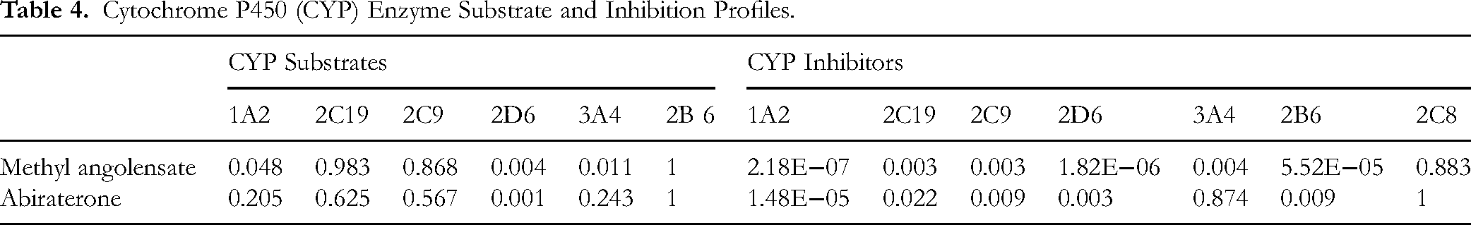
Cytochrome P450 (CYP) Enzyme Interactions
Methyl angolensate showed inhibitory potential against specific Cytochrome P450 (CYP) isoforms. In particular, the compound displayed a CYP2C9 inhibition score of 0.754 and a CYP2C19 inhibition score of 0.706. These values suggest a >70% likelihood of significant inhibition, which could affect the metabolism of co-administered drugs processed by these enzymes. This highlights the need for future pharmacokinetic studies to evaluate possible drug–drug interactions involving these isoforms (Table 4).
Cytochrome P450 (CYP) Enzyme Substrate and Inhibition Profiles.
Toxicity and Safety Profile
Toxicological risk was assessed using a suite of in silico models. The Ames mutagenicity scores were 0.132 for methyl angolensate and 0.107 for abiraterone, indicating a <15% probability of mutagenic potential, and therefore low genotoxic risk. In skin sensitization assays, methyl angolensate exhibited a sensitivity score of 0.429, which is ∼3.7-fold higher than abiraterone's score of 0.115 (Table 5). While still within an acceptable safety range, this may indicate a mild risk of skin irritation under topical exposure.
Toxicity and Safety Profile of Methyl Angolensate and Abiraterone.

Discussion
In this study, we investigated the potential of methyl angolensate as an antiandrogenic agent for the treatment of prostate cancer by examining its effects on cell viability, its binding affinity to the AR, and its pharmacokinetic properties. The inclusion of positive controls, such as metribolone and abiraterone, in the study was essential to validate the biological and pharmacological findings and provide a benchmark for comparison. Metribolone was chosen as a positive control in the docking studies with methyl angolensate and the AR due to its well-established role as a AR antagonist. Despite not being clinically approved, metribolone has been widely used in experimental studies as a reference AR antagonist due to its robust inhibitory activity on AR signaling. 32 Metribolone's binding to the AR was used to validate the docking procedure by providing a benchmark for strong AR antagonism. However, it was excluded from the pharmacokinetic study because it is not a clinically approved drug, limiting its relevance in real-world clinical applications. In contrast, abiraterone, a clinically approved CYP17A1 inhibitor used in prostate cancer therapy, was used as the positive control in the cell viability assays. This choice was driven by its documented efficacy in reducing cell viability, especially in androgen-responsive prostate cancer cell lines. 21 Abiraterone's clinical approval and its mechanism of action as an androgen biosynthesis inhibitor made it a suitable reference for comparison with methyl angolensate's cytotoxic activity.
The cell viability data presented in this study demonstrated that both the EA and its major metabolite exhibited moderate cytotoxic effects, especially in androgen-responsive cell lines such as LNCaP, 22Rv1, and VcaP whereas the activity was poor in non-androgen-dependent cells PC-3 and DU-145. These findings corroborate those of Zingue et al, 33 who reported that EA extract at optimal concentrations of 20-50 μg/mL, significantly inhibited the growth and proliferation of LNCaP, DU145, and PC3 prostate carcinoma cells. Thus, the weak activity recorded against DU145 and PC3, in our study, could be due to the low concentration used (10 µg/mL). In DU145 and PC3 prostate cancer cells, the extract was shown to up-regulate caspase-3 and down-regulates apoptosis-related proteins Akt, pAkt, and Bcl-2. 33 Our results and those reported provide credence to the ethnomedical application of E. angolense stem bark in prostate cancer management. Our results also provide the first report of the anti-prostatic carcinoma activity of the limonoid methyl angolensate and thus this compound could have contributed to the observed activity of the extract. Methyl angolensate has also been reported to show in-vitro anticancer activity against Human leukemia cell lines CEM and K562 GM00558B. 34 It induced apoptosis in the cells by binding to annexin V, fragmentation of DNA, upregulation of proapoptotic and downregulation of anti-apoptotic gene products. 35 Elsewhere, the chemotherapeutic potential of methyl angolensate was demonstrated in mice with Ehrlich ascites carcinoma where it inhibited the growth of the tumor by activating intrinsic pathway of apoptosis without affecting normal cells. 36 However, with both the extract and methyl angolensate showing less than 70% cell viability with the normal prostate epithelial cell line RWPE-1 after 48 h of treatment, suggest the need for caution in it use. We propose further studies to establish safe doses.
This activity correlates with the chemical properties of methyl angolensate, particularly its strong binding affinity to the androgen receptor. Methyl angolensate's binding free energy (ΔG = −16.5 kcal/mol) was notably stronger than that of metribolone (ΔG = −7.6 kcal/mol), suggesting that methyl angolensate may disrupt AR function more effectively than metribolone. This higher affinity for AR could explain its potent cytotoxicity in androgen-responsive prostate cancer cells. 37 Furthermore, its intermediate lipophilicity (logP = 1.977) is favorable for cellular membrane permeability, a property that may support its cellular uptake and, thus, its cytotoxic potential. The relatively higher solubility (logS = −3.899) further suggests that methyl angolensate may exhibit favorable pharmacokinetic characteristics in the gastrointestinal tract, which is critical for oral bioavailability. 38 To further validate the docking results and assess the stability of the ligand–AR complex, MD simulations totaling 100 ns were conducted, implemented as multiple 5 ns run due to technical constraints. These simulations confirmed the stability of the methyl angolensate–AR complex over time, with consistent RMSD and low root mean square fluctuation (RMSF) values across the receptor's backbone. These dynamic analyses support the hypothesis that methyl angolensate forms a stable and energetically favorable interaction with AR, which may underlie its cytotoxic activity in AR-expressing prostate cancer cells.
The pharmacokinetic evaluation of methyl angolensate highlighted several important aspects that are critical for its therapeutic potential. Methyl angolensate exhibited moderate lipophilicity and good solubility, which supports its potential as a drug-like molecule. However, the compound's relatively low permeability in MDCK and Caco-2 cell models, alongside its low VDss, suggests that methyl angolensate may have limited tissue distribution, particularly outside the bloodstream. This could be advantageous in terms of minimizing off-target toxicity, particularly to the central nervous system, as indicated by its low BBB permeability. Notably, the compound showed minimal interaction with P-glycoprotein (P-gp), reducing the likelihood of being affected by efflux mechanisms that typically limit the bioavailability of other compounds like abiraterone. 38 Moreover, methyl angolensate's interaction with CYP enzymes, particularly CYP2C9 and CYP2C19, suggests that it may have the potential to interact with other drugs metabolized by these isoforms. This underlines the importance of considering drug-drug interactions in clinical settings, as co-administration with other compounds that rely on these CYP enzymes for metabolism could alter the pharmacokinetics of either compound. 38 This aspect of methyl angolensate's pharmacokinetic profile warrants further investigation to minimize potential adverse interactions.
The role of AR inhibitors in prostate cancer therapy is well-documented. AR signaling is a critical driver of prostate cancer growth, particularly in castration-resistant prostate cancer, where androgen deprivation therapies become less effective. 37 Thus, the development of AR antagonists like methyl angolensate is an important therapeutic strategy. By binding to the AR with high affinity, methyl angolensate may block androgen-driven signaling pathways, preventing cancer cell proliferation and survival. Our results suggest that methyl angolensate has promising antiandrogenic potential, making it an attractive candidate for further clinical evaluation. The observed efficacy in reducing cell viability, coupled with its favorable pharmacokinetic profile, underscores the potential of methyl angolensate as a therapeutic agent in the treatment of prostate cancer.
Within the limitations of our resources, this research could not achieve one of its important aims to identify the antiprostate cancer compounds present in Etandrophragma angolense. Only one (methyl angolensate) of the several limonoids present in the plant could be isolated in this research. This was due to lack of reverse-phase prep-HPLC resources to afford the purification of these polar derivatives which could not be pushed down by ordinary gravity fed column chromatographic techniques. Future collaborative research shall explore the potential of some of these metabolites.
Conclusion
In conclusion, this study provides compelling evidence that Entandrophragma angolense stem bark extract and its metabolite methyl angolensate possess anti-prostatic carcinoma activity. Methyl angolensate holds promise as a novel antiandrogenic agent for prostate cancer treatment as it possesses strong binding affinity for AR, coupled with moderate cytotoxicity, favorable solubility, and and minimal toxicity, making it a viable drug candidate for oral delivery.
Supplemental Material
sj-docx-1-npx-10.1177_1934578X251384534 - Supplemental material for Anti-Prostatic Carcinoma Activity of Entandrophragma angolense and Methyl Angolensate: In Vitro and In Silico Analyses
Supplemental material, sj-docx-1-npx-10.1177_1934578X251384534 for Anti-Prostatic Carcinoma Activity of Entandrophragma angolense and Methyl Angolensate: In Vitro and In Silico Analyses by Linda Mensah Sarpong, Akwasi Acheampong, Mercy Badu, Silas Adjei, Jibira Yakubu, Kennedy Ameyaw Baah, Joseph Adusei Sarkodie and Isaac Kingsley Amponsah in Natural Product Communications
Supplemental Material
sj-docx-2-npx-10.1177_1934578X251384534 - Supplemental material for Anti-Prostatic Carcinoma Activity of Entandrophragma angolense and Methyl Angolensate: In Vitro and In Silico Analyses
Supplemental material, sj-docx-2-npx-10.1177_1934578X251384534 for Anti-Prostatic Carcinoma Activity of Entandrophragma angolense and Methyl Angolensate: In Vitro and In Silico Analyses by Linda Mensah Sarpong, Akwasi Acheampong, Mercy Badu, Silas Adjei, Jibira Yakubu, Kennedy Ameyaw Baah, Joseph Adusei Sarkodie and Isaac Kingsley Amponsah in Natural Product Communications
Supplemental Material
sj-docx-3-npx-10.1177_1934578X251384534 - Supplemental material for Anti-Prostatic Carcinoma Activity of Entandrophragma angolense and Methyl Angolensate: In Vitro and In Silico Analyses
Supplemental material, sj-docx-3-npx-10.1177_1934578X251384534 for Anti-Prostatic Carcinoma Activity of Entandrophragma angolense and Methyl Angolensate: In Vitro and In Silico Analyses by Linda Mensah Sarpong, Akwasi Acheampong, Mercy Badu, Silas Adjei, Jibira Yakubu, Kennedy Ameyaw Baah, Joseph Adusei Sarkodie and Isaac Kingsley Amponsah in Natural Product Communications
Supplemental Material
sj-docx-4-npx-10.1177_1934578X251384534 - Supplemental material for Anti-Prostatic Carcinoma Activity of Entandrophragma angolense and Methyl Angolensate: In Vitro and In Silico Analyses
Supplemental material, sj-docx-4-npx-10.1177_1934578X251384534 for Anti-Prostatic Carcinoma Activity of Entandrophragma angolense and Methyl Angolensate: In Vitro and In Silico Analyses by Linda Mensah Sarpong, Akwasi Acheampong, Mercy Badu, Silas Adjei, Jibira Yakubu, Kennedy Ameyaw Baah, Joseph Adusei Sarkodie and Isaac Kingsley Amponsah in Natural Product Communications
Footnotes
Acknowledgements
Authors are indebted to technical staff of the departments of Chemistry, Pharmacognosy and Herbal Medicine, Kwame Nkrumah University of Science and Technology for their invaluable assistance. We are also indebted to Prof Amit Pandey Lab, University of Bern (Switzerland) for making his lab available for the in vitro and molecular docking studies.
Ethical Approval
Ethical Approval is not applicable for this article.
Statement of Human and Animal Rights
This article does not contain any studies with human or animal subjects.
Statement of Informed Consent
There are no human subjects in this article and informed consent is not applicable.
Funding
The authors received no financial support for the research, authorship, and/or publication of this article.
Declaration of Conflicting Interests
The authors declared no potential conflicts of interest with respect to the research, authorship, and/or publication of this article.
Supplemental Material
Supplemental material for this article is available online.
References
Supplementary Material
Please find the following supplemental material available below.
For Open Access articles published under a Creative Commons License, all supplemental material carries the same license as the article it is associated with.
For non-Open Access articles published, all supplemental material carries a non-exclusive license, and permission requests for re-use of supplemental material or any part of supplemental material shall be sent directly to the copyright owner as specified in the copyright notice associated with the article.