
Abstract
Introduction
The versatile opportunistic pathogen Pseudomonas aeruginosa is one of the major pathogenic bacteria, capable of causing acute and chronic infections to humans and animals.1,2 It is responsible for severe nosocomial infections in humans, with a high incidence of infections occurring in immunocompromised patients, suffering from chronic obstructive lung disease 3 and is the most frequently isolated pathogen in patients with cystic fibrosis (CF) and is therefore one of the most clinically important pathogens. 4 It should be noted this flagellated bacteria easily moves and colonizes surfaces on which it establishes biofilms and makes its spread more rampant.5,6 Its pathogenicity is based on a vast and complex arsenal of soluble virulence factors (toxins, enzymes, exopolysaccharides, etc) and cellular structures (pili, secretion systems, etc) that allow it to adapt and persist in the CF respiratory tract throughout the process of infection and aiding the bacteria to survive the effects of the host's immune system. 7 These virulence factors intervene at the various stages of the process of infection and thus allow the progression of the disease by strengthening the adhesion, modifying the immune response of the patient, preventing phagocytosis, destroying host tissues and protecting the pathogen from the action of various antimicrobials. 8 The virulence factors produced by P. aeruginosa are globally controlled by an incredible, complex and interconnected control circuits and signaling systems which is triggered by the threshold population density of the microbial cells and coordinated by a communication network mechanism called quorum sensing (QS).9–11 The QS network in P. aeruginosa is therefore dependent on the density of the bacterial population and their ability to communicate with each other and it relies on the production and reception of small diffusible signal molecules mostly N-acyl-homoserine lactone (AHL) molecules.12–14 When these diffusible pheromones reach a critical threshold concentration, they bind to the type “R” transcriptional regulator. The successful linkage of autoinducers to transcriptional regulators activates the expression of “I” target genes associated with pathogenicity, ecological adaptation, control of virulence factors, the formation of biofilms and the development of antibiotic resistance. 15 Many physiological processes such as conjugation, symbiosis, antibiotic production, virulence, competence, sporulation, motility and biofilm formation are mediated by QS during which Gram-positive and Gram-negative bacteria use oligo-peptides and acylated homoserine lactones as autoinducers, respectively.2,16,17
P. aeruginosa has four self-regulating signaling systems that are hierarchically ranked and interconnected are able to modulate the activities of each other. 18 Elastase, associated with immune evasion (Las), rhamnolipid, related to the structure and dynamics of the biofilm (Rhl), Pseudomonas quinolone signal (Pqs) and integrated quorum sensing (Iqs).8,19 The Las system has a dominant role in this hierarchy since it can interfere by positively controlling the expression of the other three systems.1,20 The Las system controls the synthesis of N- (3-oxododecanoyl)-L- homoserine lactone (3-oxo C12-AHL) to activate the appropriate Las R regulator and also to trigger the expression of genes responsible for the production of Las A protease and Las B elastase as well as for the production of exotoxin-A.21,22 Similarly, the cytoplasmic receptor RhlR responds to the «N-butanoyl-L-homoserine lactone» (C4-HSL) synthesized by RhlI in order to regulate the expression of genes involved in the synthesis of elastase, rhamnolipids and alkaline proteases of sidephores and motilities.3,20,23 It also triggers the secretion of Pyocyanin which is a blue-green pigment with a crucial role in the virulence of P. aeruginosa. 15 It equally suppresses the immune response of the host cell by decreasing the frequency of ciliary beats, which can further alter the innate defense mechanisms of the respiratory tract as it induces neutrophil apoptosis and increases the production of interleukin IL-8.7,15,24,25 The third system, Pqs, uses synthesized signal molecules with a quinolone-based structure (heptyl-3 hydroxy-4 quinolone), which trigger transcription of the operon pqsABCDE-phnAB. 26 The Pqs system plays a variety of roles, including mediating intercellular signaling through QS, regulating virulence factors, iron acquisition, induction of oxidative stress and modulation of host immune responses.8,15,26 The fourth QS channel recently discovered is the Iqs which utilizes 2-(2-hydroxyphenyl)-thiazole-4-carbaldehyde as its signal molecule and contributes to regulating responses to environmental stress, inhibition of host's cell growth and stimulation of apoptosis.8,12,26
Other virulence factors are usually regulated by different systems, for example, the Las and Rhl systems are involved in the formation of biofilm. 21 Biofilms are highly structured architecture consisting of microcolonies fixed to a surface and embedded in a self-produced extracellular matrix composed of exopolysaccharides, proteins, nucleic acids and lipids.8,26,27–29 Bacteria cells use different forms of motilities such as swarming and swimming to colonize surfaces prior to biofilm establishment.30,31 In the early stages of lung infection, P. aeruginosa floats freely through the airways in planktonic form, and during the progression of the infection, the bacterial colonies transform into sessile biofilm mode. 32 There are many multifactorial dynamic processes that occur at various stages through well-established mechanisms leading to the colonization of the lungs and these steps are controlled by the QS and involve different virulence factors.19,27 Adhesion is the initial stage of biofilm formation and this stage usually requires different structural compartments. 33 This makes use of flagella that ensure the swimming motility necessary for attachment, dissemination, colonization of the host epithelium and transition from the planktonic phase to the biofilm mode.20,34 Once the attachment is achieved, P. aeruginosa passes to a more stable state of attachment and other types of surface motility, such as swarming and twitching motility, which are mediated by type IV pili and play important roles in mediating adherence to mucosal surfaces and subsequent colonization.33,35,36 Fixation of P. aeruginosa on surfaces is followed by progression towards a mature biofilm in which microcolonies develop and multiply on surfaces producing extracellular polymer substances (EPS) as a solid structural matrix for better attachement.12,15 This EPS self-secreted polymer matrix provides a scaffold and protective barrier to embedded cells against various stresses such as bacteriophages and host immune responses making them able to resist antimicrobial treatments up to 1000 times more than their planktonic counterparts.37–39 Biofilms contribute to chronic infections that are difficult to treat with ordinary antibiotics. 40 Thus, the inhibition of biofilm production and QS can disrupt the development of the protective three-dimensional structure providing a lead-way for the development of new anti-infective agents.26,33 Such new types of antimicrobials target virulence factors of the bacteria and not their vitality, and avoiding the strong pressure of conventional antibiotics which makes microbes to develop resistance.1,7,26
Humans have exploited aromatic medicinal plants as an indispensable source of therapeutic molecules, usually possessing antimicrobial activity, most especially plant essential oils (EOs). 41 EOs are a mixture of volatile compounds including fatty acids and alcohols, terpenoids and phenylpropanoids. 42 Several in vitro and in vivo studies have demonstrated interesting antimicrobial and antibiofilm effects of EOs against many pathogenic fungi and bacteria. 43 However, few studies exist on the anti-virulence and anti-quorum sensing properties of essential oils.22,44 Cistus plants are widely distributed plant in the Mediterranean area and they are very popular as herbal teas and equally used in traditional medicine, mostly for the treatment of infections.45,46 Over 111 phytochemical compounds with interesting biological activities of Cistus species have been reported. 47 Cistus munbyi is a medicinal plant that is endemic to Northwestern Algeria and Northeastern Morocco where it is mostly is used to treat some pulmonary infections. 48 This work focused on the extraction of EO from C. munbyi and determining its phytochemical composition using gas chromatography–mass spectrometry (GC–MS). Equally the effects of this EO on various virulence factors of P. aeruginosa strains were investigated and reported.
Experimental
Plant Material and Essential Oils Extraction
The whole aerial parts of C. munbyi were collected at the flowering stage in May 2022 in the coastal Region of Beni Khelad, located in the Northwest of Algeria at 35.13′48.7″ N, 1.36′23.6″ W. The specimens were taxonomically identified and authenticated by a botanist at the laboratory of Ecological Management of Natural Ecosystems of the University of Tlemcen in comparison with previous identification. 49 A voucher specimen was deposited in our laboratory under the code CS-BF250522. 500 g of the air-dried aerial parts of the collected plants were subjected to hydrodistillation for 3 h using a Clevenger-type apparatus according to the European Pharmacopoeia. The obtained essential oil (EO) which was 9.0 g, was dried over anhydrous sodium sulfate, stored in dark bottles at 4 °C until tested and analyzed.
Gas Chromatography-Flame Ionization Detector (GC-FID) and Gas Chromatography–Mass Spectrometry (GC–MS) Analyses
The essential oils were analyzed by GC-FID and GC–MS as described previously.50,51 For GC analyses, a Rxi-5Sil MS fused silica capillary non-polar column (30 m × 0.25 I.D., film thickness 0.25 µm) and FID was used. Temperatures for the injector and detector were set to 250 °C to 270 °C, respectively. Helium was used as carrier gas with a 1.4 mL/min flow rate. The sample injection volume was 0.2 µL with a split ratio of 20:1. A Class GC10 GC computer program was used to determine the percentage composition of the essential oils. The initial oven temperature was held at 60 °C for 5 min, then increased up to 240 °C with 4 °C/min increments and held at this temperature for 10 min. For GC-MS analyses, an ion trap MS and a Rxi-5Sil MS fused silica non-polar capillary column (30 m × 0.25 mm I.D., film thickness 0.25 µm) were used. The carrier gas was helium with a 1.4 mL/min flow rate. The injector and MS transfer line temperatures were 220 °C to 290 °C, respectively. The temperature of the ion source was 200 °C, and the injection volume was 0.2 µL with a split ratio of 20:1. Electronic ionization-mass spectrometry (EIMS) measurements were taken at 70 eV ionization energy. Mass range was from m/z 28 to 650 amu. Scan time was 0.5 s with 0.1 s interscan delays. The oven temperature was held at 60 °C for 5 min, then increased up to 240 °C with 4 °C/min increments and kept at this temperature for 10 min. For identification of essential oil components, co-injection with standards (whenever possible) was used, together GC retention indices determined using a homologous series of C7–C30 alkanes (Supelco), and computer matching with the Wiley, TRLIB, and NIST08 libraries, as well as a comparison of fragmentation patterns documented in the literature. 17 Quantification of each of the essential oil's individual constituents was based on internal normalization for the components.
Bacterial Strain and Growth Conditions
Twenty multi-resistant clinical strains of P. aeruginosa were selected to test the in vitro antibacterial activity of C. munbyi EO. The strains were isolated from respiratory samples of patients with pulmonary infections at the University Hospital of Tlemcen (Algeria). These isolates were sub-cultured on selective medium citremide (HIMEDIA M024) and identified by manual and automatic systems (API20NE, Vitek2). The wild type Pa01 were selected based on their ability to form biofilm, to produce pyocyanin, and to swarm and swim and two reference strains P. aeruginosa ATCC 27853 and P. aeruginosa ATCC 9027 were used as positive controls. 52 The mutant Chromobacterium violaceum CV026 and C. violaceum CV12742 were used to evaluate the inhibitory effect of C. munbyi EO on quorum sensing and violacein production respectively. All clinically isolated strains were maintained in glycerol (30%) and stored at −80 °C. For every new experiment, fresh stocks were sub-cultured. P. aeruginosa strains were grown in Brain Heart Infusion broth (BHIB) at 37 °C. Chromobacterium violaceum CV026 and CV12742 were routinely cultured aerobically in Luria–Bertani (LB) broth and the cultures were incubated at 30 °C and 37 °C, respectively, for 24 h.
Antimicrobial Activity Assay
The preliminary antimicrobial activity of C. munbyi EO was carried out by the well diffusion method. 53 Using a sterile cotton swab, fresh standardized cultures (OD600 ≈ 0.1) were extensively streaked onto petri dishes filled with Mueller-Hinton (MH) agar (Fluka®, India). Wells with 6 mm diameter were made on the MH agar using a sterile cork borer. 20 µL of C. munbyi EO was added to the wells, and the plates were incubated at 37 °C for 24 h. The diameter of the zones of inhibition was taken as a measure of the antimicrobial activity. All experiments were performed in triplicate.
Determination of Minimum Inhibitory Concentrations
The minimal inhibition concentration (MIC) value was determined for all bacteria tested in this study using the microdilution assay in 96-well microtiter plates, according to Clinical and Laboratory Standards Institute (CLSI) 2017. 54 Briefly various concentrations of EO were prepared by mixing proportions of EO (4%-0.007%, v/v) with 1% dimethyl sulfoxide (DMSO) solution. The test medium was Mueller–Hinton (MH) broth for P. aeruginosa strains and LB broth for C. violaceum CV026 and C. violaceum CV 12472, and the density of bacteria was 5 × 10⁵ colony-forming units (CFU)/mL. Serial two-fold dilutions of the EO ranging from 4% to 0.007% v/v were prepared in 96-well microtiter plate supplemented with test culture and incubated for 24 h at 37 °C. As the control, bacterial cultures were performed in DMSO at a concentration of 1% V/V. MIC concentration was estimated visually and was defined as the lowest concentration needed to inhibit the growth of each tested micro-organism in the wells. Concentrations below the MICs were considered sub-inhibitory and were further used to study the anti-QS and biofilm inhibitory properties in the above-mentioned strain.
Biofilm Formation Assays
As described by to O’toole, 55 the microtiter dish assay was used to evaluate the inhibition of biofilm formation by C. munbyi EO at two different time points during biofilm development. The EO was introduced into the medium at the beginning of the cultivation (0 h, pre-adhesion period), and after biofilm formation (24 h of bacterial culture).
Pre-Adhesion Period
A total of 180 µL of diluted overnight cultures of the tested pathogens (5 × 10⁵ CFU/mL) were dispensed into each microplate well in the absence and presence of 20 µL of varying concentrations of C. munbyi EO. DMSO was used as a control. After incubation of the microplates at 37 °C for 24 h, the plates were rinsed with water to remove planktonic cells and subsequently stained with 0.1% crystal violet for 10 min. The stained wells were rinsed, and the biofilm was dissolved in 30% acetic acid. Measurements of optical densities were spectrophotometrically performed at 490 nm in a microplate reader (Biotek Elx800 MER) using 30% acetic acid as a blank. The experiments were repeated three times.
Preformed Biofilm Eradication Assay
To determine the potency of the C. munbyi EO to inhibit mature biofilms, an assay on the preformed biofilm was also realized. Wells of a sterile 96-well microplate were filled with 200 µL of MH broth containing a 1/100 dilution of the overnight bacterial culture. Wells without EO served as controls. After 24 h incubation at 37 °C the contents of the microplates were poured out and the wells were gently washed three times with sterile distilled water. Then 200 µL of fresh medium with or without different concentrations of C. munbyi EO was added to each well. The inoculated microplates were re-incubated for an additional 24 h (48 h total) at 37 °C. After 24 h, the plates underwent the same treatment and the biofilms were quantified using crystal violet staining as previously described.
Percentage of inhibition of the tested sample was calculated using the formula in below.

In Situ Visualization of Biofilms
Visualization of the inhibition of biofilm production in situ at three different time stages is based on a previously reported methodology with some modifications.56,57 Briefly, 1% of overnight cultures of the tested pathogens (0.4 OD at 600 nm) were added in 24-well plates to 1 mL of fresh MH medium containing sterile glass coverslips in the absence and presence of different concentrations of C. munbyi EO. After an adequate incubation time (1 h for adhesion and 24 h for biofilm formation), the coverslips were rinsed with distilled water to remove planktonic cells and the biofilms formed were stained with a 0.1% crystal violet solution. The stained and air-dried coverslips were placed on slides with the biofilm facing up and the visible biofilms were visualized under a light microscope (Carl ZEISS Einbautrafo 392575-9001) at magnifications of 40×. To visualize the action of C. munbyi EO on mature biofilms, biofilms were grown on 1 cm × 1 cm glass slide placed in 24-well plates containing an overnight culture re-suspended in 1 mL of fresh medium. After 24 h incubation, the cover glasses were rinsed thrice with distilled water to remove the planktonic cells and mature biofilms were then treated with respective sub-MICs of C. munbyi EO for 24 h, biofilms were visualized by light microscope as described previously.
Visualization of Biofilm Under Scanning Electron Microscope
In order to visualize the effect of C. munbyi EO on biofilm formation under scanning electron microscope (SEM), biofilms on coverslips were grown in the treated and untreated cultures as described in light microscopic analysis. After incubations coverslips were rinsed with distilled water to remove unbound planktonic cells and biofilms were fixed with 2.5% (v/v) glutaraldehyde in PBS for 5 h at room temperature. After washing, dehydration was done in ethanol with different concentrations (30, 50, 70, 90% and 100%) as described by Bose et al.2,56 Coverslips were dried, gold coated and viewed under field. 100 TM Hitachi environmental scanning electron microscope, at pressure in microscope chamber of 4 Torr.
Antivirulance Activities
Violacein Inhibition
The inhibition potential of the violacein produced by C. violaceum CV 12472 was evaluated by quantitative analysis as described by.
58
For the experiment 10 μL of overnight fresh cultures of C. violaceum CV12472 (0.4 OD at 600 nm) were mixed with 170 μL of LB broth in sterilized microplates and 20 μL of MIC and sub-MIC concentrations of C. munbyi EO. Assay in without EO (LB broth and C. violaceum CV12472) served as positive control. After incubation at 37 °C for 24 h for test plates, the absorbances were read at 585 nm in a 96-well plate microplate reader (SpectraMax, Molecular Devices) to determine any reduction of violacein pigment with respect to the control. Violacein inhibition expressed as percentage inhibition was deduced from the formula:

Bioassay for Quorum-Sensing Inhibition (QSI) Activity Using C. violaceum CV026
The anti-quorum sensing activity of C. munbyi EO was evaluated using a modified version of a previously described method.58,59 First, 5 mL of Soft Top Agar, which consisted of 1.3 g agar, 2.0 g tryptone, 1.0 g sodium chloride, dissolved in 200 mL of deionized water, was mixed with 100 μL of an overnight culture of C. violaceum CV026 and 20 μL of 100 μg/mL acylhomoserine lactone (AHL), precisely N-hexanoylhomoserine lactone (C6-HSL; Sigma-Aldrich) as an external source of hormones. This mixture was then poured gently onto the surface of a solidified LB agar plate as an overlay. After the overlay was solidified, wells with a diameter of 5 mm were made on each plate and filled with 50 μL of C. munbyi EO at MIC and sub-MIC concentrations. Each experiment was performed in triplicate and the plates were incubated at 30 °C for 3 days. The diameter of the quorum sensing inhibition zone, which was indicated by a white or cream-colored halo around the well against a purple lawn of activated C. violaceum CV026 bacteria, was measured in millimeters.
Pyocynin Assay
Pyocyanin production in treated and untreated cultures of P. aeruginosa was determined as described by Essar et al 60 Briefly, bacterial cells were inoculated in Pseudomonas broth (PB) (20 g of Bacto-Peptone, 1.4 g of MgCl2, and 10 g of K2SO4 per liter of distilled water) with or without C. munbyi EO at their respective sub-MIC concentrations and incubated for 72 h at 37 °C. As the control, the PB was supplemented with 0.5% of DMSO. Cell free supernatants collected by centrifugation at 10 000 g for 15 min was extracted with 3 mL of chloroform and then re-extracted in 1 mL of 0.2 M HCl to get a pink to deep-red colored solution. The relative concentration of pyocyanin was subsequently spectrophotometrically quantified at 520 nm.
Swarming and Swimming Motiliies
To study the effect of C. munbyi EO on swarming motility, overnight culture of P. aeruginosa strains was point inoculated at the center of swarming medium consisting 1% w/v peptone supplemented with 0.5% w/v each of
Extraction and Quantification of Exopolysaccharide
Test strains grown in the presence and absence of sub-MICs of C. munbyi EO were centrifuged, and the resulting supernatant was filtered. Three volumes of chilled 100% ethanol were added to the filtered supernatant, which was then incubated overnight at 4 °C. 61 Precipitated EPS were collected by centrifugation at 10 000 g for 20 min and quantified by measuring sugars following the method of Dubois et al. 62
LasA Staphylolytic Assay
LasA protease activity was measured by determining the efficiency of culture supernatants of P. aeruginosa to lyse boiled Staphylococcus aureus cells. 63 A 30-mL overnight culture of S. aureus was boiled in a water bath at 100 °C for 10 min and centrifuged for 10 min at 20 683 g. Then, the pellet was resuspended in 10 mM Na2PO4 (pH 4.5) and then diluted with the same buffer to an OD of (∼0.8 at 600 nm). After that, a 100-μL aliquot of cell free supernatant of P. aeruginosa cultured with or without EO was added to 900 µL S. aureus suspension, and the OD at 600 nm was determined after 10, 20, 40, and 60 min. The percentage inhibition of protease activity was calculated with respect to the assay control (without C. munbyi EO). 64
Results
Chemical Composition of the Essential Oil
After the hydrodistillation process, it was found that from 500 g of plant material, 9.0 g of EO were obtained, making a yield of 1.8%. GC–MS was used to determine the chemical composition of C. munbyi EO and the results are presented in Table 1. The results of the analysis show the presence of 44 identified compounds (100%) as presented in Table 1. The compounds identified include a variety of monoterpenes and sesquiterpenes. Monoterpene hydrocarbons represent the largest proportion, accounting for 57.01% of total oil volume, while monoterpenoids and oxygenates account for 41.22%. Sesquiterpene hydrocarbons and sesquiterpenoids are present in smaller quantities, at 1.03% and 0.27%, respectively. The composition of C. munbyi EO is characterized by a balanced mixture of several compounds. There is no dominant compound, but rather a significant combination of Terpinen-4-ol (33.20%), Sabinene (13.20%), α-Thujene (11.30%), and p-Cymene (9.94%). It is also noteworthy that other compounds are present in significant quantities, such as γ-Terpinene (13.60%), α-Terpinene (6.81%), and p-Cymene (9.94%).
Chemical Composition of Essential oil from Cistus munbyi.

Retention index experimentally determined using homologous series of C7–C30 alkanes on Rxi-5Sil MS fused silica column.
Linear retention index taken from Adams (2007) and/or NIST 08 (2008).
Percentage concentration.
Identification methods:
Phenotypic Characterization of Clinical and References Strains
P. aeruginosa isolates selected for this study were characterized for their susceptibility to a range of antibiotics, and their ability to produce specific virulence factors, such as biofilm formation, pyocyanin production, swarming and swimming motility. The evaluation of these characteristics in the strains studied is reported in Table 2.
Phenotypic Characterization of Clinical and Reference Strains.

Biofilm production during an incubation period of 24 h without medium replacement. b Biofilm production during an incubation period of 48 h with medium replacement after 24 h.
Antimicrobial Effect of C. munbyi EO
The antibacterial effect of C. munbyi EO was verified by two laboratory tests; by determining the diameter of the inhibition zone and the MIC value. As summarized on Table 3 for the most susceptible strains, the EO demonstrated a significant inhibitory effect against all P. aeruginosa isolates and reference strains tested, with inhibition zone diameters ranging upto 22 ± 0.5 mm against the Pa3 strain and MIC values as low as 0.125% to 1% v/v against the Pa3 and Pa16 strains. Interestingly, Pa6 was most susceptible as EO had highest inhibition zone diameter of 22 ± 0.5 mm against it. In the microdilution assay, Pa3 and Pa16 were most susceptible as EO had lowest MIC values of 0.125% v/v against both of them. Antimicrobial activity of the EO against the less susceptible strains is provided in Supplemental material (Table S3).
Antimicrobial Activity of C. munbyi EO (Mean Inhibition Zone and Inhibitory Concentration (MIC) Values in mm and % v/v).

Antibiofilm Activities of C. munbyi EO
In this study, the effect of C. munbyi EO on biofilm formation was evaluated by the crystal violet test on polystyrene and glass, which are the most frequently used materials in medical devices. The percentages of biofilm inhibition against the more susceptible strains assayed at 2XMIC, MIC and sub-MIC concentrations are summarized in Table 4 (for young biofilms after 24 h) and Table 5 (for mature biofilms after 48 h). Antimbiofilm activity of the EO against the less susceptible strains are provided in the Supplemental material (Tables S4 and S5). Microtiter plate assay of the anti-biofilm activity of C. munbyi EO showed a dose-dependent reduction of biofilm biomass in the tested strains. Overall, it was found that at MIC concentration, EO removed more than 70% of the biofilm from 19 strains out of 24 tested, which represents 79.11% of all the tested strains. The most biofilm producing strains named Pa2, Pa3, Pa6, and Pa16 were more resistant to the antibiofilm effect of EO with an inhibition rate of 39.09%, 57.22%, 49.44%, and 59.07%, respectively. At MIC/2 and MIC/4 concentrations, the C. munbyi EO was less active with a rate ranging from 29.3% to 85.12% at MIC/2 and 12.6% to 77.44% at MIC/4. The strongest inhibition was observed against the Pa20 isolate, while the Pa6 strain was the most resistant. Furthermore, the inhibition rate was less than 45% at MIC/8% and 30% at MIC/16. Mature biofilms are much difficult to destroy compared with young biofilms and planktonic cells. For this purpose, pre-formed biofilms (48 h) were treated with sub-MIC of C. munbyi EO to evaluate its eradication ability. The ability of C. munbyi EO to eradicate pre-formed biofilms was found to be less important than that exerted on young biofilms.
Quantitative Measurement of the Inhibition of Young Biofilm (24 h) Estimated by the Crystal Violet Staining (Measurement at 490 nm).

The data represents mean values of three independent experiments.
Quantitative Measurement of the Inhibition of Mature Biofilm (48 h) Estimated by the Crystal Violet Staining (Measurement at 490 nm).
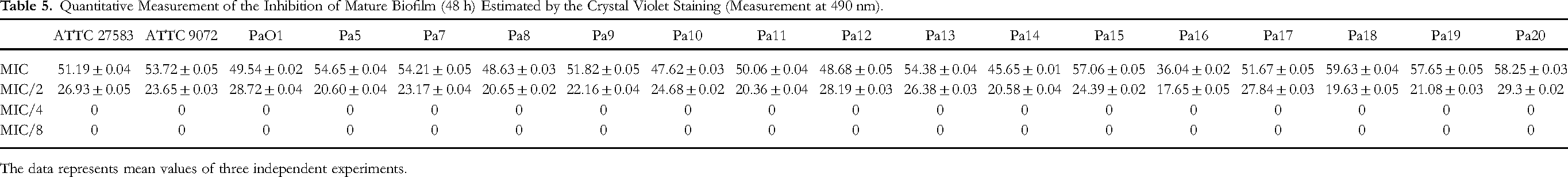
The data represents mean values of three independent experiments.
Scanning Electron Microscopy (SEM) Analysis
To demonstrate clearly the antibiofilm effect of C. munbyi EO, Microscopic investigations were conducted on glass slides in a static condition in the absence (control) and in the presence of sub-MIC concentrations of EO. Light microscopic images showed a very intensive biofilm formation on the control surface with a characteristic organization of cells in the early stages of adhesion, while a few cells were observed on the treated surfaces. SEM images taken with mature biofilms after 48 h (Figure 1a) and young biofilms after 24 h (Figure 1b) revealed that the untreated control had a well-developed biofilm while a significant reduction of young biofilms was observed with distorted architecture and isolated microcolonies upon treatment with EO in all tested strains. Compared to the control (Figure 1a and 1b), cells treated with C. munbyi EO showed slight cell shrinkage with morphology changes.

(a) SEM images of P. aeruginosa PA01 after 48 h of incubation (C = control). (b) SEM images of P. aeruginosa PA01 after 24 h of incubation (C = control).
Anti-Virulence Activities
Violacein Inhibition
AHL-mediated QS regulates biofilm formation and virulence factors such as secretion of violacein. The best studied model system for screening QS inhibitors is C. violaceum 12472 (CV12472) since it prodeuces an easily measurable pigment violacein. In this study a quantitative evaluation of the QS inhibitory activity of C. munbyi EO was performed based on the development of the biosensor organism and the decrease in violet pigment (violacein) synthesis at different concentrations compared to the untreated control. The violacein inhibition assay was performed at MIC and sub-MIC concentrations after determining the MIC value of C. munbyi EO. The obtained results showing a reduction of purple pigmentation as shown in Figure 2, indicates that C. munbyi EO inhibited the growth of C. violaceum CV12742 at MIC (0.0625%, v/v), as well as complete inhibition of AHL-mediated violacein production at MIC and MIC/2 concentrations. This inhibition was approximately 71.70%; 44.95% and 12.85% at MIC/4, MIC/8 and MIC/16 concentrations respectively. The results of violacein inhibition are given in Table 6.

Quorum-sensing inhibition plate (A); violacein inhibition plate (B).
Inhibition of Violacein Production Against C. violaceum CV12472 and Anti-QS Activity Against C. violaceum CV026 by C. munbyi EO.

Anti-Quorum Sensing Activity
C. violaceum CV026 is a mini-Tn5 mutant strain that produces purple pigmentation due to QS-dependent expression of the genes encoding violacein when supplemented externally with an inducing concentration of medium-chain AHLs. It is suitably used as a model bacterium to determine disruption of quorum sensing by determining zones of quorum sensing inhibition. 65 MIC and sub-MIC values of C. munbyi EO against C. violaceum CV026 were determined before the determination of quorum sensing inhibition zones. A measurable colorless halo against a purple lawn of produced violacein (Figure 2) observed around wells indicated QS inhibition zones. The MIC value was 0.25%v/v and showed an anti-QS activity with inhibition diameter zone of 23.0 ± 0.5 mm. Colorless halos observed around wells containing MIC/2 and MIC/4 concentrations of C. munbyi EO, indicating anti-QS activity with a diameter of 08.0 ± 1.0 mm and 0.7 ± 0.5 mm, respectively, whereas MIC/8 concentration was inactive.
Effect of C. munbyi EO on Motility Phenotypes
P. aeruginosa possesses a unique polar flagellum involved in adhesion to respiratory epithelial cells and participates in both swarming and swimming motility which play an important role in the early stages of biofilm development and formation. To verify the ability of C. munbyi EO at different concentrations to reduce swarming and swimming motility, tests were performed on different motility media by spot inoculation of P. aeruginosa isolates pre-selected for the assay. The strains denoted Pa01, Pa ATCC9027, Pa16, Pa2, and Pa3 were chosen according to their high capacity to invade agar plates with diameters exceeding 30 mm. The swimming and swarming motility plates are given on Figure 3 and the reduction in swim or swarm fronts can be seen on plates. The results demonstrate that all sub-MIC doses used exerted a concentration-dependent inhibitory effect on swarming and swimming motilities in the plates compared to control plates with untreated cells. However, major migration inhibition of all tested strains was achieved at MIC and MIC/2 with good percentage inhibitions and in both models, Pa3 was most susceptible while Pa01 was least susceptible. The results of motility inhibition of C. munbyi EO are given on Table 7.
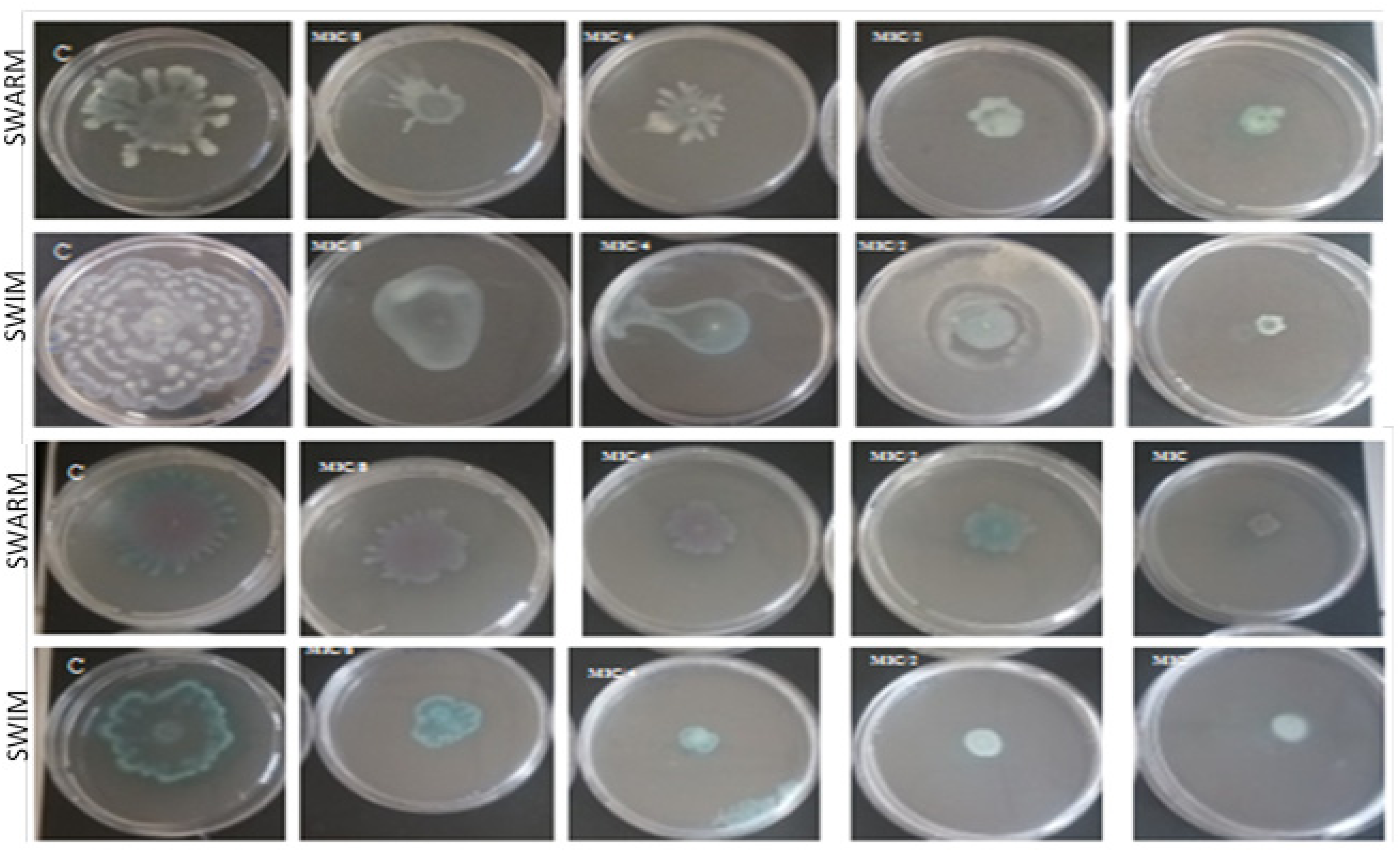
Swimming and swarming motility plates (C = control).
Effect of C. munbyi EO on Swarming and Swimming Motility.

Effect of C. munbyi EO on Pyocyanin Production
Among the many QS-controlled virulence factors produced by P. aeruginosa during infection, pyocyanin is a redox-active blue-green phenazine pigment involved in many pathogenic mechanisms. 66 Therefore, inhibition of pyocyanin production is important for disease progression. As summarized in Table 8 for the most susceptible strains, pretreatment with C. munbyi EO at MIC and sub-MIC concentrations showed that the inhibitory effect on pyocyanin production was positively correlated with EO concentration of with compared to that of untreated control. At the MIC concentration, a maximum of more than 80% inhibition of pyocyanin production was observed at all tested strains, while at concentrations below the MIC, EO was still able to significantly inhibit pyocyanin production in all strains tested, with inhibition rates ranging from 54.34% (Pa12 strain) to 86.66% (Pa14 strain) at MIC/2, from 24.32% (Pa8 strain) to 56.93% (Pa1 strain) at MIC/4. Whereas the inhibition rate was less than 19% for all strains tested at MIC/8. The inhibition of pyocyanin production of the EO against the less susceptible strains is provided in Supplemental material (Table S8).
The Quantitative Assessment of Pyocyanin Inhibition (%).

The data represents mean values of three independent experiments.
* P ≤ .05.
** P ≤ .005.
Inhibition of EPS Production by C. munbyi EO
The biofilm confers resistance to different antimicrobial agents on the bacteria embedded within it. This resistance is due to the EPS matrix which plays a crucial role in the formation and maintenance of the biofilm architecture. The study of the inhibitory effect of EPS production was performed on four selected clinical strains of P. aeruginosa, namely Pa2, Pa3, Pa6, and Pa16 and the model strain Pa01. These strains were selected based on their high capacity to produce biofilms. In P. aeruginosa Pa01, a maximum reduction of 90.62% was observed at MIC concentration (Figure 4). At MIC/2, MIC/4 and MIC/8 concentrations C. munbyi EO reduced significantly (P < 0.05) EPS production by 64.21%, 42.17% and 15.32% respectively compared to the untreated control. The tested strains at MIC concentration showed maximum reduction (70-90%) in the concentration of EPS, according to the spectrometric examination at 490 nm. The oils tested decreased EPS production by (45-56%) and (30-40%) at MIC/2 and MIC/4 concentrations, respectively, while MIC/8 was almost inactive with less than 11%.

Quantitative analysis of EPS inhibition by measuring the absorbance at 490 nm.

Inhibition of LasA staphylolytic activity by different concentrations of C. munbyi EO. The percentage of LasA inhibition was calculated with respect to control OD at 600 nm.
Inhibition of Las A Staphylolytic Activity by C. munbyi EO
QS in P. aeruginosa controls the secretion of a range of extracellular protease enzymes, such as protease IV, alkaline protease, elastase A and elastase B, which are associated with the virulence of this opportunistic pathogen. Las A elastase (also called staphylolytic protease or staphylolysin) is a serine endopeptidase able to cleave the pentaglycine bridge present in the peptidoglycan of S. aureus cell walls. 63 The percentage inhibition of LasA staphylolytic activity by different concentrations of C. munbyi EO were evaluated at different times and summarized on Table 9 for the most susceptible strains. The plots of percentage inhibition of LasA staphylolytic activity against time are given on Figure 5. The supernatant from EO-treated cultures showed a significant reduction in Las A staphylolytic activity in a concentration-dependent manner compared to that of the control. Complete data of Table 9 for Las A staphylolytic activity is provided in the Supplemental material.
Inhibition of LasA Staphylolytic Activity by Different Concentrations of C. munbyi EO.

Data are reported as the percentage of residual pyocyanin inhibition after the C. munbyi EO treatment in comparison with untreated controls.
The data represents mean values of three independent experiments.
Discussion
P. aeruginosa is an opportunistic bacterium known to cause lung infections, particularly in patients with CF and other chronic lung diseases. 25 The effectiveness of antibiotics that act directly on bacteria can be compromised by biofilm formation, particularly under conditions of stress. As a result, current therapeutic options for combating various pathogens are inadequate and pose significant health problems. Biofilms provide a protective environment for bacteria and increase their resistance to antibiotics and the immune system. This therefore poses a serious problem for human health, making it important to research new therapies that directly target or disrupt biofilm formation and attenuate virulence factors controlled by QS to overcome this resistance and improve clinical outcomes. Compounds derived from natural sources, especially secondary plant metabolites, are valuable resources in the context of QS inhibition.67,68 Consequently, C. munbyi was chosen in this study as it has been used to alleviate lung infections for many years in folk medicine. Although some biological activities of C. munbyi have been tested in many aspects, its QSI properties against P. aeruginosa have not yet been explored.
In the first part of the work, the chemical composition of C. munbyi EO was established by GC-MS analysis. Our results indicate that Terpinene-4-ol (33.2%), γ-Terpinene (13.6%), Sabinene (13.2%), α-Thujene (11.3%), p-Cymene (9.94%), and α-Terpinene (6.81%) are the main compounds. These results agree with some quantitative and qualitative differences with the work of Benbelaid et al
48
in which terpinen-4-ol was also dominant but with a percentage of (23.75%). A significant difference lies in the both the class and amount of certain compounds identified in this study compared to other reports. In our work, p-Cymene and α-Terpine were detected in significant concentrations, whereas the study by Benbelaid et al
32
highlighted the presence of other compounds such as trans-Sabinene hydrate (
The main objective of this study was to focus on the attenuation of virulence factors phenotypically expressed by P. aeruginosa strains isolated from patients suffering from pulmonary infections. Firstly, the antimicrobial activity of C. munbyi EO was explored, and the MIC was determined to select sub-MICs in order to study their effects on growth and the inhibition of QS-regulated functions. The MIC of C. munbyi EO was found to be between 0.125% and 1%, consistent with previous studies and suggesting that the chemical compounds present in C. munbyi EO account for the antimicrobial effects. 48 The anti-biofilm effect of C. munbyi EO against P. aeruginosa evaluated on polystyrene and glass which are materials commonly used in medical devices, indicated interesting results. The findings showed a dose-dependent reduction in biofilm biomass for most of the strains tested and the inhibition percentages at same dose varied from one strain to another. This observation was equally valid at all stages of biofilm formation, whether in the pre-adhesion test by adding EO at the start of the microbial culture or after one hour of adhesion, or on the mature biofilm with the application of EO 24 h after its formation. However, EO was found to be less effective at eradicating mature biofilms than young biofilms, consistent with the fact that mature biofilms are known to be more resistant and difficult to remove than young biofilms and planktonic cells. These findings were confirmed by optical and electron microscopic observations. Optical microscope images showed very intensive biofilm formation on the control surface with the characteristic organization of cells in the early stages of adhesion, while only a few cells were observed on EO-treated surfaces. Scanning electron microscopy results revealed a significant reduction in young biofilms with distorted architecture and isolated microcolonies upon EO treatment for all strains tested. In addition, cells treated with C. munbyi EO showed slight cell shrinkage with changes in morphology compared to the untreated control. These effects may be attributed to the chemical composition of C. munbyi essential oil, and in particular to its major compound Terpinen-4-ol. A previous study, such as that carried out by Noumi et al 2018, 52 has demonstrated that Terpinen-4-ol disrupts biofilm production and simultaneously inhibits the adhesion of S. aureus to polystyrene and glass surfaces. Another more recent study carried out by Bose and co-workers also highlighted the potential of Terpinen-4-ol in the attenuation and formation of biofilms in P. aeruginosa. 3 This observed variability may be due to the individual sensitivity of bacterial strains to different compounds present in EOs. Different compounds may act synergistically or complementarily to disrupt biofilm formation, interfere with the extracellular matrix or inhibit cell communication.70,71 Therefore, the ability of C. munbyi EO to reduce biofilm formation by inhibition, as shown in this study, is a good indication of its possible application in the elimination of resistance and virulence during infections.
It is well established that bacterial communication and secretion of virulence factors are related to QS-regulated phenotypes. In order to further understand the mechanism of attenuation of cell communication signals in P. aeruginosa called QS, by C. munbyi EO, an in vitro study based on competition with appropriate biosensor bacterial strains was carried out. C. violaceum CV12742, which produces purple pigmentation in response to QS, and C. violaceum CV026, which requires the addition of a specific hormone AHL to trigger a QS-mediated process were used and they represent signal emission and signal reception, respectively. It was observed that the sub-MIC of C. munbyi EO showed a pronounced interference in the production of violacein in C. violaceum CV12742 up to the MIC/16 concentration. Similarly, C. munbyi EO exerted anti-quorum sensing activity against C. violaceum CV026, reflected by a creamy halo around the wells, while violacein inhibition was assessed by observing the absence of violet pigment. The ability of EOs to disrupt quorum sensing in bacteria may be a promising target for attenuating virulence and countering the progression of resistance. According to previous studies, anti-quorum sensing activity has been observed in several EOs and their main components, 72 including Mentha piperita, 73 Syzygium aromaticum,74,75 Rosmarinus officinalis, 76 Piper bredemeyeri, P. bogotense, P. brachypodom, 77 and Lippia alba. 78 Many EOs have demonstrated good ability to inhibit QS activity on C. violaceum, even at their sub-MIC. 79 In a study conducted by Ramírez-Rueda and Salvador in (2020), the anti-quorum sensing activity of 24 EOs was evaluated on C. violaceum out of which 17 showed positive effects on violacein inhibition. 18 It is interesting to note that previous studies have already highlighted the anti-quorum sensing activity of certain EOs that share similar major compounds with C. munbyi EO namely Citrus limon (γ-terpinene 10.1%), Eucalyptus polybractea (p-cymene 25.5%), 80 Citrus clementina (sabinene 31.4%), Melaleuca alternifolia (Terpinene-4-ol 40.4%, γ-terpinene 19.5%, and p-cymene 4.7%) 52 M. alternifolia (Terpinene-4-ol 45.6%, γ-Terpinene 19.4%, and p-Cymene 7.6%). 50 Extensive studies have highlighted the potential of terpinene-4-ol to attenuate QS pathways through inhibition of long-chain AHLs.52,57 These findings reinforce the idea that these compounds may play a key role in inhibiting QS and suppressing bacterial virulence activities.
Among the many colonization strategies employed by P. aeruginosa is the use of the flagellum to generate a rotational movement that enables it to swim in liquids such as water, culture media or lung secretions, moving towards sites of infection and colonizing surfaces. Swarming, on the other hand, is a form of movement that occurs on solid surfaces, such as culture plates or cell surfaces. 8 Swarming and flagellum-mediated swimming are QS-dependent virulence functions (regulated by the Rhl system) that play a crucial role in the initiation of cell/surface attachment during biofilm development in lung infections. 25 The attenuation and inhibition of these strategies may therefore guide the development of new therapeutic approaches and more effective preventive measures against lung infections caused by P. aeruginosa. The results in this study suggest that C. munbyi EO may have the ability to interfere with the QS system mediated by Las and Rhl, as well as with flagellum functions. A study by Ramírez-Rueda & Salvador showed swarming inhibition in the range of 85.7% to 100% compared to the control. Coridothymus capitatus EO also strongly affected the swarming and swimming motility of P. aeruginosa in CF patients.7,18 A maximal reduction in swarming motility was observed upon treatment with sub-MIC of Cinnamomum tamala EO. 81 Similar results were obtained with clove oil, where different concentrations significantly reduced swimming motility compared to the untreated control. 73 The antimotility effect of Terpinen-4-ol has already been described and the percentage inhibition of PA01 swarming was about 25% at a concentration of 100 g/mL of Terpinen-4-ol. 52 A significant reduction in swarming (33.3%), twitching (50%) and swimming (25%) movements compared to the untreated control to the presence of sub-MIC (0.06%) of Terpinen-4-ol have been reported as well. 3
Pyocyanin production also contributes to P. aeruginosa virulence factors. One of the main mechanisms by which pyocyanin contributes to virulence is its ability to interfere with numerous cellular functions in host cells via an oxidative process; this causes oxidative damage to cellular components, cell membrane damage, DNA degradation, protein inactivation. 82 It can also alter cellular signaling pathways, leading to an exacerbated inflammatory response in the respiratory tract. 24 Furthermore, this green pigment, whose production is controlled by QS, is also involved in suppressing the ciliary activity of respiratory epithelial cells, thus compromising mucociliary clearance, an important pulmonary defense mechanism against pathogens. This promotes the persistence of P. aeruginosa in the airways, leading to more severe colonization and infection. 83 The results of our study undoubtedly demonstrated a significant decrease in pyocyanin production for all strains tested in the presence of C. munbyi EO. This observation strongly suggests a possible interference of EO with the activation and regulation of the QS system in P. aeruginosa. Several previous studies have reported a reduction in pyocyanin production following the use of EOs and extracts from various plants. The results of a previous study showed a 75% reduction in pyocyanin production in P. aeruginosa strains treated with Micromeria thymifolia EO, which contains numerous phytochemicals, including Terpinen-4-ol. 17 More recently, Terpinen-4-ol was shown to be able to inhibit pyocyanin production by 33% to 96% efficiency. 3
EPS is one another virulence factor of P. aeruginosa, which facilitates the formation of biofilms and promoting the chronicity of infections.33,84 In the present study, C. munbyi EO significantly inhibited the EPS production of P. aeruginosa PA01 and clinical strains. Decreased EPS production can potentially disrupt biofilm architecture and consequently, lead to good antibiotic penetration. Our results are comparable to some reported data which indicated that M. piperita EO at a concentration of 3% v/v exhibits 76% decrease in EPS production in PA01. 37 Similarly, Sankar and co-workers showed a reduction in the EPS production of pathogens tested in P. aeruginosa PA01 by M. koenigii EO and T. bellerica plant extract. 57 Interestingly, C. munbyi EO was also able to reduce the Las A staphylolytic activity of P. aeruginosa isolates. The Las A protease is an endopeptidase secreted under the control of the QS and has the ability to cleave the pentaglycine bridge present in the peptidoglycan of S. aureus cell walls and degrade infected tissues, thereby promoting bacterial invasion. 63 Previous studies have demonstrated that certain EOs and plant extracts can have a neutralizing effect on Las A protease activity in P. aeruginosa. Similarly, M. koenigii EO and C. tamala EO showed a 63.1% and 76% reduction respectively in LasA protease staphylolytic activity in Pa01.22,81
This study may involve some limitations. This include the detailed genetic identification of clinical strains and the resistant genes that may be involved. Equally, the poor solubility of essential oil in water can affect the bioassays.
Conclusion
Microbial infections and resistance to antibiotics is a serious health problem that needs urgent response. The research for new antibiotics with different mechanisms of action attracts the attention of many researchers. Pseudomonas aeruginosa is a highly pathogenic bacterium, resistant to several antibiotics and causes infections in the lungs (pneumonia), blood, urinary tract and many other organs. It is also involved in various nosocomial infections and expresses virulence factors and forms resistant biofilms. In this study, essential oil from endemic plant Cistus munbyi from Algeria was obtained by hydrodistillation in good yields and characterized by GC–MS. α-Thujene, Sabinene, p-Cymene, γ-Terpinene, and Terpinen-4-ol were found to be the major constituents. The essential oil had good antimicrobial, antibiofilm and anti-QS effects against various clinical strains of P. aeruginosa. Given the promising results obtained from clinical strains of P. aeruginosa isolated from patients with pulmonary infections, C. munbyi EO could be considered as a candidate for the development of new therapeutic approaches and more effective preventive measures against this type of infection. Therefore, further research is needed to better understand the mechanisms of action of C. munbyi EO compounds and their efficacy in inhibiting quorum sensing pathways, reducing virulence and preventing biofilm formation in P. aeruginosa, in order to develop targeted and more effective approaches to deal with this pathogen.
Supplemental Material
sj-docx-1-npx-10.1177_1934578X241245234 - Supplemental material for Biofilm Disruption and Virulence Attenuation Effects of Essential Oil From Endemic Algerian Cistus munbyi (Cistaceae) Against Clinical Strains of Pseudomonas aeruginosa
Supplemental material, sj-docx-1-npx-10.1177_1934578X241245234 for Biofilm Disruption and Virulence Attenuation Effects of Essential Oil From Endemic Algerian Cistus munbyi (Cistaceae) Against Clinical Strains of Pseudomonas aeruginosa by Asma Benaissa, Abdelmounaim Khadir, Alfred Ngenge Tamfu, Selcuk Kucukaydin, Nawel Latti, Fethi Benbelaïd, Sameh Boudiba, Busra Eroglu, Mourad Bendahou and Ozgur Ceylan in Natural Product Communications
Footnotes
Acknowledgments
This work is supported by the General Directorate for Scientific Research and Technological Development (DGRSDT), Algeria.
Declaration of Conflicting Interests
The authors declared no potential conflicts of interest with respect to the research, authorship, and/or publication of this article.
Ethical Approval
Ethical approval is not applicable for this article.
Funding
The authors received no financial support for the research, authorship, and/or publication of this article.
Statement of Human and Animal Rights
This article does not contain any studies with human or animal subjects.
Statement of Informed Consent
There are no human subjects in this article and informed consent is not applicable.
Supplemental Material
Supplemental material for this article is available online.
References
Supplementary Material
Please find the following supplemental material available below.
For Open Access articles published under a Creative Commons License, all supplemental material carries the same license as the article it is associated with.
For non-Open Access articles published, all supplemental material carries a non-exclusive license, and permission requests for re-use of supplemental material or any part of supplemental material shall be sent directly to the copyright owner as specified in the copyright notice associated with the article.