
Abstract
Keywords
Introduction
Renal tubulointerstitial fibrosis (RTF) is the progressive and irreversible typical pathological changes of various chronic kidney diseases (CKDs) progressing to end-stage renal failure, 1 and is the best predictor of renal survival. CKD refers to diseases that have adverse effects on human health for more than 3 months due to abnormal kidney function or structure. CKD can be divided into primary (glomerulonephritis) and secondary (hypertensive nephropathy and diabetic nephropathy) patterns. 2 CKD has low awareness rate, high prevalence rate, high treatment cost, poor prognosis, high morbidity and mortality in the population. It usually occurs in people with diabetes and hypertension, and is considered as a disease that deeply affects people's quality of life besides circulatory system diseases, cancers, and diabetes. It is predicted that the burden of CKD of residents is still rising in China, even worldwide.3,4 However, the present management of CKD is still limited, focusing only on symptomatic treatment with pharmacologic approach or nonpharmacologic approach.2,5 Therefore, there is an urgent need for new therapeutic targets to preserve kidney function, and prevent or reverse the progress of CKD. 6
Caffeic acid (CA), with chemical name 3,4-dihydroxycinnamic acid, is a common plant-derived phenolic acid. It is widely distributed in edible fruits and vegetables (such as tomatoes, olives, coffee beans, potatoes, carrots), propolis and medicinal plants such as wormwood, eucommia, artichoke, honeysuckle, dandelion. 7 CA and its metabolic derivatives in vivo, such as CA phenylethyl ester, have a wide range of biological activities, for instance, anti-inflammatory, antioxidant, antitumor, anticell proliferation, liver antifibrotic effect.7,8 CA is mainly absorbed through the intestine, with a small burden on the liver and kidney, and has a high distribution in the blood and kidney. 9 This is the basis for its therapeutic effect in the kidney. CA has been widely used as an alternative therapy against microbial pathogenic mechanisms and chronic infections induced by microorganisms such as bacteria, fungi, and viruses.7,10 As in kidney-related diseases, it has been reported that CA derivatives, such as CA phenethyl ester or rosmarinic acid, have a protective effect from ischemia/reperfusion injury in kidney 11 and can ameliorate RTF. 12 Veeren et al 13 also reported that oral administration of CA performs nephroprotective effects, but no more detail molecular mechanism has been explored. It is necessary to explore the molecular mechanism of CA in RTF, in order to find out reliable therapeutic targets, reduce and even reverse the damage.
Network pharmacology is an interdisciplinary product of classical pharmacology, chemical biology, and biochemistry, which makes it possible to discover potential interactions between active compounds and targets. 14 In this study, unilateral ureteral obstruction (UUO) was performed to construct a rat model of RTF to investigate the nephroprotective effect of CA. UUO results in subacute renal injury marked by tubular cell damage, interstitial inflammation, and fibrosis. 15 UUO-induced kidney fibrosis mimics CKDs, making it a suitable model for studying fibrosis mechanisms. 16 Then, a network pharmacologic analysis was performed to identify the core targets of CA in RTF. After that, the obtained core targets above were further selected and verified for the sake of exploring the detail molecular mechanism.
Results
CA Alleviated Renal Injury in UUO-induced RTF Rats
To investigate the potential nephroprotective effects of CA, it was administered via intraperitoneal injection over a period of 5 days following UUO. On the eighth day, marking the conclusion of the experimental protocol, renal tissues were harvested for subsequent analyses (Figure 1A). The kidney weight/body weight (KW/BW) ratio exhibited a significant increase in the UUO group compared to the Sham group, whereas CA treatment led to a notable decrease in the UUO group (P < .01; Figure 1B). Elevated levels of serum creatinine (Scr), blood urea nitrogen (BUN), and 24 h urine protein (UP) are indicative of worsening renal function in RTF. In comparison to the sham group, the UUO group showed significantly higher levels of Scr, BUN, and 24 h UP (P < .01). Treatment with CA effectively reduced these parameters in UUO-induced RTF rats (P < .01; Figure 1C). Hematoxylin and eosin (HE) staining showed that the UUO group displayed multiple pathological alterations, such as vacuolar degeneration in tubular epithelial cells, enlarged tubular lumens, and epithelial cell atrophy, necrosis, and shedding. Additionally, there was an increase in interstitial space, monocyte and lymphocyte infiltration, fibroblast proliferation, and RTF (P < .01). Treatment with CA notably alleviated these changes, leading to diminished inflammation and a renal tissue structure more akin to normal histology (P < .01; Figure 1D).

CA treatment alleviated renal injury in UUO-induced RTF rats. (A) Experiment design. (B) The KW/BW ratio of rats. (C) Levels of Scr, BUN, and 24 h UP in rats. (D) HE staining (scale bar = 20 µm). **P < .01. Abbreviations: RTF, renal tubulointerstitial fibrosis; CA: caffeic acid; UUO, unilateral ureteral obstruction; KW/BW, kidney weight/body weight; Scr, serum creatininel; BUN, blood urea nitrogen; UP, urine protein; HE, hematoxylin and eosin.
CA Attenuated Renal Fibrosis in UUO-induced RTF Rats
Masson's trichrome staining, used to evaluate RTF, is depicted in Figure 2A. The Sham group displayed minimal pathological alterations, characterized by scant blue-stained collagen tissue linearly arranged along the renal tubular basement membrane. In stark contrast, the UUO group showed a marked increase in blue-stained collagen, accompanied by a faint red staining in tubular epithelial cells (P < .01). Relative to the UUO group, the UUO + CA group demonstrated a notable decrease in blue-stained collagen, indicating reduced fibrosis (P < .01). Immunohistochemistry was employed to detect the expression levels of fibrosis-promoting factors connective tissue growth factor (CTGF), platelet-derived growth factor (PDGF), and transforming growth factor-β1 (TGF-β1) in renal tissue (Figure 2B). The expressions of CTGF, PDGF, and TGF-β1 (indicated by brown deposits) in the UUO group were higher than those in the Sham group. However, upon CA treatment, these factors exhibited lower expression levels compared to the UUO group. α-SMA, collagen1, fibronectin, and E-cadherin are key biomarkers of RTF, reflecting the balance between myofibroblast activation, ECM deposition, and tissue integrity, which ultimately impact kidney function. Compared to the sham group, the UUO group exhibited significantly increased expression levels of α-SMA, collagen1, and fibronectin, whereas E-cadherin protein expression was significantly decreased (P < .01). In contrast, the UUO + CA group showed significantly lower expression levels of α-SMA, collagen1, fibronectin proteins compared to the UUO group, and a significantly higher expression level of E-cadherin protein (P < .01; Figure 2C).

CA attenuated renal fibrosis in UUO-induced RTF rats. (A) Masson staining (scale bar = 20 µm). (B) Immunohistochemistry indicated that the expression levels of TGF-β1, CTGF, and PDGF in renal tissue (scale bar = 20 µm). (C) The protein expression levels of α-SMA, collagen1, fibronectin, and E-cadherin in renal tissue of rats were assessed by western blotting. **P < .01. Abbreviations: RTF, renal tubulointerstitial fibrosis; CA: caffeic acid; UUO, unilateral ureteral obstruction; CTGF, connective tissue growth factor; PDGF, platelet-derived growth factor; TGF-β1, transforming growth factor-β1.
Identification of Co-target Genes and Functional Enrichment Analysis for CA Treating RTF
From public databases, we identified 6645 target genes associated with RTF and 366 target genes related to CA. To pinpoint genes linked to both RTF and CA, we eventually selected 309 co-target genes located at the intersection of the disease-related and drug-related genes (Figure 3A and Table S1). The top 20 significantly enriched terms in gene ontology (GO)-BP, CC, and MF categories were shown in Figure 3B-D, including positive regulation of MAPK cascade (BP), collagen-containing extracellular matrix (CC), and DNA-binding transcription factor binding (MF). In Kyoto Encyclopedia of Genes and Genomes (KEGG) pathway enrichment analysis, the top 20 results were presented in Figure 3E, including PI3K-Akt and MAPK signaling pathways.

Identification of co-target genes and functional enrichment analysis for CA treating RTF. (A) The Venn diagram shows the selection process for co-target genes associated with both RTF and CA. (B-D) Top 20 significantly enriched terms in BP, CC, and MF categories based on GO enrichment analysis. (E) Top 20 significantly enriched pathways based on KEGG pathway enrichment analysis. Abbreviations: RTF, renal tubulointerstitial fibrosis; CA: caffeic acid; GO, gene ontology; KEGG, Kyoto Encyclopedia of Genes and Genomes.
Construction of Protein-protein Interaction Network to Explore Core Targets and Pathway
A total of 309 intersection targets were analyzed using the STRING database to predict their interacting proteins. The resulting protein-protein interaction (PPI) visualization network is displayed in Figure 4A. To further elucidate the molecular mechanism by which CA alleviates RTF, a PPI-pathway network was constructed. This network incorporated all interacting proteins of TGFB1 and PDGF (PDGFRA) as well as enriched targets associated with renal cell carcinoma and the MAPK signaling pathway. The network consists of 28 nodes, with the color intensity representing the correlation between targets; the redder the node, the higher the correlation (Figure 4B). Among the 28 genes screened in PPI-pathway network, 13 genes (EGFR, MAPK14, HIF1A, IL1B, NFKB1, PDGFRA, PIK3CA, PIK3R1, PTPN11, TGFB1, TGFB2, TNF, VEGFA) were significantly differently expressed between the renal fibrosis group and the normal group of GSE216376 dataset (P < .01; Figure 5). The signaling pathways involved in these 13 differently expressed genes (DEGs) are shown in Figure 6.

Construction of PPI network to explore core targets and pathways. (A) The PPI visualization network of 309 intersection targets predicted by the STRING database. (B) The PPI-pathway network incorporated all interacting proteins of TGFB1 and PDGF (PDGFRA), as well as enriched targets associated with renal cell carcinoma and the MAPK signaling pathway. Abbreviations: PPI, protein-protein interaction; PDGF, platelet-derived growth factor; STRING, Search Tool for the Retrieval of Interacting Gene.

Differential expression of 13 DEGs screened in PPI-pathway network. EGFR, MAPK14, HIF1A, IL1B, NFKB1, PDGFRA, PIK3CA, PIK3R1, PTPN11, TGFB1, TGFB2, TNF, VEGFA were significantly differently expressed between the renal fibrosis and control groups of GSE216376 dataset. *P < .05, **P < .01, ***P < .001, and ****P < .0001. Abbreviations: PPI, protein-protein interaction; DEGs, differently expressed genes.

Signaling pathways involved by 13 differently expressed genes (DEGs).
CA Downregulated the Expression of EGFR and MAPK14 in UUO-Induced RTF Rats
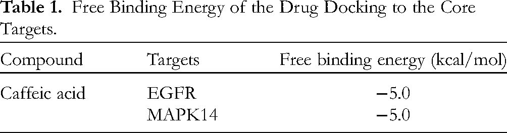
Molecular docking is a prevalent theoretical simulation technique that primarily examines the recognition mechanism between multiple molecules and anticipates their binding configurations and affinities. 17 The 13 DEGs were prepared for molecular docking with CA, with the binding energy <−5.0 kcal/mol and hydrogen bonds could be formed between receptor ligands as the screening criteria. 18 EGFR and MAPK14 exhibited a strong affinity for CA, with free binding energies of −5.0 kcal/mol (Figure 7A and Table 1). The protein expression levels of EGFR and MAPK14 in renal tissue of rats were assessed by western blotting (Figure 7B). Compared to the sham group, the UUO group exhibited significantly increased expression levels of EGFR and MAPK14 proteins (P < .01). CA treatment significantly decreased the expression levels of EGFR and MAPK14 proteins in UUO rats (P < .05).

CA downregulated the expression of EGFR and MAPK14 in UUO-induced RTF rats. (A) Molecular docking. (B) The protein expression levels of EGFR and MAPK14 in renal tissue of rats were assessed by western blotting. *P < .05, **P < .01. Abbreviations: RTF, renal tubulointerstitial fibrosis; CA: caffeic acid; UUO, unilateral ureteral obstruction.
Free Binding Energy of the Drug Docking to the Core Targets.
Discussion
RTF is the common histological manifestation of the late stage of various CKDs, which usually belongs to irreversible changes. 19 As a wide-distributing phenolic acid compound in nature, CA is well known because of its excellent antitumor activity and antioxidant activity. 8 However, the antifibrotic effect and underlying mechanism has not been well investigated. To further unveil the potential molecular mechanism of CA in RTF, a “CA-Targets-Renal fibrosis” pharmacology network combined with experimental validation were used in this study. Based on the network pharmacology approach, a totally 28 fibrosis-related target genes were screened out, and of them, 13 targets were further selected using GSE38117 data set. Finally, 3 targets fitted well with CA according to drug-target molecular docking. Moreover, the in vivo study demonstrated that CA could alleviate RTF in rats, and EGFR and MAPK14 may performs critical roles in the process.
In this study, CA treatment alleviated renal injury and dysfunction in UUO-induced RTF rats. This improvement was indicated by reduced levels of KW/BW ratio, Scr, BUN, and 24-h UP. Crucially, profibrotic factors, notably TGF-β1, CTGF, and PDGF, which originate from tubular epithelial cells, are instrumental in the progression of renal fibrosis by stimulating nearby fibroblasts.20–22 The CA treatment effectively reduced the expression of these profibrotic factors in the renal tissues of UUO-induced RTF rats. Furthermore, the markers E-cadherin, an epithelial cell indicator, and α-SMA and fibronectin, key molecular markers of myofibroblasts, along with collagen deposition, a significant hallmark of renal fibrosis, were also investigated.23,24 The treatment notably downregulated α-SMA, fibronectin, and collagen I, while upregulating E-cadherin in these rats. These findings collectively suggest that CA possesses nephroprotective properties, contributing to the alleviation of renal injury and fibrosis. These results align with those reported by Veeren et al. 13
In the following network and bioinformatics analysis, totally 309 targets related with both CA and RTF were screened out. The GO analysis states that they participated in many biological processes including positive regulation of MAPK cascade. The pathway enrichment analysis indicates that the nephroprotective effect of CA also acts on MAPK signaling pathway. MAPK signaling pathway is a kind of intercellular signal transduction pathway, responsible for a variety of cellular activities including proliferation, differentiation, survival, and death. 25 MAPK signaling pathway has been reported referring to RTF.26,27 Thus, it is reasonable to suppose that CA may alleviate RTF through MAPK signaling pathway.
Moreover, targets with high connectivity in PPI network, such as VEGFA, JUN, IL1B, EGFR, TNF, TP53, and AKT1, were also enriched in the PPI-pathway network related to RTF. Further preparation by screening with GSE216376 data set indicates that EGFR, MAPK14, HIF1A, IL1B, NFKB1, PDGFRA, PIK3CA, PIK3R1, PTPN11, TGFB1, TGFB2, TNF, and VEGFA may be the key targets in the nephroprotective effect of CA. Then, the results of the molecular docking simulation for the identification of the interaction between the key targets and CA showed that EGFR and MAPK14 fit well with free binding energy <−5.0 kcal/mol. Of them, EGFR and MAPK14 are both distributed in MAPK signaling pathway. EGFR is responsible for cellular activities like proliferation and differentiation in fibrosis. 28 It has been reported that MAPK signaling pathway is the downstream of EGFR. 29 MAPKs orchestrate various physiological processes in response to a diverse array of stimuli, and are also known as cytokine-suppressive anti-inflammatory drug-binding proteins. 30 A preceding network pharmacology analysis also identified MAPK14 and the MAPK pathway as pivotal targets associated with renal fibrosis, glomerulosclerosis, and nephrotic syndrome. 31 In vivo experiments found that CA treatment downregulated the protein expression of EGFR and MAPK14 in UUO-induced RTF rats. As a result, we can draw a conclusion that CA may mitigate RTF through MAPK signaling pathway, and EGFR and MAPK14 may be the critical factors.
In summary, our study identified the nephroprotective effects and mechanisms of CA against RTF. However, given that this study was conducted using a rat model and has not yet been extended to human subjects, the applicability of these key targets to human conditions remains uncertain. Therefore, further validation of these core targets in both animal models and human cases of RTF is imperative.
Conclusions
This study demonstrates that CA treatment significantly ameliorates renal injury and fibrosis in UUO-induced RTF rats. Based on the critical targets identified through network pharmacology prediction, EGFR and MAPK14 appear to play pivotal roles in the therapeutic action of CA in RTF. The nephroprotective effects of CA are mediated through the modulation of MAPK signaling pathway.
Materials and Methods
Animal Model
Totally 18 healthy male Sprague-Dawley rats (Beijing HFK Bioscience Co. Ltd, China) aged 6 to 8 weeks with a weight of 190 to 210 g were selected. The animals were housed in accordance with guidelines of Laboratory Animal Requirements of Environment and Housing Facilities and the Committee on the Ethics of Animal Experiments. Rats were then subjected to UUO to construct a model of RTF. All rats were randomly divided into Sham group, UUO group (model group) and UUO + CA group (treatment group), with 6 rats in each group. All rats were anesthetized by intraperitoneal injection of ketamine (100 mg/kg) and xylazine (5 mg/kg), with an incision in the midline of the abdomen and exposure of the left ureter. Rats in the UUO group and UUO + CA group underwent 2-point ligation of the ureter, while the sham group did not undergo ligation. After surgery, the UUO + CA group was given 25 mg/kg of CA (C8990, Solarbio) daily by intraperitoneal injection, while the sham group and the UUO group were treated with an equal volume of 0.3% sodium carboxymethylcellulose. The dose and method for CA treatment were selected according to a previous study. 13 After 7 days, rats were weighted and sacrificed under anesthesia, meanwhile the blood, urine, and kidneys were collected. The ratio of KW/BW in each group was calculated. The levels of Scr, BUN, and 24-h UP were quantitatively assessed using assay kits acquired from Nanjing Jiancheng (C011-2-1, C013-2-1) and Shanghai Yuanye (R21592). The rest samples were stored at −80 °C for further use. This study was approved by the Institutional Animal Care and Use Committee of Xiamen University (XMULAC20220034-15).
Histopathological Examination
Renal specimens from the kidneys in every group were dissected and fixed in 4% formalin buffer. Paraffin-embedded blocks were prepared and sections at 4 to 7 μm thickness were cut. Then, the tissue sections were subjected to HE staining, Masson's trichrome staining for histopathological examination according to the instructions of the detecting kits (C0105S, Beyotime; G1340, Solarbio). After that, all sections were observed and photographed using a digital camera (SC180, Olympus).
Immunohistochemical Assay
The expression levels of the profibrotic factors TGF-β1, CTGF, PDGF in renal tissues were determined by immunohistochemistry. The sections at 4 to 7 μm thickness were first repaired by high pressure (126 °C, 2 min), and blocked by endogenous peroxidase blocking agent, followed by incubation with anti-TGF-β1(1:500, ab215715, Abcam), anti-CTGF (1:500, ab125943, Abcam) and anti-PDGF (1:200, AF0240, Affinity) antibodies overnight at 4 °C. Then, all the slices were incubated with the corresponding secondary antibodies: Goat anti-rabbit IgG antibody (1:2000, ab205718, Abcam) for 1 h at room temperature. Then, each slice was treated with 100 μL fresh-prepared DAB solution (P0202, Beyotime) for 10 min. After stained by hematoxylin for 3 min, the slices were observed and photographed using a light microscope (SC180, Olympus).
Identification of Disease-drug Co-associated Target Genes
The “Renal fibrosis” was set as a keyword and searched in the databases of GeneCards 32 and DisGeNET, 33 and the results were combined without duplicate genes to obtain the disease-related target genes. Then, “caffeic acid” was set as the keyword and searched using HIT, 34 GeneCards, SEA, 35 Super-PRED 36 to further obtain the drug-related target genes. After that, the screening of co-associated target genes was carried out using VennDiagram 37 (version1.7.3).
Identification of Key GO Terms and KEGG Pathways
To elucidate the potential relationships among these co-associated target genes, the clusterProfiler 38 (version 4.4.2) was used to perform biological function enrichment analysis, including GO enrichment analysis and KEGG pathway analysis. P-value < .01 was considered significant.
The Construction of PPI Network
The PPI network analysis of co-associated target proteins was performed using the Search Tool for the Retrieval of Interacting Genes (STRING) 39 database to further explore the related protein interactions. The screening conditions were limited to score_cutoff > 0.4 and size_cutoff < 10. The results were visualized by Cytoscape v3.4.0 software (http://www.cytoscape.org/).
Target-pathway Network Construction
TGFB1 40 and PDGF (PDGFRA) 41 were reported to be involved in the formation of RTF, in which TGFB1 depended on MAPK signaling pathway. 42 Therefore, the interacting proteins of TGFB1 and PDGF (PDGFRA) were extracted, as well as the enriched genes in KEGG pathways involving kidney related diseases, including MAPK signaling pathway. PPI-pathway network was constructed to screen the co-associated genes and to comprehensively understand the molecular mechanisms of CA in RTF. We then retrieved the mouse kidney fibrosis model dataset, GSE216376, from the GEO database. A t-test was conducted to identify PPI-pathway-associated genes that showed significant differential expression between the kidney fibrosis and normal groups. The selected genes were considered as candidates for molecular docking analysis.
Drug-target Molecular Docking
Molecular docking is a theoretical simulation method to study the interaction between molecules, such as ligand and receptor, and to predict their binding mode and affinity. In this study, molecular docking was proceeded to validate the interaction between the selected disease targets and CA. First, the crystal structure of the target proteins was downloaded from the RCSB protein database (http://www.pdb.org/). The structure file of CA in MOL2 format was then downloaded from ZINC (https://ZINC.docking.org/). The downloaded protein and drug files were converted to PDBQT format using Open Babel GUI software. Then Pymol and AUTODOCK1.5.6 software were used to optimize the removal of ligand, dewatering, addition of hydrogen and amino acids, and calculate the charge of protein. 43 Finally, molecular docking was carried out using AUTODOCK1.5.6, and the results were visualized by Pymol.
Western Blot Detection
Western blot detection was carried out to obtain the protein expression levels of ɑ-SMA, collagen1, E-cadherin, Fibronectin, EGFR, and MAPK14 in renal tissues. Renal tissue was first lysed (200 μL lysate per 60 mg tissue) for 30 min, and then centrifuged at 12 000 r/min for 10 min at 4 °C to collect the lysate supernatant. Then, the supernatant underwent protein quantification using the BCA method. After denaturation at 95 °C for 10 min, samples were separated on a 5% SDS-PAGE gel (start from 60 V, and after the protein sample entered the separation glue, changed to 90 V). After that, the proteins were transferred to PVDF membranes under the constant pressure state (65 V, 2 h), following by being blocked in 5% nonfat milk solution for 1 h at room temperature. Then, the PVDF membranes were incubated with primary anti-ɑ-SMA, anticollagen1, anti-E-cadherin, antifibronectin, anti-EGFR, anti-MAPK14 monoclonal antibodies (1:1000, Abcam) and anti-GAPDH polyclonal antibody (1:3000, ab9485, Abcam) overnight at 4 °C. Then, the membranes were washed by TBST buffer for 10 min, 3 times and incubated with HRP-labeled secondary antibodies (1:2000) for 1 h at room temperature. After proceeding with the enhanced chemiluminescence kit (P1000, Pulilai), the PVDF membranes were visualized and photographed using a chemiluminescence imaging system (Tanon).
Statistical Analysis
All statistical analyses were processed with the GraphPad prism V7.0 software. Data were expressed as the mean ± standard deviation and analyzed using one-way ANOVA. P < .05 was statistically significant.
Supplemental Material
sj-doc-1-npx-10.1177_1934578X241237894 - Supplemental material for Uncovering the Nephroprotective Mechanism of Caffeic Acid in Renal Tubulointerstitial Fibrosis through Network Pharmacology Analysis
Supplemental material, sj-doc-1-npx-10.1177_1934578X241237894 for Uncovering the Nephroprotective Mechanism of Caffeic Acid in Renal Tubulointerstitial Fibrosis through Network Pharmacology Analysis by Wenbo Sun, Haojie Liu, Baoqiao Wu, Limiao Dai, Yan Ren and Danna Zheng in Natural Product Communications
Supplemental Material
sj-docx-2-npx-10.1177_1934578X241237894 - Supplemental material for Uncovering the Nephroprotective Mechanism of Caffeic Acid in Renal Tubulointerstitial Fibrosis through Network Pharmacology Analysis
Supplemental material, sj-docx-2-npx-10.1177_1934578X241237894 for Uncovering the Nephroprotective Mechanism of Caffeic Acid in Renal Tubulointerstitial Fibrosis through Network Pharmacology Analysis by Wenbo Sun, Haojie Liu, Baoqiao Wu, Limiao Dai, Yan Ren and Danna Zheng in Natural Product Communications
Footnotes
Data Availability Statement
The datasets used and/or analyzed during the current study are available from the corresponding author on reasonable request.
Declaration of Conflicting Interests
The authors declared no potential conflicts of interest with respect to the research, authorship, and/or publication of this article.
Ethical Approval
Ethical approval was obtained from Institutional Animal Care and Use Committee of Xiamen University (XMULAC20220034-15).
Funding
The authors received no financial support for the research, authorship, and/or publication of this article.
Informed Consent
There are no human subjects in this article and informed consent is not applicable.
Statement of Human and Animal Rights
All procedures in this study were conducted in accordance with the Institutional Animal Care and Use Committee of Xiamen University (XMULAC20220034-15) approved protocols.
Supplemental Material
Supplemental material for this article is available online.
References
Supplementary Material
Please find the following supplemental material available below.
For Open Access articles published under a Creative Commons License, all supplemental material carries the same license as the article it is associated with.
For non-Open Access articles published, all supplemental material carries a non-exclusive license, and permission requests for re-use of supplemental material or any part of supplemental material shall be sent directly to the copyright owner as specified in the copyright notice associated with the article.