
Abstract
Background
This study compared the contents of extracts of Chenopodium formosanum (Djulis) originating from 2 aboriginal areas (Kaohsiung and Taitung, Taiwan) and evaluated their efficacy in ameliorating insulin resistance in vitro.
Methods
First, we analyzed the anti-oxidant compositions and activity, and α-glucosidase, α-amylase inhibitory effects of 2 Djulis and their extracts, and then evaluated the ability of the 2 extracts to ameliorate insulin resistance in HepG2 cell. To do this, we incubated the cells in DMEM medium spiked with insulin and TNF-α.
Results
Taitung Djulis extract (TDE) had higher concentrations of flavonoids and polyphenols than the Kaohsiung Djulis extract (KDE). However, KDE had a greater anti-oxidative effect and greater ability to inhibit α-glucosidase and α-amylase inhibitory than TDE. In the cellular experiment, insulin promoted cell glucose uptake, and increased the protein expression of glucose transporter-2 (GLUT-2). However, when adding TNF-α with insulin, TNF-α worsened insulin-stimulated glucose metabolism. This might be associated with TNF-α induced mitogen-activated protein kinases (MAPKs) expression. Treating the insulin resistant cells with either TDE or KDE significantly reversed the effect of TNF-α on glucose uptake and related protein expressions.
Conclusion
Although both TDE and KDE reduced TNF-α-induced insulin resistance, KDE was found to be more potent. KDE might have more bioactivity or other undetected components that lead to more efficiency than TDE. Taken together, Djulis extract could potentially be used to control insulin resistance.
Introduction
The prevalence of type 2 diabetes mellitus (T2DM) and cardiovascular disease, both associated with insulin resistance and metabolic syndrome, have increased substantially worldwide. 1 Long-term high carbohydrate and fat intake, fatty tissue accumulation, and obesity can cause chronic inflammation, gradually reducing insulin activity and function. This chronic inflammation affects the processing of glucose in the major peripheral organs, such as the liver, skeletal muscle, and adipose tissue, leading to hyperglycemia and hyperlipidemia as well as T2DM. This chronic inflammation is caused by the secretion of such cytokines as tumor necrosis factor-α (TNF-α) or interleukin-6 (IL-6) from adipose tissue and the immune system. The activated inflammatory pathways interfere with insulin function and ultimately lead to insulin resistance. 2
One way to prevent insulin resistance and metabolic syndrome is adjust eating habits, limiting oneself to foods with low glycemic indices, foods rich in dietary fiber and antioxidants. Although many staple foods such as white rice and flour have high glycemic indexes, there are healthier staple foods, including brown rice and pre-germinated brown rice (PGBR), which has an endosperm, aleuronic layer, bran layer, and germ that slow down the digestion and absorption of carbohydrates. 3 In fact, PGBR has been found to have antioxidants that give it the additional ability to ameliorate metabolic syndrome. 4 There may be other staples that have the same benefits. For example, there is an abundance of a grain staple called Chenopodium formosanum (also known as Djulis) growing in Taiwan's aboriginal areas. Taiwan aborigines have cultivated and eaten Djulis for many centuries (Figure 1). It is suitable for growing in middle and low altitudes, with short sunshine, and good drainage areas. Djulis was originally the staple food of Taiwan's aborigines. In recent years, it has gradually become popular under Taiwanese people's pursuit of a healthy diet, and the planting area has gradually increased. Taking the cultivation of Djulis in southern Taiwan, most of them are planted in the aboriginal tribes of Kaohsiung (located in Southwest Taiwan) and Taitung (located in Southeast Taiwan). The weather and the soil conditions are different between 2 places, which may lead to differences in the quality of Djulis. Studies have been conducted to assess the anti-metabolic syndrome effects of Djulis, but not its related pharmacological mechanisms.5,6 Few studies have evaluated the effect of Djulis extract on insulin resistance or compared the relative efficacy of Djulis grown in different regions in treating this dysfunction.
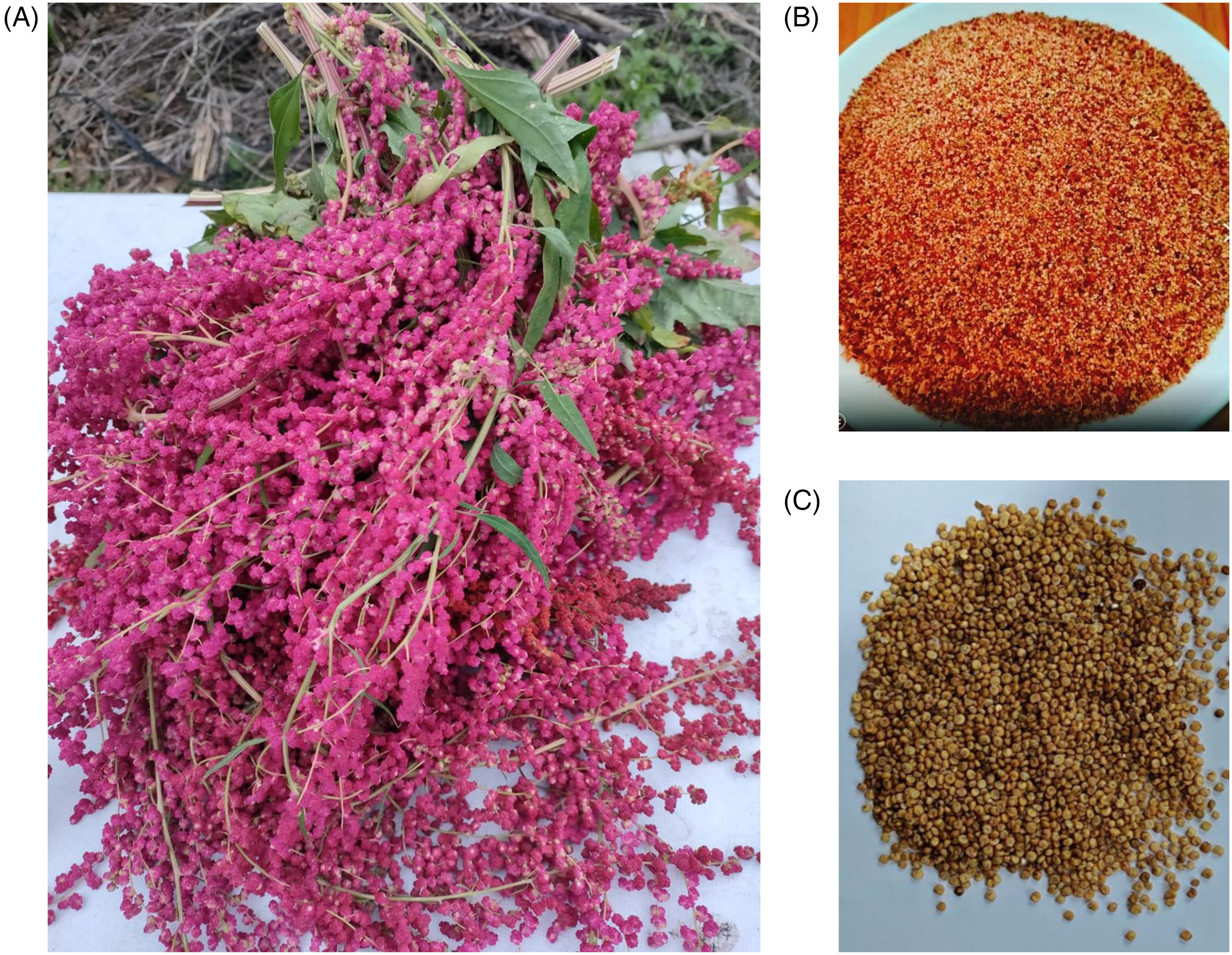
Introduction of Chenopodium formosanum (Djulis). The whole plant (A). Provided edible seed (B). Shelled seed (C).
Hence, in this study, we investigated content differences between extracts of Djulis collected from aboriginal areas in Kaohsiung and Taitung; and evaluated their relative effect on the prevention of TNF-α-induced insulin resistance and the mechanism underlying this effect.
Materials and Methods
Djulis Extraction and Composition Analysis
Djulis were obtained from local farmers in the regions of origin, Kaohsiung and Taitung. Fifty grams of dried and shelled Djulis were sonicated in 250 mL 95% alcohol for 2 h and left to settle overnight. After filtering, the solution was evaporated in vacuo to remove the alcohol. The stock solution was used 95% alcohol to make up and used DMSO as the solvent to dilute, and then performed the following experiments. By the way, DMSO solution was also evaluated in the following experiments separately, and DMSO solution did not have any effects (data not shown) and therefore would not affect the efficacy of Dijulis extracts.
Polyphenol concentrations were quantified as described by Liu et al. 7 Briefly, 0.1 mL Djulis extract was mixed with 5.6 mL pure water, 4 mL of 2% Na2CO3 solution, and 0.1 mL of 37% formaldehyde. The mixture was maintained in darkness for 20 min at room temperature. The absorbance of resulting color was measured by spectrophotometer at 765 nm. Gallic acid was used to calculate the standard curve.
Anthocyanidin concentrations were measured according to Goufo and Trindade. 8 Briefly, 0.1 mL extract was mixed with 2.5 mL of 1% vanillin methanol solution and 2.5 mL of 9N HCl. The mixture was maintained in darkness for 30 min at room temperature. The absorbance of resultant color was measured by spectrophotometer at 500 nm. Catechin was used as the standard.
The total flavonoids content was quantified using the aluminum chloride colorimetric method. 9 Briefly, 0.5 mL sample was mixed with 1.5 mL of 95% ethanol, 0.1 mL of 10% aluminum chloride, 0.1 mL of 1 M potassium acetate, and 2.8 mL of pure water. The mixture was maintained in darkness for 60 min at room temperature. The absorbance of resultant color was measured by spectrophotometer at 415 nm. Quercetin was used as the standard.
Measurement of Anti-Oxidative Activity
Antioxidants were determined following the procedures for assessing DPPH free radical scavenging activity described by Yeh et al. 10 In brief, 100 μL of the extract was mixed with 400 μL methanol and 500 μL DPPH. The mixture was kept in darkness for 20 min at room temperature. The absorbance of the resulting color was assessed by spectrophotometer at 517 nm. Ascorbic acid was used as the standard.
Total antioxidant capacity was assessed following Nilsson et al. 11 Briefly, 0.1 mL of the extract was mixed with 0.9 mL of an ABTS−+ solution consisting of 0.25 mL ABST, 0.25 mL hydrogen peroxide, 0.25 mL glutathione peroxidase, and 1.5 mL pure water. The mixture was stored in darkness for 10 min at room temperature. The absorbance of the resulting color was measured by spectrophotometer at 734 nm. Trolox was used as the standard.
We followed a method of reducing power previously reported by Wu et al. 12 In brief, 5 mL various concentrations of the standard (25, 50, 100 μm) or Djulis extract were mixed with 2.5 mL of 200 mM sodium phosphate buffer (pH 6.6) and 2.5 mL of 1% potassium ferricyanide. The mixture was incubated at 50 °C for 2 min. About 2.5 mL of 10% tricloroacetic acid was added and the mixture was centrifuged at 3000 rpm for 10 min. The upper layer (2.5 mL) was mixed with 2.5 mL of deionized water and 0.5 mL of 0.1% ferric chloride, and the absorbance was measured by spectrophotometer at 700 nm. Ascorbic acid and gallic acid were used as standards.
Inhibition Effects of α-Glucosidase and α-Amylase
Inhibition of α-glucosidase was analyzed using a method modified from Kwon et al. 13 In brief, 250 μL sample was mixed with 500 μL of α-glucosidase solution (1U/mL) and 250 μL of 5 mM p-nitrophenyl-β-glucopyranoside. After incubation at 37 °C for 20 min, 500 μL of 0.1 M Na2CO3 solution was added to the mixture to stop the reaction. Absorbance was then assessed by spectrophotometer at 405 nm. Acarbose was used as the standard.
Inhibition of α-amylase was analyzed using a modification of the method described by Wickramaratne et al. 14 In brief, 50 μL sample was mixed with 100 μL of α-amylase solution (1U/mL) at 37 °C for 20 min, and then added 50 μL of 1% starch solution. After incubation at 37 °C for 30 min, the mixture was then added with 200 μL of 3,5-dinitrosalicylic acid solution and incubated at 100 °C for 10 min. Absorbance was measured by spectrophotometer at 540 nm, using α-amylase as the standard.
Cell Culture
HepG2 cells, obtained from the National Development Center of Biotechnology (Taipei, Taiwan), were maintained as a monolayer culture in complete medium at 37 °C in a humidified 5% CO2 incubator with medium changed every 2 days. Initially, HepG2 cells were seeded in 6-well plates at a density of 1 × 10⁵ cells/well, and treated 100 ng/mL TNF-α for 5 h. Afterward, changing medium containing 10 μM insulin only, 10 μM insulin and 45 ng/mL TNF-α, or 10 μM insulin, 45 ng/mL TNF-α and TDE or KDE (20, 25, 50, or 100 μg/mL) stimulated the cells for 16 h. We referred to and modified the method of Kim et al 15 and Lin et al 4 in the cell experiment.
Glucose Uptake Assay
After treating the agents for 16 h, the medium and cells were collected. 50 μL of the medium was withdrawn and centrifuged at 500g for 5 min. 5 μL of the supernatant was mixed with 95 μL of assay buffer (Glucose Colorimetric Assay Kit II, BioVision) in a 96-well plate and incubated at 37 °C for 30 min. The absorbance was then measured at 450 nm under spectrophotometer.
Western Blot Analysis
The homogenized HepG2 cells were identified by AMP-activated protein kinase (AMPK), insulin receptor (IR), glucose transporter-2 (GLUT-2), glucokinase (GCK), pyruvate kinase (PK), glucose 6-phosphatase (G6Pase), phosphoenolpyruvate carboxykinase (PEPCK), ERK and JNK antibodies (Santa Cruz Biotechnology, Santa Cruz, CA; 1:500 dilution) and IgG conjugated antibody (Santa Cruz Biotechnology, Santa Cruz, CA; 1:10 000 dilution). The relative expression of those proteins in each tissue was quantified by densitometric scanning of the western blots using Image-pro plus software (Media Cybernetics, MD) as previously described. 4
Statistical Evaluation
Results are expressed as mean ± SE. Statistical differences were determined by independent and paired Student's t-test in unpaired and paired samples, respectively. If a significant difference was found, we used Dunnett's or Student-Newman-Keuls test for further analysis. A P-value <.05 was considered significant in all experiments. All statistical operations and plotting of table and figure were performed using SigmaStat: Version 2.03 and SigmaPlot: Version 8.0 (Systat Software, Point Richmond, CA, USA).
Results
The Composition of Djulis Extract
In our analysis of the contents of Taitung Djulis and Kaohsiung Djulis, we found them to be clearly different. Taitung Djulis was found to have higher concentrations of polyphenols than Kaohsiung Djulis (Table 1). One gram of Taitung Djjulis extract (TDE) was found to have 8233.3 ± 626.3 ppm of flavonoid, 1654.8 ± 3.1 ppm of polyphenol, and 374.3 ± 34.0 ppm of anthocyanidin. The respective concentrations in 1 g Kaohsiung Djulis extract (KDE) were 5955.7 ± 818.1 ppm, 1054.0 ± 16.7 ppm, and 377.7 ± 4.7 ppm, respectively. Thus, TDE had more flavonoids and polyphenols than KDE, but not more anthocyanidin (Table 1).
Estimated the Functional Bioactive Substances of Total Flavonoid, Polyphenols and Anthocyanidin per Gram of Djulis and Djulis Extract.

Kaohsiung (K) Djulis (D) was 50 g, and KD extract (KDE) could be obtained 3.3 g approximately.
Taitung (T) Djulis (D) was 50 g, and TD extract (TDE) could be obtained 2.8 g approximately.
Each value represents the mean ± S.E. (n = 3). *P < .05 versus K Djulis; #P < .05 versus KDE.
The Anti-Oxidation, and α-Glucosidase, α-Amylase Inhibition Effects of Djulis Extract
TDK and KDE had similar clearance rates of DPPH, while KDE had a greater reducing power and trolox equivalent antioxidant capacity (TEAC) than TDK (Table 2). Both extracts inhibited α-glucosidase and α-amylase. KDE's inhibition of α-glucosidase was twice as potent as that of TDE, and its inhibition of α-amylase was 4 times as potent (Table 3).
The Anti-Oxidative Ability of Djulis Extract.

KDE, Kaohsiung Djulis extract; TDE, Taitung Djulis extract; DPPH, α,α-diphenyl-ß-picrylhydrazyl; TEAC, Trolox equivalent antioxidant capacity.
Each value represents the mean ± S.E. (n = 3). *P < .05 versus KDE.
The Digesting Carbohydrate Enzyme Inhibition Rate of Djulis Extract.

KDE, Kaohsiung Djulis extract; TDE, Taitung Djulis extract.
Each value represents the mean ± S.E. (n = 3). *P < .05 versus KDE.
The Effect of Djulis Extract in Glucose Uptake Assay
The concentration of glucose in the medium decreased when 10 μM insulin was added, indicating that glucose uptake was higher in the medium with added insulin than in the medium with no added insulin. When 30 ng/mL TNF-α was added along with the insulin, glucose concentration remained high indicating that glucose uptake was clearly inhibited. When KDE or TDE (20, 25, 50, or 100 μg/mL) was added into the medium, glucose concentrations decreased. Both extracts ameliorated TNF-α-induced insulin resistance, KDE more efficiently than TDE (Table 4).
KDE and TDE Ameliorated TNF-α Inhibited Insulin Stimulated Glucose Uptake in HepG2 Cells.

KDE, Kaohsiung Djulis extract; TDE, Taitung Djulis extract.
Each value represents the mean ± S.E. (n = 6). *P < .05 versus control; #P < .05 versus insulin; ‡P < .05 versus insulin + TNF-α.
The Effect of Djulis Extract on Protein Expressions of Glucose Metabolism
When HepG2 cells were stimulated by insulin only, protein level of GLUT-2 was increased, compared to control group. After adding TNF-α with insulin, we found decreased protein expression of IR, GLUT-2, GCK, PK; and increased expression of PEPCK. These findings confirmed that TNF-α influenced the glucose uptake, glycolysis, and gluconeogenesis to induce insulin resistance. The addition of either TDE or KDE improved those protein (IR, GLUT-2, GCK, PK, G6Pase, PEPCK) levels of glucose metabolism including AMPK, with no obvious differences between the 2 extracts (Figures 2 and 3).

Effects of KDT and TDE on AMPK, IR, GLUT-2, GCK, and PK protein expressions in HepG2 cell. The abbreviation “I” refers to the 10 μM insulin group, “I + T” refers to the insulin 10 μM and 45 ng/mL TNF-α group, and KDE or TDE (20, 25, 50, and 100 μg/mL) refers to treating group for 16 h. Each value represents the mean ± S.E. (n = 6). *P < .05 versus control group; #P < .05 versus insulin group; ‡P < .05 versus insulin and TNF-α group.

Effects of KDT and TDE on G6Pase and PEPCK protein expressions in HepG2 cell. The abbreviation “I” refers to the 10 μM insulin group, “I + T” refers to the insulin 10 μM and 45 ng/mL TNF-α group, and KDE or TDE (20, 25, 50, and 100 μg/mL) refers to treating group for 16 h. Each value represents the mean ± S.E. (n = 6). *P < .05 versus control group; #P < .05 versus insulin group; ‡P < .05 versus insulin and TNF-α group.
The Effect of Djulis Extract on Protein Expressions of Inflammation Signals
TNF-α significantly induced the inflammation signals ERK and JNK protein expressions, except p38, compared to control and insulin groups. Either KDE or TDE decreased ERK and JNK protein levels in insulin resistance cell (Figure 4). According to the above results, TNF-α not only worsened the glucose metabolism, but also promoted the inflammation to induce insulin resistance. Two Djulis extracts ameliorated insulin resistance might be related with possessing the effects of diminishing inflammation.

Effects of KDT and TDE on p38, ERK and JNK (MAPKs pathway) protein expressions in HepG2 cell. The abbreviation “I” refers to the 10 μM insulin group, “I + T” refers to the insulin 10 μM and 45 ng/mL TNF-α group, and KDE or TDE (20, 25, 50, and 100 μg/mL) refers to treating group for 16 h. Each value represents the mean ± S.E. (n = 6). *P < .05 versus control group; #P < .05 versus insulin group; ‡P < .05 versus insulin and TNF-α group.
Discussion
This study is the first to examine the effect of Djulis extract on glucose metabolism, and found that the Djulis grown in 2 different places (Taitung and Kaohsiung Counties, Taiwan) had different concentrations of the same contents and different bioactivities. Djulis extract was found able to ameliorate TNF-α induced insulin resistance in HepG2 cell, the KDE more potently so than the TDE. The experimental results show that the content of antioxidant substances in KDE is lower, but no matter in the analysis of antioxidant effects, α-glucosidase and α-amylase inhibitory activity, and anti-insulin resistance in the cell experiment, KDE is obviously more effective than TDE. Except from the perspective of biological activity, we cannot rule out the possibility that there are other undiscovered active ingredients in KDE, which still need follow-up research to discover.
Djulis, which is native to Taiwan, is rich in dietary fiber, protein, vitamins, and minerals. It contains various antioxidants, including betalains, flavonoids, phenolic acid, and saponins. 16 Because the saponins present on its shell are slightly toxic, the shells should be removed when eating. 17 In this study, we used the shelled Djulis. Only polyphenols, not flavonoids and anthocyanidin, were found in higher concentrations in shelled Taitung Djulis than in shelled Kaohsiung Djulis. After extraction, flavonoids and polyphenols were found to be higher in TDE than in KDE (Table 1). Although TDE has higher concentrations, KDE had a higher antioxidant capacity (Table 2). These findings suggest that environmental conditions had an effect on Djulis content and biological activity.
α-Glucosidase is an enzyme that catalyzes the hydrolysis of glycoside bonds in polysaccharides and glycoconjugates. It plays important roles in various biological processes, including glycoconjugate catabolism, glycoprotein post-translational modification, and carbohydrate digestion. In mammalians, α-glucosidase exists in the mucosal brush border of the small intestine where it contributes greatly to the digestion of dietary starch and disaccharides. α-Glucosidase inhibitors, such as acarbose, are widely used to interrupt the metabolism of carbohydrates in the small intestine and moderate the postprandial blood glucose, and they have been used to develop therapeutic agents against diabetes, obesity, metastatic cancer, and viral infection. 18 α-Amylase, which is an enzyme found in pancreatic juice and salvia, degrades starches and hydrolyzes them into oligosaccharide. One strategy for decreasing glucose absorption and maintaining blood glucose is to inhibit α-amylase activity. Some studies have reported that flavonoids, polyphenols, anthocyanins, etc in fruits, vegetables, or mushroom extracts can inhibit α-glucosidase and α-amylase, which could make them potentially useful in the control of insulin resistance, diabetes, and obesity.19–23 In this study, both KDE and TDE were found able to inhibit α-glucosidase and α-amylase activity. KDE, in particular, was found to have an inhibitory effect on α-glucosidase activity similar to that of acarbose, and inhibitory ratio of α-amylase was 65% (Table 3). These findings suggest that more studies might be performed to study the possible application of Djulis to the control metabolic syndrome and DM.
In obesity, adipose tissue is thought to be an inflammatory tissue that produces several reactive oxygen species (ROS) and cytokines to induce chronic inflammation. ROS and cytokines as well as the stimulated immune cells affect the peripheral tissues, including pancreatic islets, liver, adipose tissue, and skeletal muscle to reduce the insulin secretion and function. 2 TNF-α is the main inflammatory factor contributing to the impairment of insulin signaling, glucose uptake and glucose metabolism, and eventually insulin resistance. 24 According to the results of our in vitro experiments, insulin significantly decreased glucose concentrations in the culture medium, indicating that insulin levels could stimulate glucose uptake into the cells. When TNF-α was added to insulin-treated cells, glucose concentrations were not reduced, indicating that TNF-α inhibited insulin function and caused insulin resistance. After adding TDE or KDE to medium containing TNF-α and insulin, the insulin resistance was reversed. KDE had a stronger effect than TDE (Table 4). Based on the above results, although KDE had less antioxidant content (flavonoid, polyphenols) than TDE, KDE had exerted greater biological activity.
We analyzed the protein expressions of AMPK, IR, GLUT-2, GCK, PK, G6Pase, and PEPCK to explore possible mechanisms underlying their effect on TNF-α worsening of insulin-promoting glucose uptake and metabolism. AMPK is an enzyme that plays several roles in the regulation of glucose metabolism. AMPK stimulates glucose transporter (GLUT) expression to promote glucose uptake and glycolysis (related enzyme such as IR, GCK, PK) and regulate glycogen synthesis and gluconeogenesis (related enzyme such as G6Pase and PEPCK).25,26 In those with obesity-related long-term inflammation, the expressions of AMPK and GLUT are suppressed, which influences glucose metabolism leading to insulin resistance. Subsequently, gluconeogenesis is initiated to generate energy. Hyperglycemia and DM eventually occur in this vicious circle. 27 In this study, insulin increased GLUT-2 expressions, but when adding TNF-α with insulin, the levels of IR, GLUT-2, GCK, and PK were diminished, and the level of PEPCK was increased by TNF-α. It demonstrated that TNF-α deteriorated glucose uptake and glucose metabolism. Both TDE and KDE were found able to reverse those changes. Besides, 2 extracts could increase AMPK levels. There was no obvious difference between the 2 extracts with regard to their ability to reverse the changes (Figures 2 and 3).
The activity of nuclear factor-κB (NF-κB) through mitogen-activated protein kinases (MAPKs) pathway is closely related to the inflammatory response. As we know, inflammation is a major risk factor to impair cellular function, insulin secretion and sensitivity, and energy metabolism, which eventually lead to insulin resistance. 25 According to the results, ERK and JNK levels were increased by TNF-α. Combining the above results of glucose uptake and metabolism, we suggested that TNF-α induced ERK and JNK levels that impaired the effects of insulin and lead to insulin resistance. However, 2 Djulis extracts significantly reduced ERK and JNK levels to inhibit inflammation (Figure 4). In summary, the ability of KDE and TDE to ameliorate TNF-α induced insulin resistance might be related to their effect on anti-inflammation and inflammation worsened glucose uptake, glycolysis, and gluconeogenesis.
In conclusion, both TDE and KDE reduced TNF-α-induced insulin resistance, but KDE was found to be more potent. KDE might have more bioactivity or other undetected components that lead to more efficiency than TDE. The same crops from different places possessed different potencies. We suggested that environmental conditions could affect the biological activity of the same crops. The results could provide the references for follow-up research. KDE and TDE not only assumed antioxidant activity but they also possessed the activities of α-glucosidase and α-amylase inhibition. Meanwhile, KDE and TDE reversed the adverse effect that TNF-α had on glucose uptake and metabolism (Figure 5). Taken together, these findings suggest that Djulis extract could potentially be developed into therapeutics that can be used to control insulin resistance, metabolic syndrome, and DM.

Properties of Djulis extract and its action mechanisms in ameliorating TNF-α worsened insulin resistance and inflammation.
Footnotes
Acknowledgements
We would like to thank Ming Zhun Business Company East Taiwan Djulis for providing high-quality agricultural products.
Author Contributions
Conceptualization: YHT, HLL, and KPS. Methodology: CLH, YCT, and BCY. Biochemical analysis: YHT, HLL, and PWC. Original draft writing & editing: HLL, PWC, and KPS.
Declaration of Conflicting Interests
The author(s) declared no potential conflicts of interest with respect to the research, authorship, and/or publication of this article.
Ethical Approval
Ethical Approval is not applicable for this article.
Funding
The author(s) disclosed receipt of the following financial support for the research, authorship, and/or publication of this article: This work was supported by grants PS111024-1 to Dr Kuo-Ping Shen from Pingtung Christian Hospital, Pingtung, Taiwan, as well as 108-2320-B-276-002-MY3 to Dr Kuo-Ping Shen from National Science and Technology Council, Taiwan.
Statement of Human and Animal Rights
This article does not contain any studies with human or animal subjects.
Statement of Informed Consent
There are no human subjects in this article and informed consent is not applicable.