
Abstract
The brown seaweed Sargassum carpophyllum J. Agardh is an unused warm-temperate species in the family Sargassaceae that has been expanding its distribution along the coastal areas of Japan in recent years. In this study, 3 types of phlorotannins were identified from the EtOAc fraction of the 80% MeOH extract of S. carpophyllum. From the spectroscopic (1H NMR, 13C NMR, and HMBC) and ESI/MS data and comparison with those of prior literature, it was demonstrated that the compounds are oligomers of phlorethol, which is one of the subclasses of phlorotannins, that is triphlorethol B (phloroglucinol trimer), tetraphlorethol C (phloroglucinol tetramer), and pentaphlorethol A (phloroglucinol pentamer). Among the phlorethols, tetraphlorethol C and pentaphlorethol A were isolated and identified for the first time from a brown seaweed collected from the East China Sea, including the coastal areas of Japan. The identified phlorethols were tested for their antioxidant properties. In the antioxidant assay using liposomes, the phlorethols showed comparable inhibitory effects to epigallocatechin gallate (tea polyphenol) and α-tocopherol (liposoluble vitamin) on lipid peroxidation by 4 mM 2,2′-azobis(2-methylpropionamidine) dihydrochloride. In addition, it was revealed that pentaphlorethol A has a superoxide anion scavenging activity (50% effective concentration: 21 μM) higher than that (50% effective concentration: 46 μM) of ascorbic acid (hydrosoluble vitamin).
Introduction
Phlorotannins are polyphenols contained only in brown seaweeds as secondary metabolites, and are presumed to be biosynthesized via the acetate-malonate pathway. 1 Based on the means of linkage between phloroglucinol (1,3,5-trihydroxybenzene) units, phlorotannins can be classified into the following subclasses of compounds: those with an ether linkage (fuhalols and phlorethols), those with a phenyl linkage (fucols), those with ether and phenyl linkages (fucophlorethols), and those with a dibenzodioxin linkage (eckols and carmalols).2,3 As a result of recent pharmacological research, it has been demonstrated that phlorotannins, especially eckols, have various physiological functions, e.g., antioxidant,4–8 anti-glycation,9–13 anti-diabetic,14,15 anti-hypertension, 16 anti-inflammatory,6,17,18 and suppression of melanogenesis.8,19 Based on these scientific findings, phlorotannins are attracting attention as novel functional polyphenols derived from seaweed.
In the case of research on phlorotannins targeting brown seaweeds distributed in the East China Sea, including coastal areas of Japan, most of the scientific findings at present are related to compounds derived from Lessoniaceae. The Lessoniaceae distributed in this area are only Eisenia bicyclis, Eisenia nipponica, Ecklonia cava, Ecklonia kurome, Ecklonia stolonifera, and Ecklonia radicosa. Many types of eckol oligomers (e.g., fucofuroeckols, phlorofucofuroeckols, dieckol, and bieckols) have been isolated and identified from all 5 species,4,20–22 with exception of E. radicosa. Of brown seaweeds, the family Sargassaceae is the most abundant, and more than 500 species have been confirmed around the world.23,24 In the coastal areas of Japan, it has been clarified that more than 60 species of Sargassaceae are distributed. 24 In addition, with the recent rise in seawater temperature, expansion of the distribution area of subtropical and warm-temperature Sargassaceae (e.g., Sargassum carpophyllum, Sargassum ilicifolium, and Sargassum alternato-pinnatum) have also been observed in the coastal areas of Japan.24–26 In the research on phlorotannins targeting Sargassaceae distributed in the region of Japan, Kawamura-Konishi et al. 27 identified triphlorethol A (phloroglucinol trimer) from the EtOH extract of Sargassum patens as an inhibitor of α-amylase. Tsukamoto et al. 28 isolated a diphlorethol (phloroglucinol dimer) from Sargassum thunbergii and found that it promotes larval metamorphosis in ascidians. In addition to these seaweeds, there are reports on the analysis of chemical structures and physiological functions of phlorotannins from Sargassum fusiforme, 29 Sargassum hemiphyllum, 30 and Sargassum ringgoldianum. 31 So far, the scientific findings regarding phlorotannins contained in Sargassaceae have been limited to such reports, and it can be concluded that the research findings are extremely limited considering the abundant amount of biomass.
We have searched for phlorotannins in Japanese subtropical and warm-temperate Sargassaceae using a previously developed TLC-screening method,5,32 and as a result, prominent bands were detected in the hydrophilic fraction of S. carpophyllum. In this study, we report on the chemical structures of phlorotannins isolated from S. carpophyllum and their antioxidant properties.
Results and Discussion
As a result of extraction, approximately 0.30 g of crude phlorotannins was obtained from 100 g of S. carpophyllum powder. The crude phlorotannins were analyzed using TLC with vanillin-H2SO4 and paprika pigment as detection reagents. In a strong acid solution, vanillin reacts at a free ring carbon position of 1,3- or 1,3,5-oxy-substituted benzonid units to produce covalently bonded reddish-brown-colored products.2,33 Hence, vanillin-H2SO4 is able to specifically detect polyphenols, including phlorotannins. Nakamura et al.
34
reported that the combination of UV bleaching and spraying of paprika pigment (mainly capsanthin) can clearly detect antioxidant substances on a TLC plate. The TLC analysis suggested that 3 phlorotannins (compounds
Further analysis using LC/MS with an Inertsil ODS-2 column revealed the properties of each compound detected on the TLC plate. Within 40 minutes a distinct separation of each of the compounds was obtained by isocratic elution with water:ACN:EtOAc (89.5:4.5:2, v/v). Each retention time was 10.2 minutes for compound
From the agreement between the obtained and literature NMR data,35–37 3 phlorethols were identified as follows: triphlorethol B (compound

Structures of the phlorethols isolated from Sargassum carpophyllum. Compound
It is well-known that polyphenols, which are secondary metabolites of plants, have antioxidant properties41–43 as a typical physiological function, similar to vitamins such as ascorbic acid and α-tocopherol. The antioxidant properties of the 3 phlorethols isolated from S. carpophyllum were evaluated. It has been pointed out that cell membranes rich in unsaturated fatty acids are susceptible to oxidative damage caused by free radicals and reactive oxygen species (ROS) generated in vivo. Liposomes are widely used as cell membrane models in studies of lipid peroxidation in vitro and the antioxidant properties of test compounds in the lipid/aqueous system.44–46 Therefore, the inhibitory effects of the identified phlorethols against lipid peroxidation were tested using unilamellar liposomes. Figure 2 shows the antioxidant activities of the phlorethols against lipid peroxidation caused by 4 mM 2,2′-azobis(2-methylpropionamidine) dihydrochloride (AAPH) as a radical initiator. As a result of testing at a final sample concentration of 1 μM, each identified phlorethol suppressed lipid peroxidation by approximately 30% (Figure 2). It was demonstrated that the phlorethols have comparable antioxidant properties to α-tocopherol and epigallocatechin gallate (EGCG), which are known as typical antioxidants (Figure 2). In previous research,
47
it has been reported that catechins of tea polyphenols have a strong interaction with the lipid bilayer in the liposomal system, and effectively scavenge peroxyl radicals derived from AAPH. Like catechins, it may be that the phlorethols have high affinity for lipid bilayers and inhibit phospholipid peroxidation. The excessive amount of superoxide anion may lead to damage of the architecture of biomolecules via the formation of highly reactive secondary oxidants, such as hydrogen peroxide and hydroxyl radical.
48
Similar to superoxide dismutase in vivo, the substances with superoxide anion scavenging activity are expected to scavenge ROS, resulting in delayed aging and prevention of lifestyle-related diseases. For phlorethols, the superoxide anion scavenging activity was measured, and the 50% effective concentration (EC50) values of each are shown in Table 1. Among the identified phlorethols, pentaphlorethol A (compound

Antioxidant activities of the identified phlorethols in the liposomal system. Lipid peroxidation was induced by the addition of 4 mM AAPH. EGCG, resveratrol, and α-tocopherol were used as positive controls. Each value is the mean ± SD (n = 5).

The viability of each cell treated with the identified phlorethols. Cell viability was determined using an MTT assay. Each value is the mean of triplicate cultures. (a) HeLa cells; (b) 3T3-L1 cells.
EC50 Values of Phlorethols on Superoxide Anion Scavenging Activity.
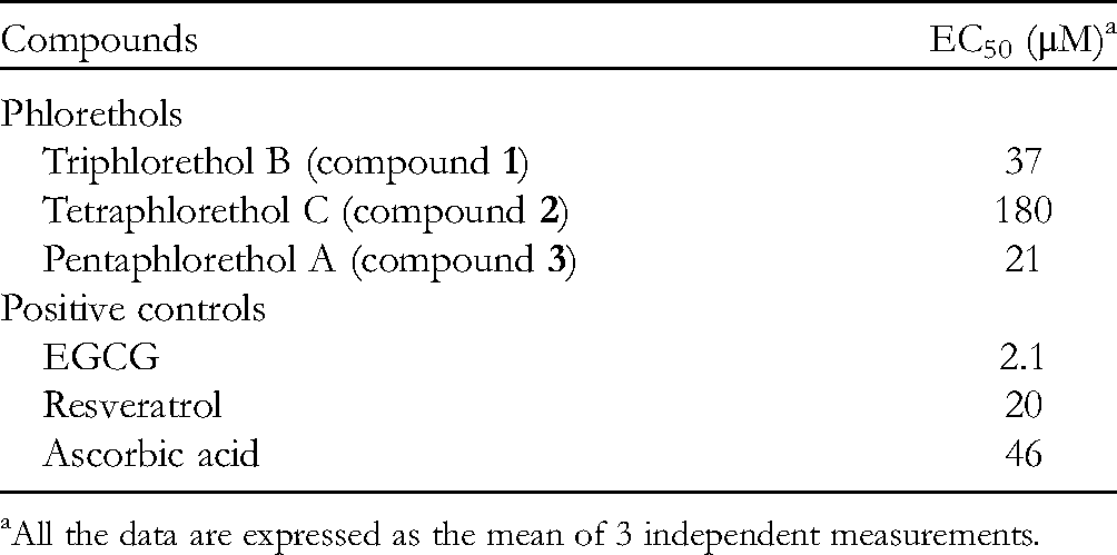
All the data are expressed as the mean of 3 independent measurements.
S. carpophyllum is an unused marine biomass, whose distribution is rapidly expanding in the coastal areas of Japan.24–26,49 In this study, it was revealed for the first time that phlorethol oligomers (phlorotannins) with antioxidant properties are contained in Japanese Sargassaceae. Tang et al. 50 reported 2 novel sterols and 5 known steroids from the EtOH extract of S. carpophyllum. In that study, 50 it was shown that many of the identified compounds were cytotoxic to several cancer cell lines, including HL-60 and P-388. Thus, the findings obtained in the current and previous studies suggest that S. carpophyllum can be used as a new source of bioactive substances, similar to Japanese Lessoniaceae.
Experimental
Materials
The brown seaweed S. carpophyllum J. Agardh was collected from the coast of the Nishisonogi Peninsula (32°81′N, 129°77′E) in Nagasaki prefecture, Japan. The species was verified by Dr. Tadao Yoshida, an emeritus professor at Hokkaido University. The collected seaweed was air-dried in the shade and pulverized using a pulverizing mill (ABS-W, Osaka Chemical). The powder was stored at −30 °C until use.
Extraction and Isolation of Phlorotannins
The extraction of phlorotannins from seaweed powder was carried out according to the method of Nakamura et al., 4 with slight modification. MeOH (80%, 200 mL) was added to the seaweed powder (40 g) having a particle size of 3 µm or less, and the mixture was shaken overnight using a shaker (KS260 control, IKA) at room temperature. After filtration with a qualitative filter, 240 mL of CHCl3:MeOH (2:1, v/v) was added to the residue and shaken overnight at room temperature. The filtrate was mixed, and the mixture partitioned between upper (hydrophilic fraction) and lower (lipophilic fraction) layers by the addition of CHCl3 (320 mL) and ultrapure water (140 mL). After removing the MeOH from the hydrophilic fraction using a rotary evaporator (N-1000, Eyela), the fraction was extracted twice with EtOAc (200 mL). The EtOAc fraction was evaporated in vacuo using a rotary evaporator and the obtained residue was used as crude phlorotannins.
Isolation and purification of phlorotannins was carried out using a preparative HPLC system (Prominence, Shimadzu) consisting of a solvent delivery unit (LC-20 AR), a UV-visible detector (SPD-20A), a sample injector (7725i, Rheodyne), a system controller (CBM-20A), and an analytical data processing system (Labsolutions). The preparative column was used in combination with an Inertsil ODS-2 column (7.6 mm I.D. × 250 mm, GL Sciences) and an Inertsil ODS-2 preparative PREP guard cartridge (7.6 mm I.D. × 30 mm, GL Sciences). Elution was performed at a flow rate of 2.7 mL/min with water: ACN: EtOAc (89.5:4.5:2, v/v). The UV-visible detector was set to a wavelength of 280 nm.
TLC
The TLC plate (silica gel 60 F254, Merck) was developed with CHCl3:MeOH:water:acetic acid (65:25:4:3, v/v), as in previous reports.5,32 After removal of the developing solvent, vanillin-H2SO4 for detecting polyphenols,2,5,33 and paprika pigment solution for detecting antioxidant substances5,32,34 were used as spray reagents.
Structural Analysis of Phlorotannins
ESI/MS was obtained on an Agilent 6120 quadrupole LC-MS consisting of a 6120 quadrupole MS, a 1260 Infinity LC, and an OpenLAB chromatography data system (Agilent) with an Inertsil ODS-2 column (4.6 mm I.D. × 250 mm, GL Science). Elution was performed at a flow rate of 1.0 mL/min with the same eluent described in the section on extraction and isolation of phlorotannins. Each phlorotannin was monitored by ESI/MS measurement in negative mode. Other experimental conditions were as follows: dry gas, 12.0 L/min; nebulizer, 35 psi; dry temperature, 250 ˚C; vaporizer, 200 ˚C; scan range from m/z 100 to m/z 1500.
Each of the purified phlorotannins was dissolved in (CD3)2SO (Cambridge Isotope Laboratories) and used as a sample for analysis. 1H NMR (500 MHz), 13C NMR (125 MHz), and HMBC spectra of each phlorotannin were measured on an ECP-500 NMR spectrometer (JEOL).
Measurement of Antioxidant Activity in a Liposomal System
The large unilamellar vesicle was prepared by the extrusion method using LipoFast-Basic (Avestin), as in a previous report. 5 The antioxidant activity of each phlorotannin in the liposomal system was examined according to the method of Esaki et al. 44 The test samples were dissolved in 100 μL of EtOH. In the control test, 100 μL of EtOH was used instead of the sample solution. The amount of thiobarbituric acid reacting substance was measured by the absorbance at 535 nm using a UV–visible spectrophotometer (U-1900, Hitachi High-Tech). Terrestrial polyphenols (EGCG and resveratrol) and liposoluble vitamin (α-tocopherol) were used as positive controls.
Measurement of Superoxide Anion Scavenging Activity
The activity was determined using the SOD assay Kit-WST (Fujifilm Wako). The samples were dissolved in 20 μL of EtOH. In the control test, 20 μL of EtOH was used instead of the sample solution. The change in absorbance at 450 nm due to the formation of a water-soluble tetrazolium salt resulting from the reducing action of the superoxide anion was measured using a microplate reader (Infinite F50, Tecan). The EC50 value was defined as the amount of sample required to scavenge 50% of superoxide anion. Terrestrial polyphenols (EGCG and resveratrol) and hydrosoluble vitamin (ascorbic acid) were used as positive controls.
Cytotoxicity Assay
Similar to the previous reports,10,51 cell viability was measured using MTT (Dojindo) as an indicator. Each sample (phlorethols) was dissolved in Dulbecco's phosphate buffered saline without calcium and magnesium (DPBS, Nacalai Tesque) at concentrations ranging from 1 μg/mL to 100 µg/mL. The cell lines (Hela or 3T3-L1) were cultured in 96-well microplates (BioLite, Thermo Scientific) at a density of 5 × 10³ cells per well. After cultivation for 24 hours in a CO2 incubator (CPE-2601, Hirasawa) with 5% CO2 at 37 °C, the cell lines were washed with fresh medium (Dulbecco's modified Eagle's medium with glucose and pyruvate, containing 10% fetal bovine serum and 1% antibiotic-antimycotic, Gibco) and treated with each sample solution (10 µL) for 24 hours in the incubator. The cell lines were then rewashed with the medium, treated with 10 μL of MTT solution, and cultured at 37 °C for 4 hours in the incubator. The MTT solution was prepared by dissolving MTT (25 mg) in DPBS (5 mL). After removing the medium containing MTT solution, 200 μL of DPBS was added to each well and maintained at 37 °C for 1 minute. In order to dissolve the formed formazan salt, dimethyl sulfoxide (200 μL, Fujifilm Wako) was added to each well from which DPBS had been removed. The absorbance of each well was measured at 535 nm using a microplate reader (Infinite 50, Tecan). The cell viability (%) was calculated using the following formula:
Cell viability (%) = [(absorbance of sample well – absorbance of blank well) / (absorbance of control well – absorbance of blank well)] × 100.
Footnotes
Acknowledgments
We gratefully thank Dr. Takanari Kiriyama of Nagasaki Prefectural Institute of Fisheries for providing S. carpophyllum. We acknowledge that the English was revised by Mr. Andrew Rother (English Step, Tsu, Japan).
Declaration of Conflicting Interests
The authors declared no potential conflicts of interest with respect to the research, authorship, and/or publication of this article.
Funding
The authors disclosed receipt of the following financial support for the research, authorship, and/or publication of this article: This research was supported in part by the New Energy and Industrial Technology Development Organization (NEDO) in Japan.