A new lobane-type diterpenoid, loba-8,10,13(15)-triene-14,17,18-triol 17-acetate (1), and 5 known compounds, lobatriene (2), (17R)-loba-8,10,13(15)-triene-17,18-diol (3), loba-8,10,13(15)-triene-14,17,18-triol 14,17-diacetate (4), loba-8,10,13(15)-triene-14,17,18-triol 14-acetate (5), and lobatrienetriol (6), were isolated from a population of Okinawan soft coral Lobophytum sp. The structures of these compounds were elucidated by spectroscopy, including nuclear magnetic resonance and high-resolution mass spectrometry. The cytotoxicity and genotoxicity of these compounds were tested using human lymphoblastoid TK6 cells.
Soft corals are well recognized as a rich source of terpenoids and steroids with unique structural diversity.1-3 These compounds protect them from predators,4 and they have been reported to show various biological potentials, such as cytotoxic, anti-inflammatory, and repellent activities.5-7 Specifically, lobane diterpenoids have been recognized as one of the most prolific components of the genera Lobophytum and Sinularia.8-11 The main island of Okinawa is located in the subtropical part of Japan and is well known for its rich marine biodiversity. Our research on bioactive secondary metabolites from Japanese Lobophytum sp. from Okinawa has led to the isolation of a new lobane diterpenoid, loba-8,10,13(15)-triene-14,17,18-triol 17-acetate (1), and 5 known compounds, lobatriene (2),12 (17R)-loba-8,10,13(15)-triene-17,18-diol (3),13 loba-8,10,13(15)-triene-14,17,18-triol 14,17-diacetate (4),13 loba-8,10,13(15)-triene-14,17,18-triol 14-acetate (5),13,14 and lobatrienetriol (6)14 (Figure 1). The cytotoxic and genotoxic effects of these compounds in human cells have been evaluated using the Counting Kit-8 (CCK-8) assay to determine cytotoxicity, the Comet assay to assess DNA damage, and the micronucleus (MN) test as an index of chromosomal aberrations. Here, we report the isolation, structural elucidation, and biological properties of these compounds.
Structures of compounds 1 to 6.
Compound 1 was obtained as a colorless oil with an [α]D23 of + 4.0 (c 0.1, CHCl3). The IR absorptions at 1721 and 3408 cm−1 indicate a carbonyl ester and an alcohol OH stretch, respectively. The molecular formula was determined to be C22H36O4 based on the pseudomolecular ion [M + H]+ at m/z = 365.2696, suggesting 5 degrees of unsaturation. The 1D NMR spectra of 1 (Table 1) showed that 4 olefin groups were present: vinyl [δC 150.3 (d), 110.0 (t); δH 5.83 (dd, J = 17.9, 10.4Hz), 4.88 – 4.94 (m)], exomethylene [δC 147.7 (s), 112.2 (t); δH 4.60 (br s), 4.84 (t, J = 1.7Hz)], trisubstituted olefin [δC 146.3 (s), 123.0 (d); δH 5.40 (t, J = 7.7Hz)], and acetoxy [δC 170.8 (s)]. These facts suggested that one ring was present in the structure of 1. The vinyl and exomethylene groups with a tertiary methyl [δH 1.02 (s)] and an olefinic methyl [δH 1.72 (br s)], which were long-range coupled with exomethylene protons, was reminiscent of the 3-isopropenyl-4-methyl-4-vinylcyclohexane-1-yl group that was included in 1. 1H–1H COSY experiment revealed the sequences of the correlations depicted by the bold lines in Figure 2a. The key HMBC cross peaks (Figure 2a) facilitated the connection of partial structures and the establishment of a lobane-type skeleton of 1. Accordingly, the spectroscopic data of 1 were similar to those of 6, except that the acetoxy group at C-17 in 1 was replaced by a hydroxy group in 6. Based on these findings, the planar structure of 1 was determined, as shown in Figure 2a.
(a) 1H–1H COSY and key HMBC and (b) selected NOESY correlations of 1.
1H and 13C NMR Spectroscopic Data (500 and 125 MHz, CDCl3) for 1 (δ in ppm, J in Hz).
Compound 1
Position
δC
δH (mult., J in Hz)
1
39.8 (s)
2
52.9 (d)
2.03 (1H, dd, 12.4, 3.6)
3
33.5 (t)
1.49 – 1.55 (1H, m)
1.58 – 1.62 (1H, m)
4
44.2 (d)
2.14 (1H, dt, 11.8, 3.7)
5
27.5 (t)
1.42 – 1.50 (1H, m)
1.58 – 1.62 (1H, m)
6
40.0 (t)
1.45 – 1.55 (2H, m)
7
16.7 (q)
1.02 (3H, s)
8
150.3 (d)
5.83 (1H, dd, 17.9, 10.4)
9
110.0 (t)
4.88 – 4.94 (2H, m)
10
147.7 (s)
11
112.2 (t)
4.60 (1H, br s)
4.84 (1H, t, 1.7)
12
24.9 (q)
1.72 (3H, br s)
13
146.3 (s)
14
59.7 (t)
4.17 (2H, br s)
15
123.0 (d)
5.40 (1H, t, 7.7)
16
28.1 (t)
2.50 –2.56 (2H, m)
17
79.3 (d)
4.88 – 4.91 (1H, m)
18
72.4 (s)
19
25.7 (q)
1.27 (3H, s)
20
26.7 (q)
1.26 (3H, s)
OAc
170.8 (s)
OAc
21.1 (q)
2.09 (3H, s)
The relative configurations of the 3 successive chiral centers at C-1, C-2, and C-4 in 1 were determined by NOESY (Figure 2b) and by comparison with other known analogs. The key NOESY correlations between H-2/H-4, H-2/H-8, and H3-7/H2-11 indicated that the H-2, H-4, and vinyl groups were located on the same face of the molecule, while H3-7 and isopropenyl groups were located on the opposite face. Considering the known absolute configuration of the lobane diterpenes previously isolated from soft coral, it was proposed that they have the same absolute configuration.14,15 To determine the absolute configuration at C-17, we carried out acetylation experiments on 1 and 6 to generate acetylated compounds, followed by comparison of their NMR spectra and optical rotation with those of 4. The results showed that these spectra of acetylated 1 and 6 were identical to those of 4 ([α]D23: + 15.0). Therefore, the absolute configuration of 1 is shown in Figure 1.
The cytotoxicity of the 5 compounds, excluding compound 4, was determined using the CCK-8 assay in TK6 cells (Figure 3). Compounds 2 and 5 dose-dependently decreased cell survival with significant differences (P < .05). Compound 3 showed significant cytotoxicity at a concentration of 50 µg/mL (P < .1). Compounds 1 and 6 did not show cytotoxicity at concentrations of 4, 8, and 16 µg/mL and concentrations of 25, 50, and 75 µg/mL, respectively.
Cytotoxicity of compounds 1 to 3, 5, and 6. Each symbol represents the mean value of 3 experiments. An asterisk (*), 2 asterisks (**), and 3 asterisks (***) indicate P < .1, P < .05, and P < .01, respectively.
The comet assay was conducted to assess the induction of DNA damage, observed as DNA migration, in TK6 cells (Figure 4). All compounds induced significant differences in DNA damage in a dose-dependent manner (P < .05).
DNA damage induction by treatment with compounds 1 to 3, 5, and 6. Each symbol represents the mean value of 3 experiments. An asterisk (*), 2 asterisks (**), and 3 asterisks (***) indicate P < .1, P< .05, and P < .01, respectively.
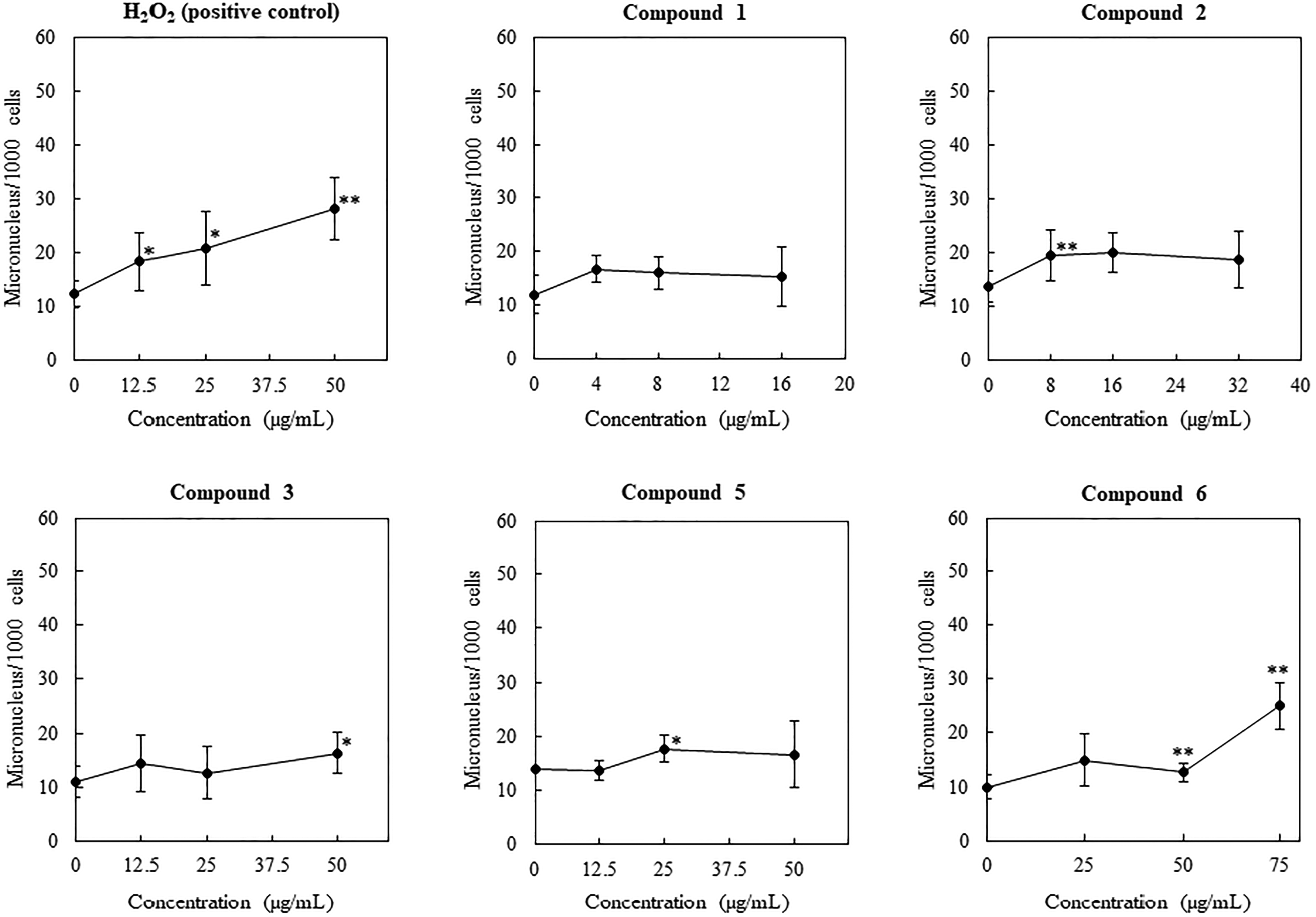
The MN assay was conducted to evaluate chromosomal aberrations in TK6 cells (Figure 5). The treatment of the TK6 cells with compound 1 did not increase the number of MN cells at concentrations of 4, 8, and 16 µg/mL. Compounds 2 and 6 exhibited significant differences in MN induction (P < .05). Compounds 3 and 5 showed significant increases in MN cells (P < .1). The treatment of the TK6 cells with compound 1 did not increase the number of MN cells.
Micronucleus induction resulting from treatment with compounds 1 to 3, 5, and 6. Each symbol represents the mean value of 3 experiments. An asterisk (*), 2 asterisks (**), and 3 asterisks (***) indicate P < .1, P < .05, and P < .01, respectively.
In conclusion, as a part of our chemical investigation of the Okinawan soft corals, a new diterpenoid, loba-8,10,13(15)-triene-14,17,18-triol 17-acetate (1), was isolated from a Lobophytum sp. collected from Itoman city, Okinawa, Japan. Compounds 1 to 3, 5, and 6 were screened for cytotoxicity and genotoxicity, including DNA and micronucleus induction, in the human lymphoblastoid cell line TK6.
Experimental
General: Optical rotations were measured using a JASCO P-1010 polarimeter, and NMR spectra on either a 500MHz Bruker AVANCE III spectrometer or a JEOL ECA 400 FT-NMR spectrometer using CDCl3 with tetramethylsilane as an internal standard. IR spectra were recorded on a JASCO FT/IR-6100. High-resolution mass spectra were acquired using a Waters SYNAPT HDMS. Semi-preparative high-performance liquid chromatography (HPLC) was performed on a Shimadzu HPLC system (CBM-20A system controller, LC-20AT binary pump, and SPD-20A UV/vis detector) with a COSMOSIL πNAP column (250mm × 10mm I.D.). Preparative thin-layer chromatography (TLC) was performed using silica gel glass plates (Merck, Kieselgel 60 F254). Column chromatography (CC) was performed using silica gel (Merck, Kieselgel 60, 70-230 mesh). TLC spots were visualized by UV light and/or by spraying with a 5% phosphomolybdic acid–ethanol solution, followed by heating at 90°C. Without purification, commercially available dehydrated pyridine (WAKO) and acetic anhydride (WAKO) were used for acetylation.
Biological material: A specimen of Lobophytum sp. was collected from Odo Coast, Itoman city, Okinawa, on April 23, 2017. The voucher specimen was deposited at the Faculty of Agriculture, University of the Ryukyus.
Extraction and isolation: Fresh soft coral (2.5 kg wet weight) was homogenized and extracted in MeOH at room temperature for 2 days. The resulting MeOH extract was concentrated in vacuo and partitioned into EtOAc/H2O. The EtOAc fraction (1.0 g) was subjected to CC, eluting with a gradient of n-hexane and EtOAc with increasing polarity. Fraction 2 (150.0 mg) was subjected to repeated preparative TLC with n-hexane–EtOAc (95:5) and toluene to yield compound 2 (21.3 mg). Fraction 4 (100.6 mg) was subjected to preparative TLC with n-hexane–EtOAc (2:1) to yield compound 3 (6.3 mg). The residue was further purified using chloroform–MeOH (99:1) to obtain compounds 4 (3.1 mg) and 5 (6.2 mg). Fraction 5 (150.6 mg) was subjected to repeated preparative TLC with n-hexane–EtOAc (1:1) and chloroform–MeOH (95:5) to yield compounds 1 (7.4 mg) and 6 (10.8 mg). Compound 1 was purified using a πNAP column, detection at a UV wavelength of 210 nm, and isocratic elution with 90% MeOH (0-60 min).
HRESIMS: m/z 365.2696 [M + H]+ (calculated for C22H37O4, 365.2692).
Acetylation: To compounds 1 (2.0 mg, 5.5 mmol) and 6 (1.8 mg, 5.6 mmol) in dehydrated pyridine (2.0 mL), acetic anhydride (2.0 mL, 21.2 mmol) was added. The mixtures were stirred at r.t under N2 for 12 h and then 10.0 mL of toluene was added to each one. The mixtures were poured into n-hexane (100 mL) and evaporated to yield acetylated compounds 1 (quant.) and 6 (quant.), respectively.
Cell culture: Human lymphoblastoid cell line TK6 was grown in an RPMI1640 medium supplemented with 100 U/mL penicillin, 100 µg/mL streptomycin, 0.25 µg/mL amphotericin B, 10% heat-inactivated fetal bovine serum, and 200 µg/mL sodium pyruvate. TK6 was incubated at 37°C in a 5% CO2 atmosphere at 100% humidity.
Cell survival: The cytotoxicity of the 5 compounds, excluding compound 4, on human lymphoblastoid TK6 cells were assessed by CCK-8 assay. Briefly, TK6 cells, after treatment with each compound, were seeded at a density of 5 × 10⁴ cells/well in 96-well plates. Afterwards, CCK-8 solution was added to each well. After 12 h of incubation, the optical density (OD) at 450 nm was measured.
Comet assay: The alkaline comet assay (single-cell gel electrophoresis) was conducted as previously described.16 TK6 cells were treated with various concentrations of each compound for 4 h. The treated cell suspension was centrifuged, washed, and suspended in 1.0 mL of phosphate-buffered saline (PBS). Thereafter, a 10 µL aliquot of the cell solution was mixed with 0.5% low-melting point agarose gel and spread on a MAS-coated microscope slide. Afterward, the agarose gel was allowed to solidify for 10 min at room temperature. The cells packaged in the agarose gel were lysed at 4°C overnight in 2.5 M NaCl, 100 mM ethylenediaminetetracetic acid disodium (EDTA–2Na), 10 mM Tris, 10% dimethyl sulfoxide (DMSO), and 1% TritonX-100 at pH 10. After cell lysis, the slides were transferred to an electrophoresis chamber filled with an ice-cold solution of 300 mM NaOH and 1 mM EDTA–2Na at a pH higher than 13 for 20 min to allow the DNA to unwind. Next, electrophoresis was performed for 15 min at 300 mA. Finally, the slides were neutralized for 5 min in 0.4 M Tris buffer (pH 7.5), rinsed with 99% EtOH for 5 min, and then air-dried. Subsequently, the cells were stained with ethidium bromide, and 100 cells were analyzed per concentration using a fluorescence microscope. The entire length of the comet (“migration”) was measured.
MN test: The MN test was conducted as previously described. Briefly, TK6 cells were treated with each compound at various concentrations for 4 h. After 48 h of treatment, approximately 1 × 10⁶ cells were suspended in 0.075 M KCl and incubated for 10 min at room temperature. The suspended cells were fixed in ice-cold methanol containing 25% acetic acid. Afterwards, the fixed cells were centrifuged and resuspended in ice-cold methanol containing 25% acetic acid. In the final fixation, the fixed cells were suspended in ice-cold methanol containing 1% acetic acid. A drop of fixing cell solution was spotted on glass slides, and the glasses were air-dried. The fixed cells were stained with acridine orange and analyzed under a fluorescence microscope.
Supplemental Material
sj-doc-1-npx-10.1177_1934578X221075978 - Supplemental material for New Marine Diterpenoid from the Okinawan Soft Coral, Lobophytum sp.
Supplemental material, sj-doc-1-npx-10.1177_1934578X221075978 for New Marine Diterpenoid from the Okinawan Soft Coral, Lobophytum sp. by Kosuke Sato, Shinnosuke Ishigami, Masaki Koike, Haruto Takegahara, Ayumi Yamamoto, Kensuke Kaneko, Kazuki Tani, Takahiro Ishii and Takashi Kamada in Natural Product Communications
Footnotes
Acknowledgments
This study was supported by JSPS KAKENHI (grant number 21K14904). The authors would like to thank Mr. Mirai Shigetake and Mr. Haruki Kakino (Shizuoka Institute of Science and Technology) for their kind support during the pretreatment experiments. We would like to thank Editage () for English language editing.
Declaration of Conflicting Interests
The authors declared no potential conflicts of interest with respect to the research, authorship, and/or publication of this article.
Funding
The authors disclosed receipt of the following financial support for the research, authorship, and/or publication of this article: This work was supported by the JSPS KAKENHI (grant number 21K14904).
Ethical Approval
Not applicable, because this article does not contain any studies with human or animal subjects.
Informed Consent
Not applicable, because this article does not contain any studies with human or animal subjects.
ORCID iD
Takashi Kamada
Trial Registration
Not applicable, because this article does not contain any clinical trials.
Supplemental Material
Supplemental material for this article is available online.
References
1.
PhanCSYeeCSVairappanCSIshiiTKamadaT. Sinulaflexiolide P, a cembrane-type diterpenoid from Bornean soft coral Sinularia flexibilis. Chem Nat Compd. 2019;55(2):285-288.
2.
KamadaTZanilIIPhanCSVairappanCS. A new cembrane from soft coral genus Sarcophyton in Borneo. Nat Prod Commun. 2018;13(2):123-124.
3.
NgocNTHanhTTHQuangTH, et al.Polyhydroxylated steroids from the Vietnamese soft coral Sarcophyton ehrenbergi. Steroids. 2021;176:108932.
4.
ChangyunWHaiyanLChanglunSYananWLiangLHuashiG. Chemical defensive substances of soft corals and gorgonians. Acta Ecol Sin. 2008;28(5):2320-2328.
5.
PhanCSKamadaTIshiiTHamadaTVairappanCS. A new guaiane-type sesquiterpenoid from a Bornean soft coral, Xenia stellifera. Nat Prod Commun. 2018;13(1):15-16.
6.
YinCTWenZHLanYHChangYCWuYCSungPJ. New anti-inflammatory norcembranoids from the soft coral Sinularia numerosa. Chem Pharm Bull. 2015;63(9):752-756.
7.
IshiiTKamadaTPhanCSVairappanCS. Chabrolene, a novel norditerpene from the Bornean soft coral Nephthea sp. Sains Malays. 2018;47(2):319-322.
8.
ChangCHAhmedAFYangTS, et al.Isolation of lobane and prenyleudesmane diterpenoids from the soft coral Lobophytum varium. Mar Drugs. 2020;18(4):223.
9.
EdradaRAProkschPWrayVWitteLvan OfwegenLV. Four new bioactive lobane diterpenes of the soft coral Lobophytum pauciflorum from Mindoro, Philippines. J Nat Prod. 1998;61(3):358-361.
10.
WrightADNielsonJLTapiolasDMLiptrotCHMottiCA. A Great Barrier Reef Sinularia sp yields two new cytotoxic diterpenes. Mar Drugs. 2012;10(8):1619-1630.
11.
PhanCSNgSYKamadaTVairappanCS. Two new lobane diterpenes from a Bornean soft coral Sinularia sp. Nat Prod Commun. 2016;11(7):899-900.
12.
KusumiTHamadaTIshitsukaMOOhtaniIKakisawaH. Elucidation of the relative and absolute stereochemistry of lobatriene, a marine diterpene, by a modified Mosher method. J Org Chem. 1992;57(3):1033-1035.
13.
DunlopRWWellsRJ. Isolation of some novel diterpenes from a soft coral of the genus Lobophytum. Aust J Chem. 1979;32(6):1345-1351.
14.
HamadaTKusumiTIshitsukaMOKakisawaH. Structures and absolute configurations of new lobane diterpenoids from Okinawan soft coral Sinularia flexibilis. Chem Lett. 1992;21(1):33-36.
15.
OhtaniIKusumiTKashmanYKakisawaH. High-field FT NMR application of Mosher’s method. The absolute configuration of marine terpenoids. J Am Chem Soc. 1991;113(11):4092-4096.
16.
YamamotoANakashimaKKawamoritaS, et al.Protective effects of raw and cooked blackcurrant extract on DNA damage induced by hydrogen peroxide in human lymphoblastoid cells. Pharm Biol. 2014;52(6):782-788.
Supplementary Material
Please find the following supplemental material available below.
For Open Access articles published under a Creative Commons License, all supplemental material carries the same license as the article it is associated with.
For non-Open Access articles published, all supplemental material carries a non-exclusive license, and permission requests for re-use of supplemental material or any part of supplemental material shall be sent directly to the copyright owner as specified in the copyright notice associated with the article.